Downloaded 205 times


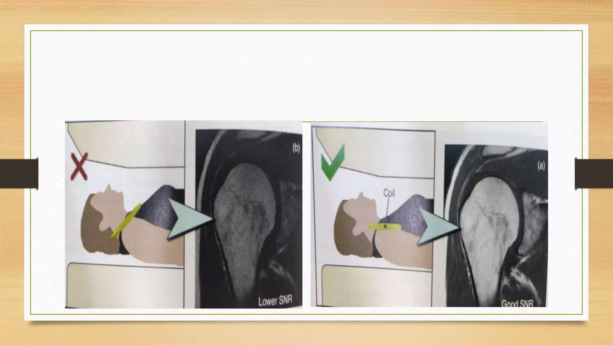


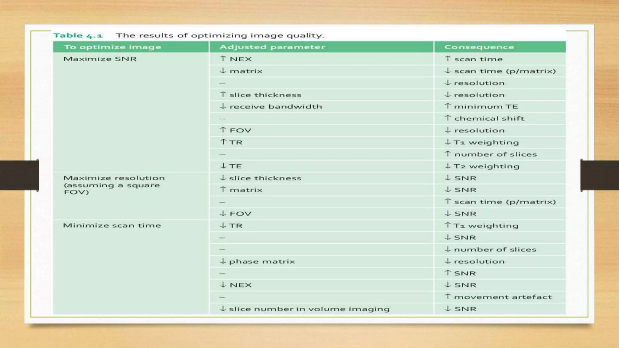
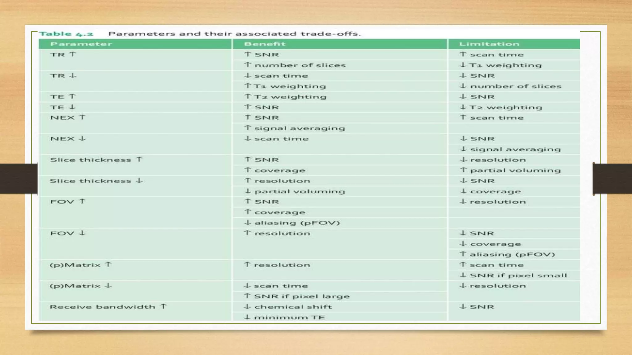
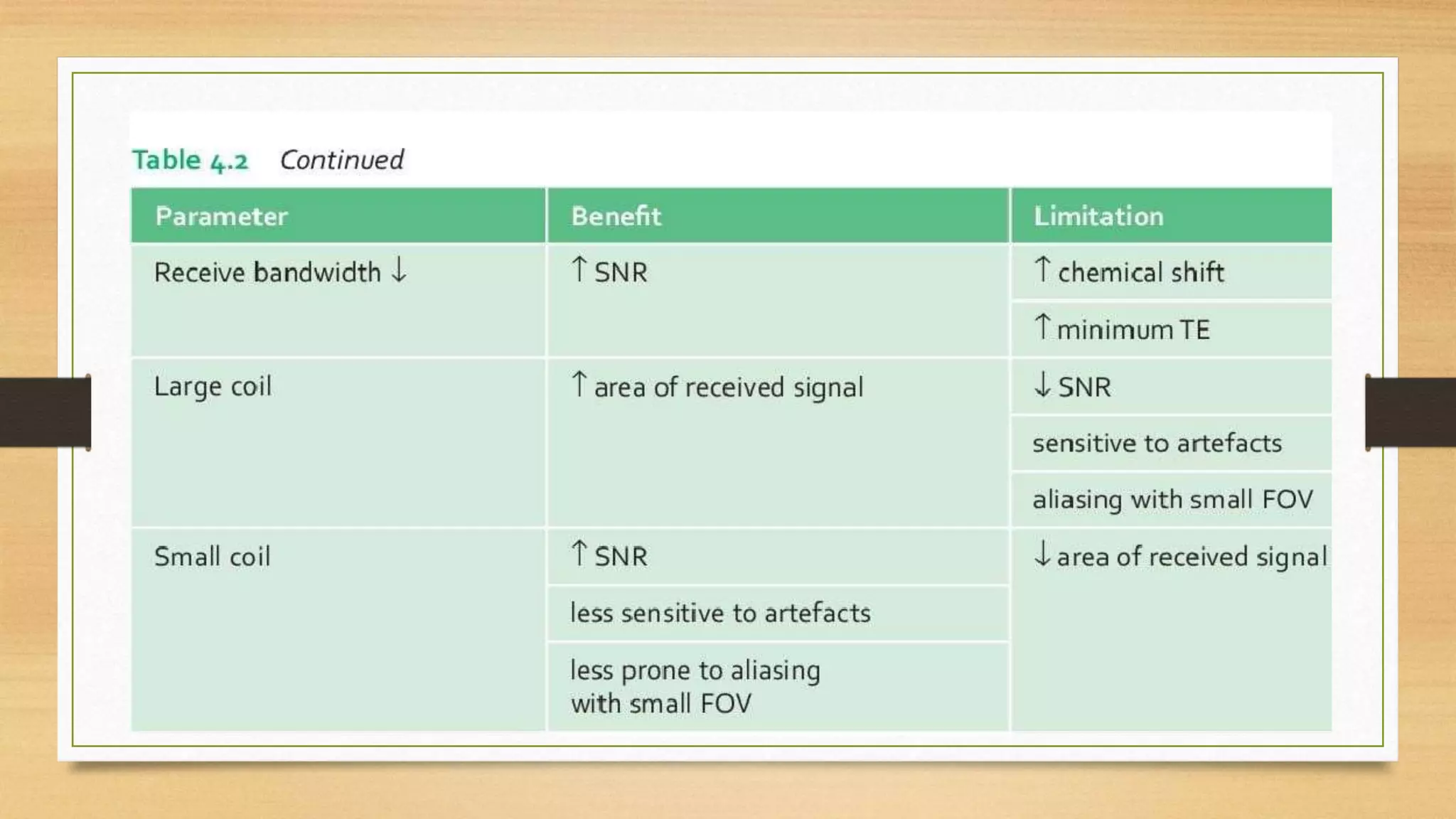
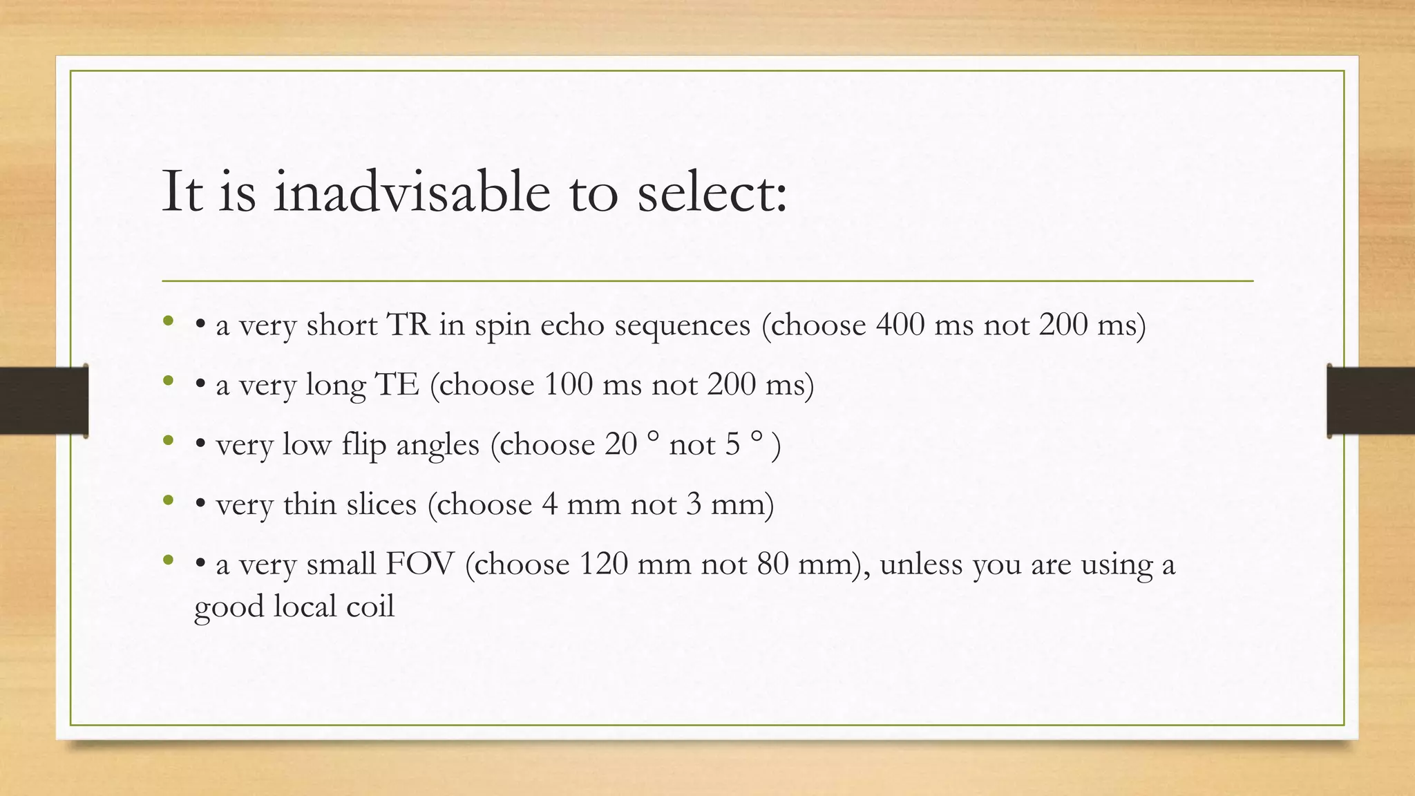


Optimizing MRI image quality requires balancing trade-offs between signal-to-noise ratio (SNR), contrast-to-noise ratio (CNR), spatial resolution, and scan time. SNR can be increased by using a long TR, short TE, large flip angle, and proper coil selection. CNR is improved by increasing signal from pathology or decreasing normal tissue signal. Spatial resolution is affected by slice thickness, field-of-view, and image matrix. Scan time depends on TR, phase matrix, and number of signal averages. No single parameter can be optimized alone without impacting others.


