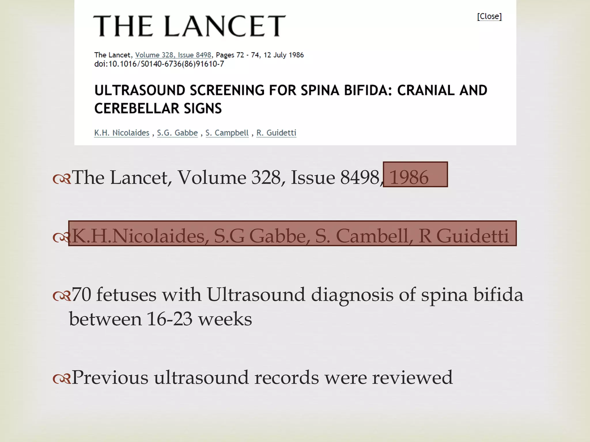
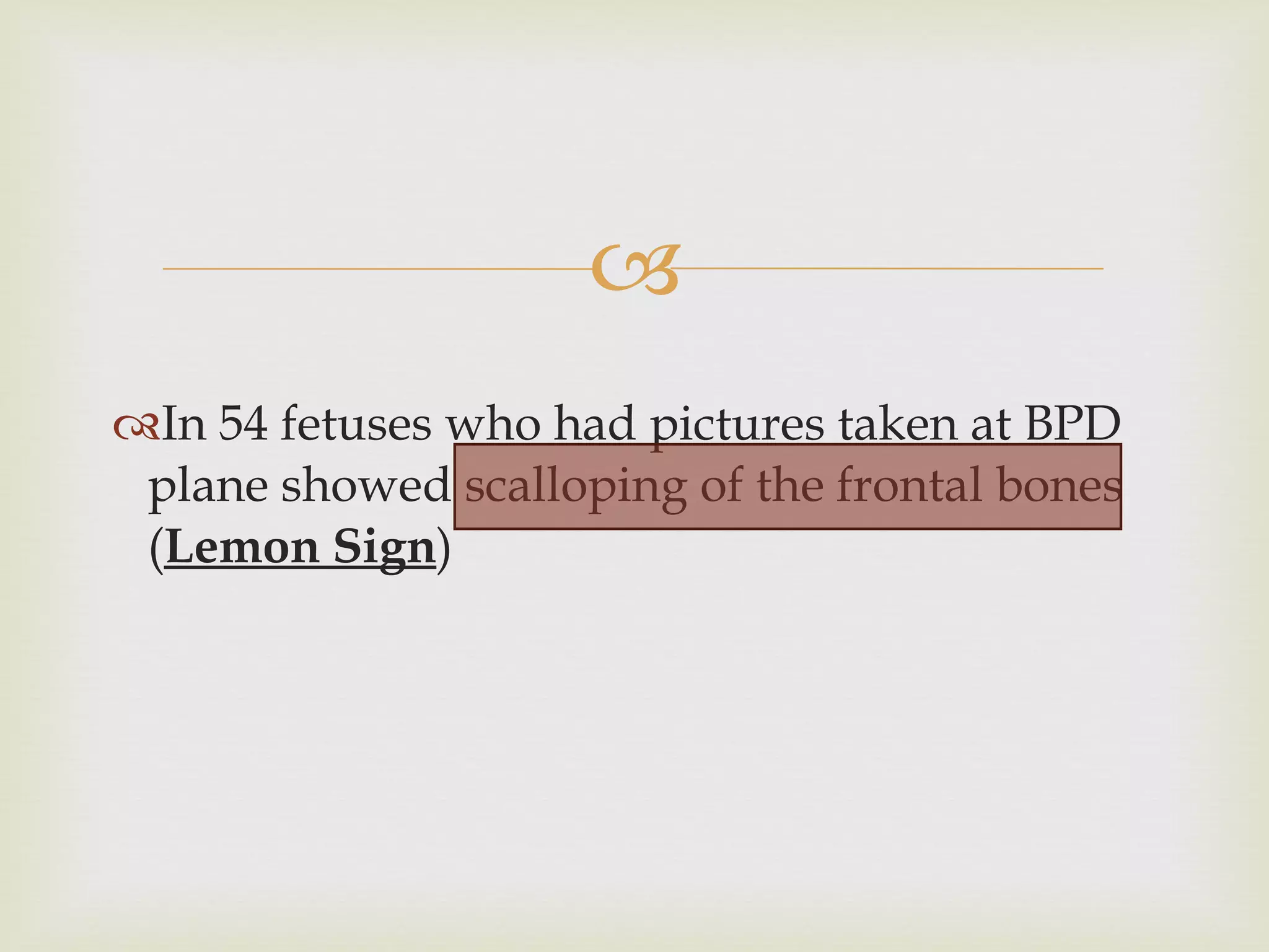
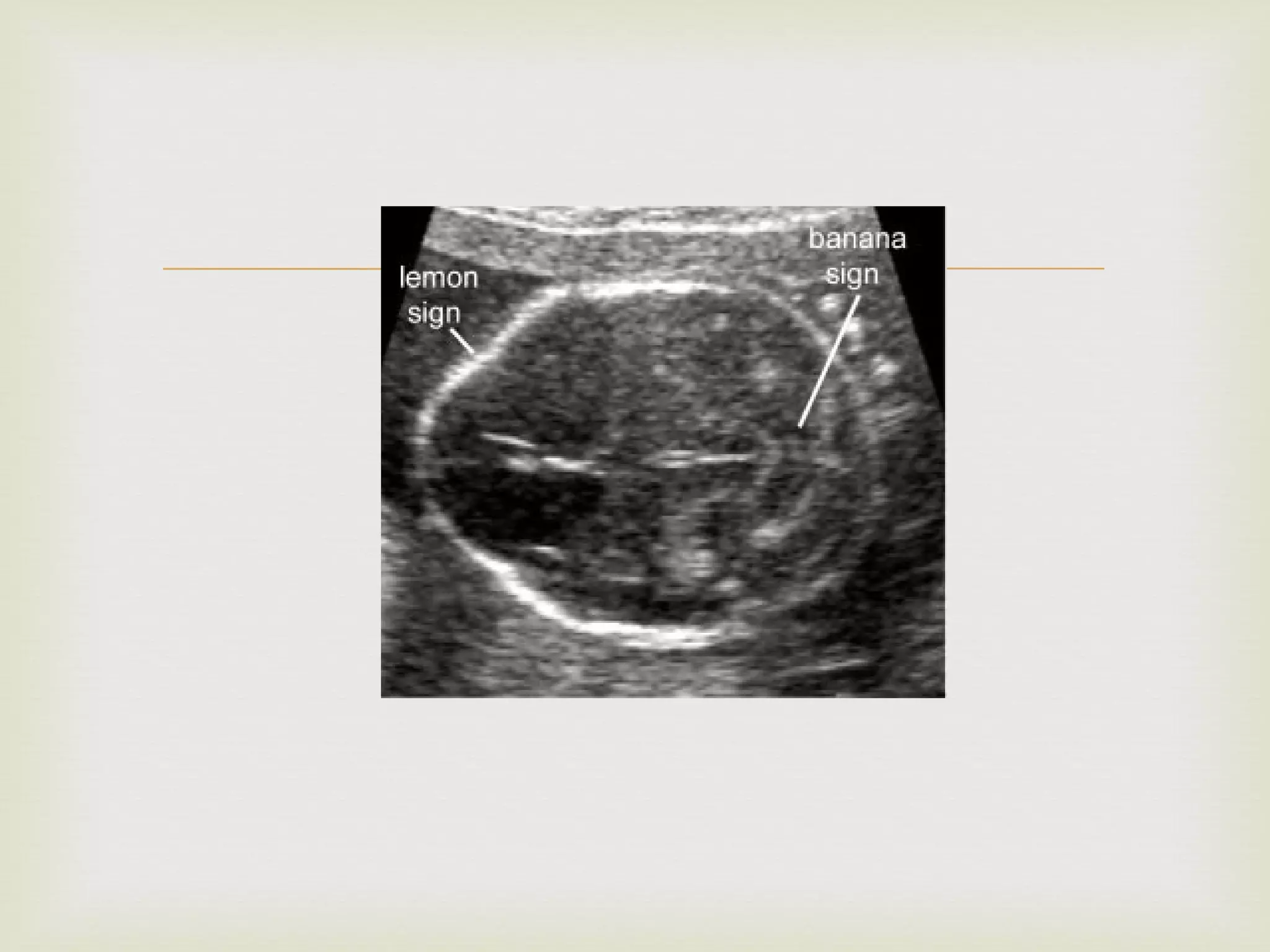
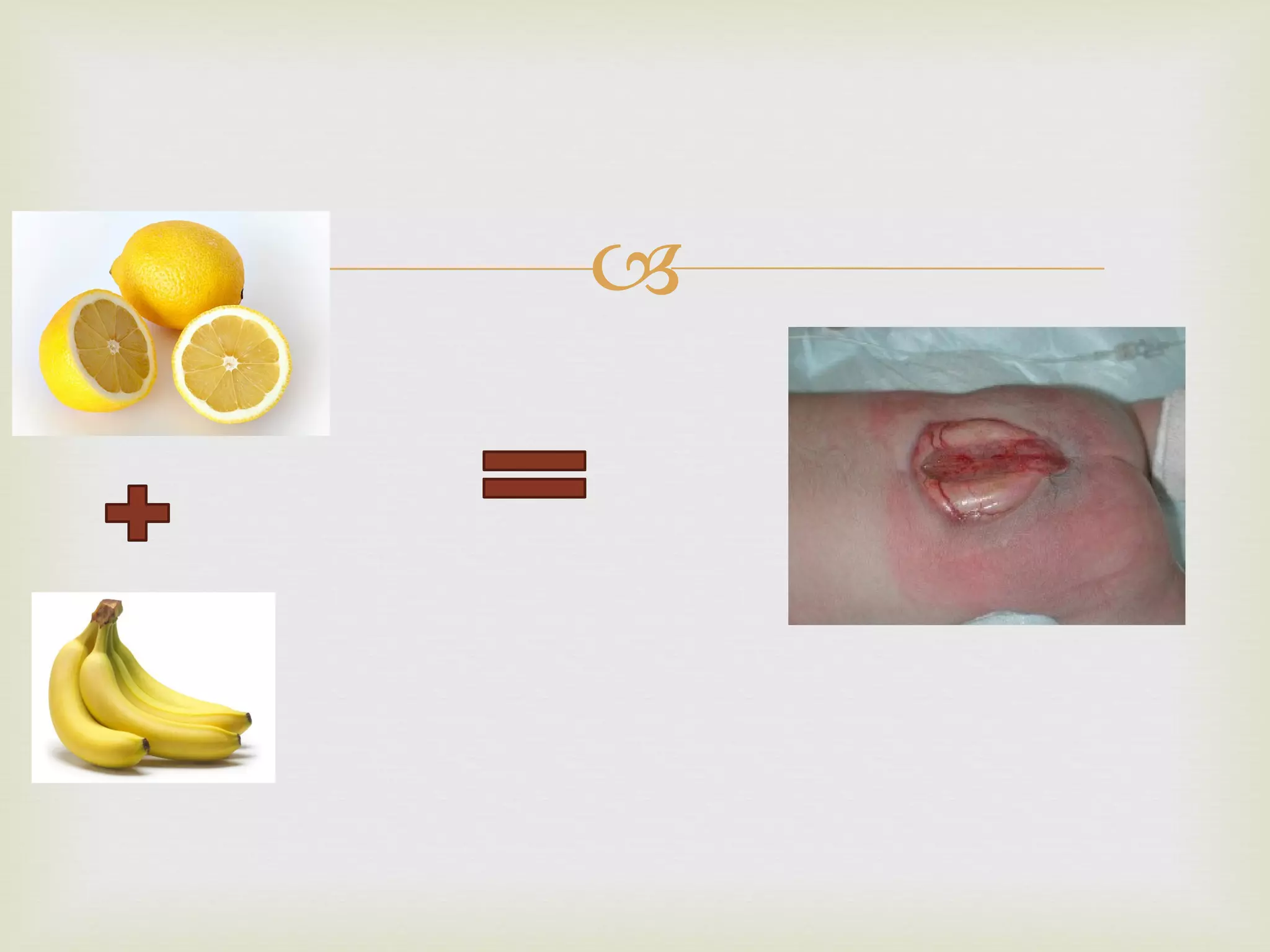
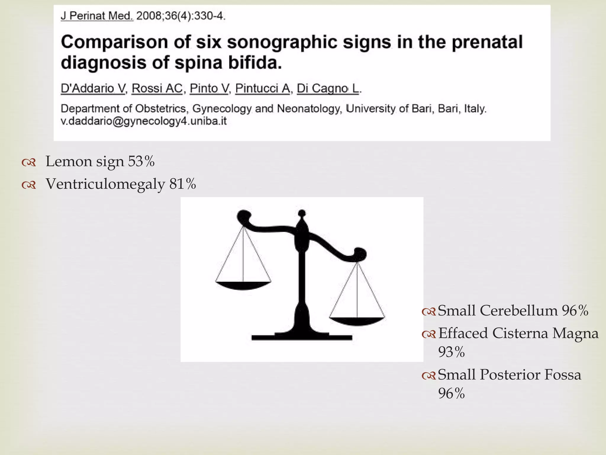
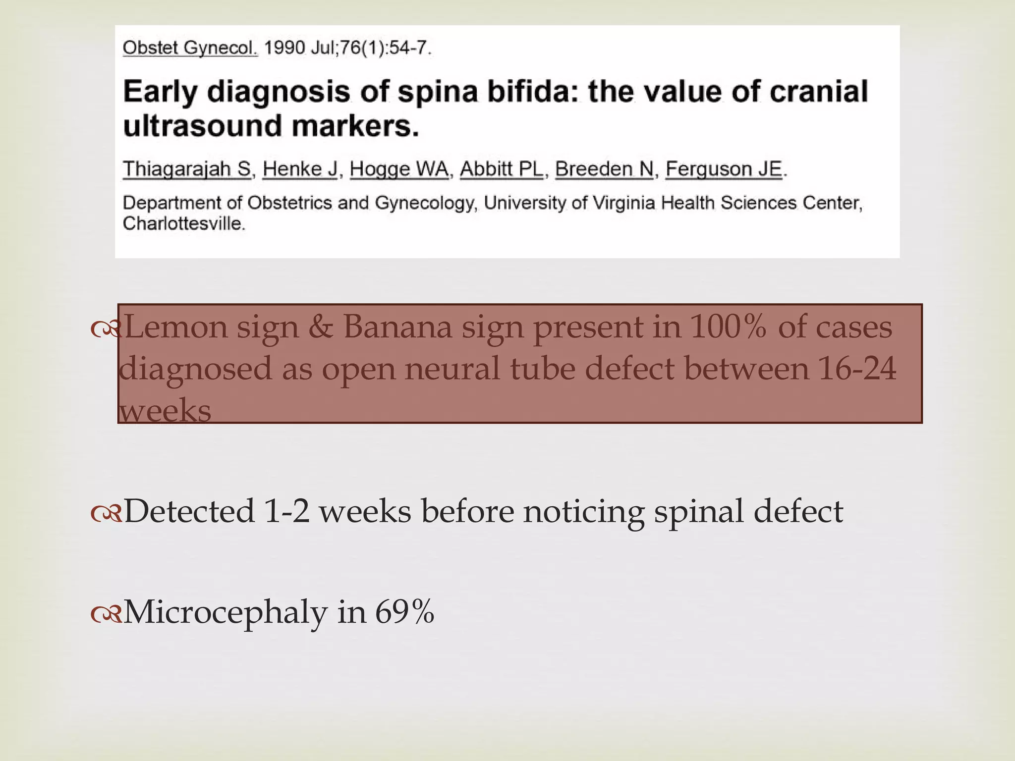
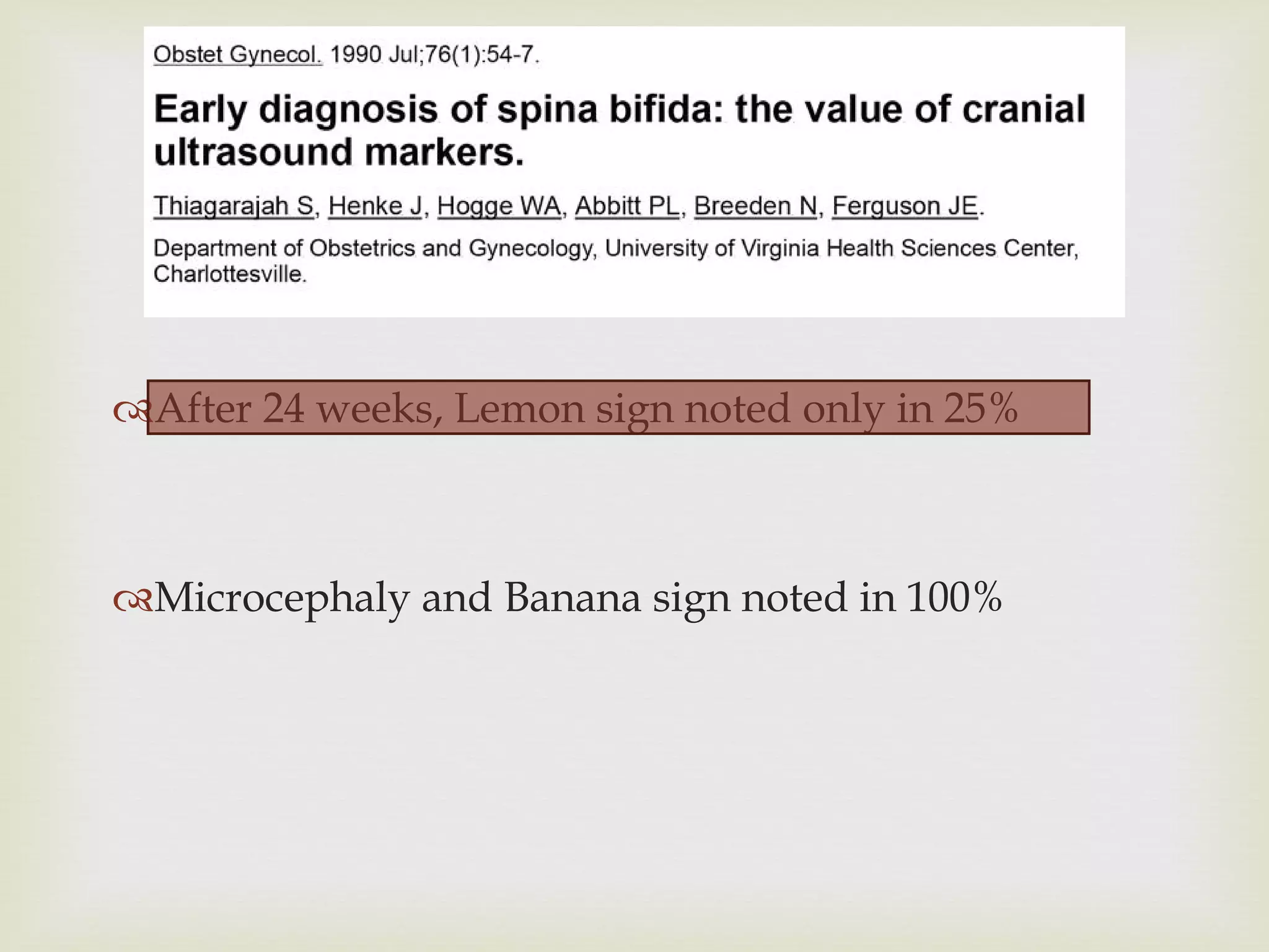
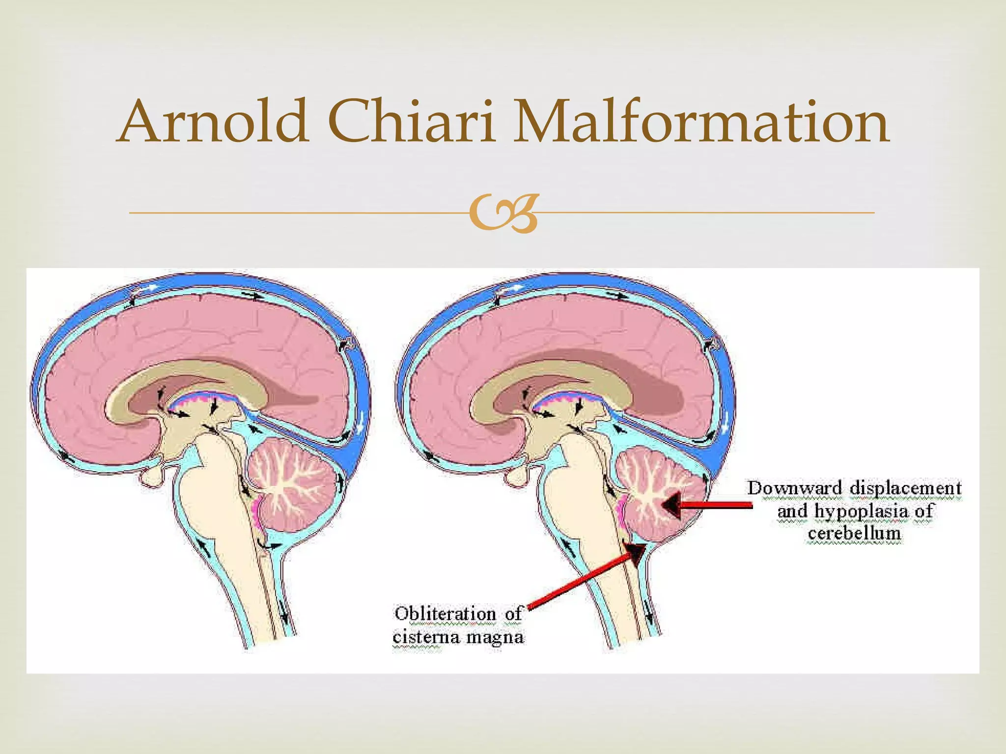
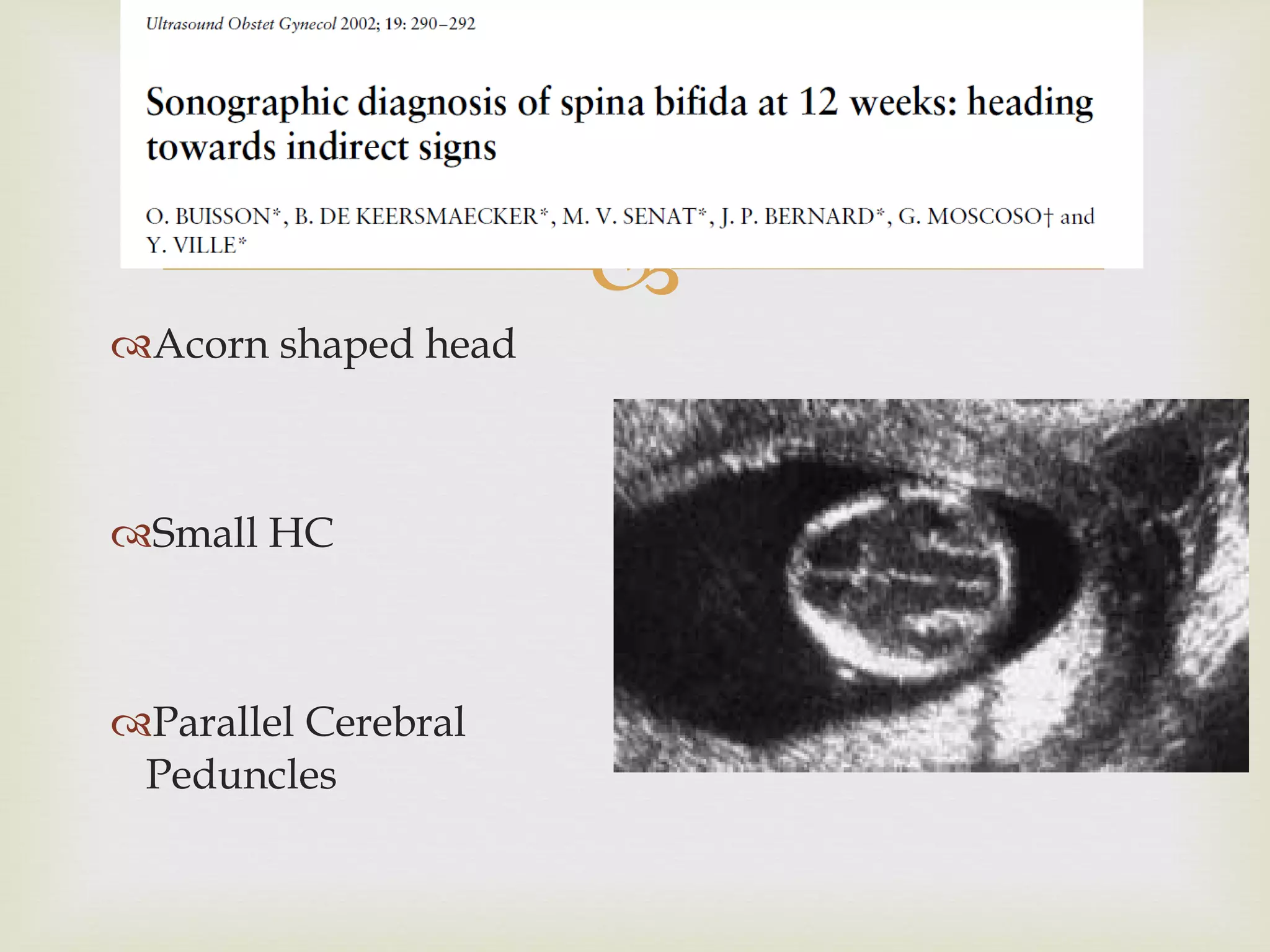
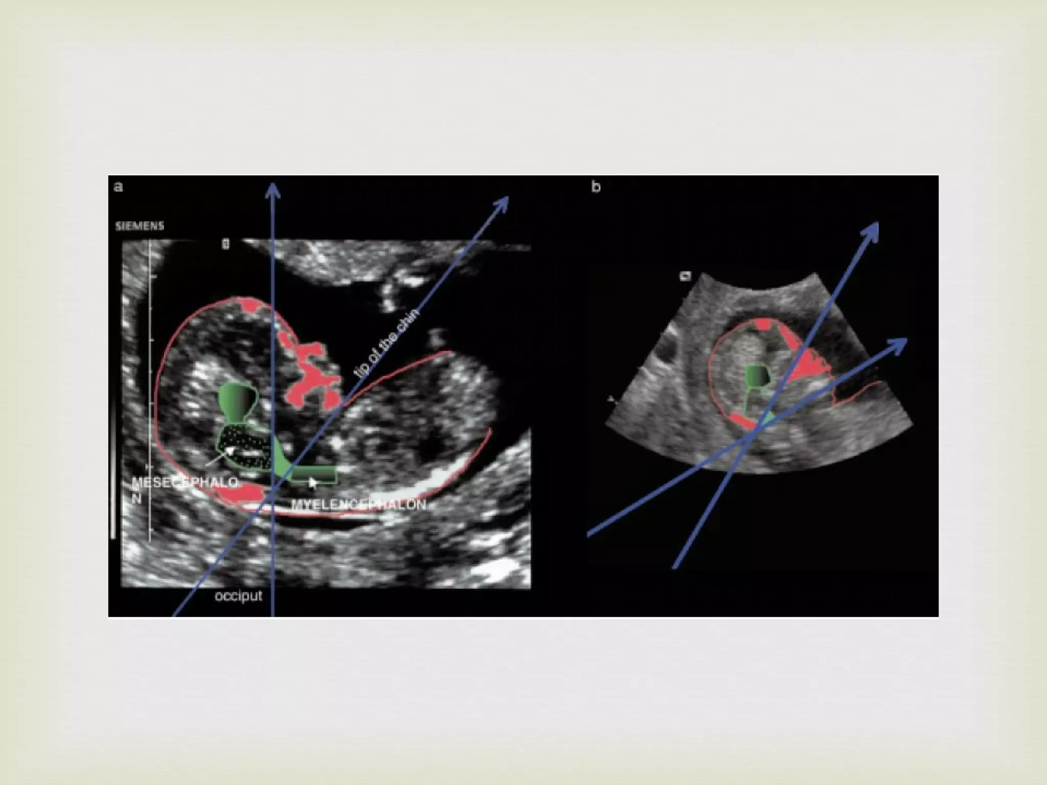
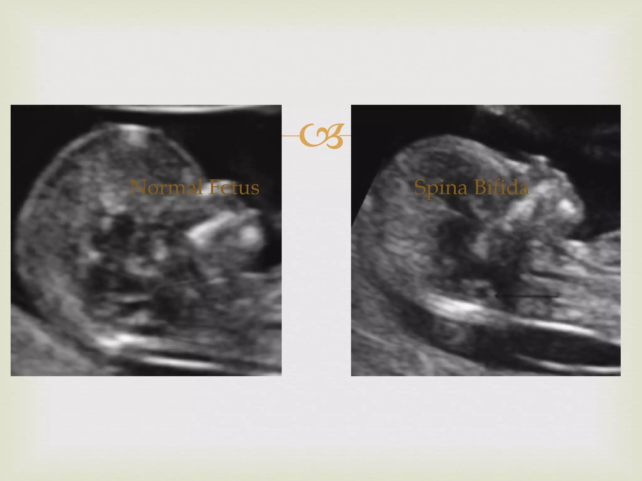
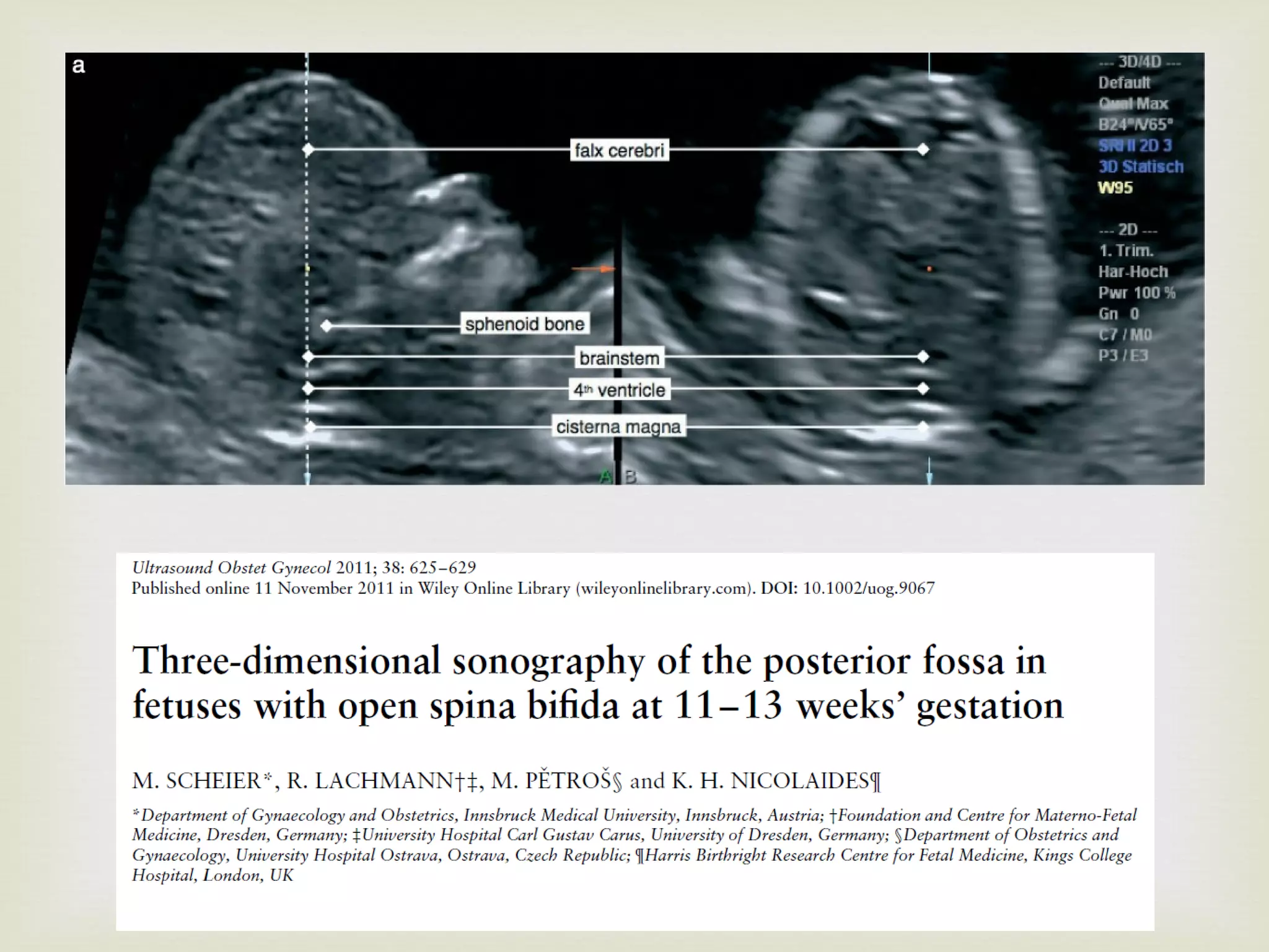
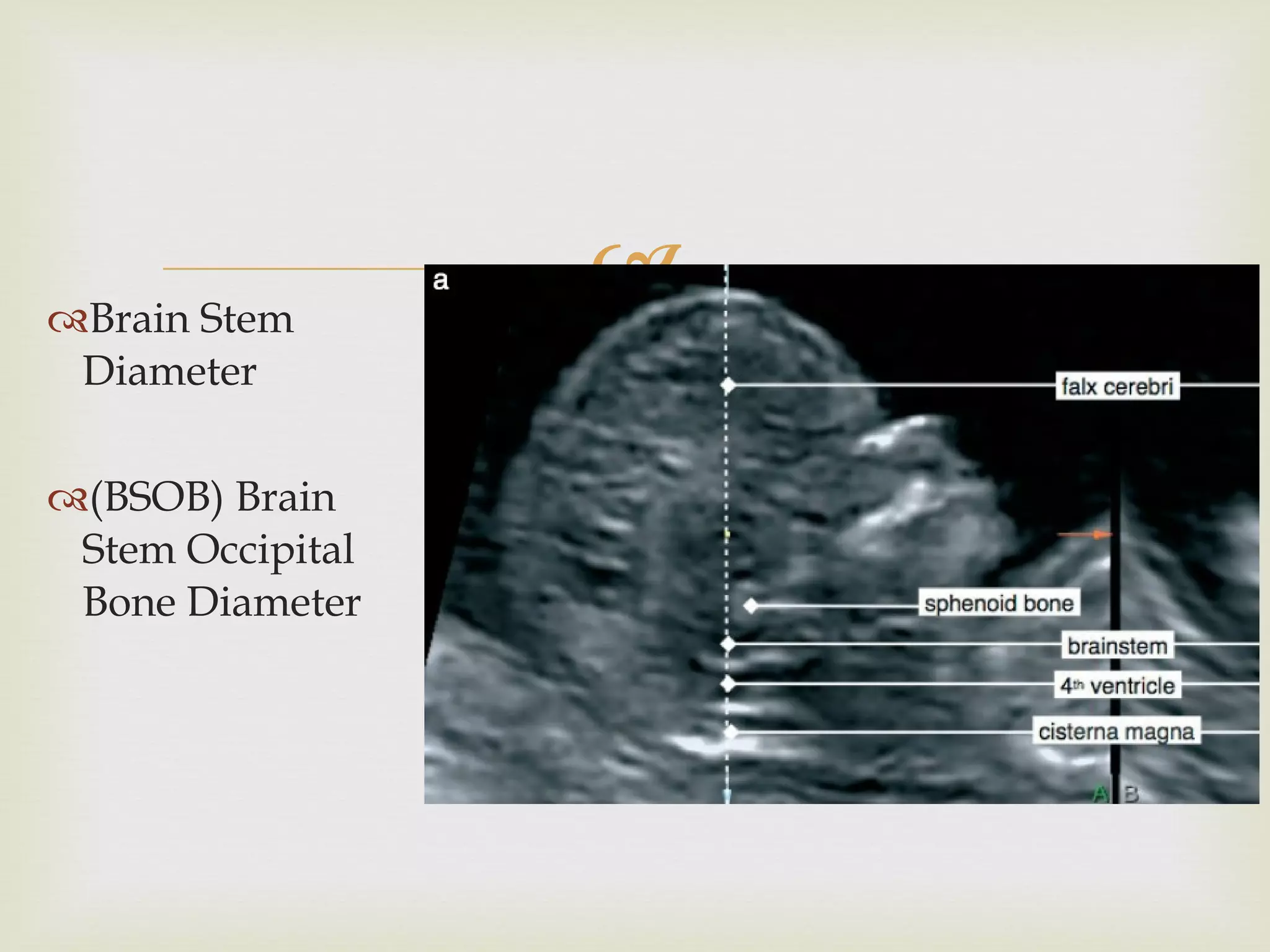
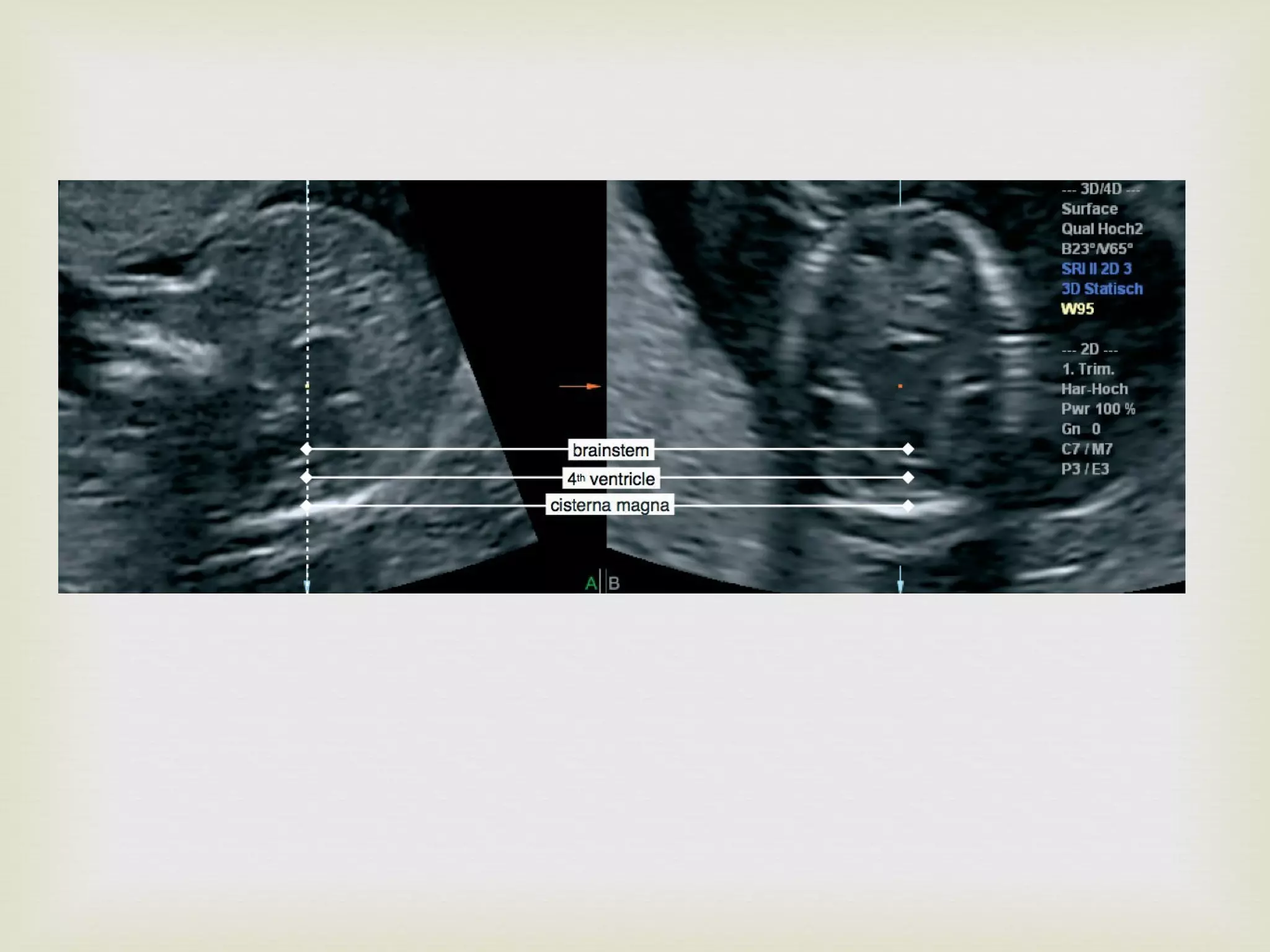
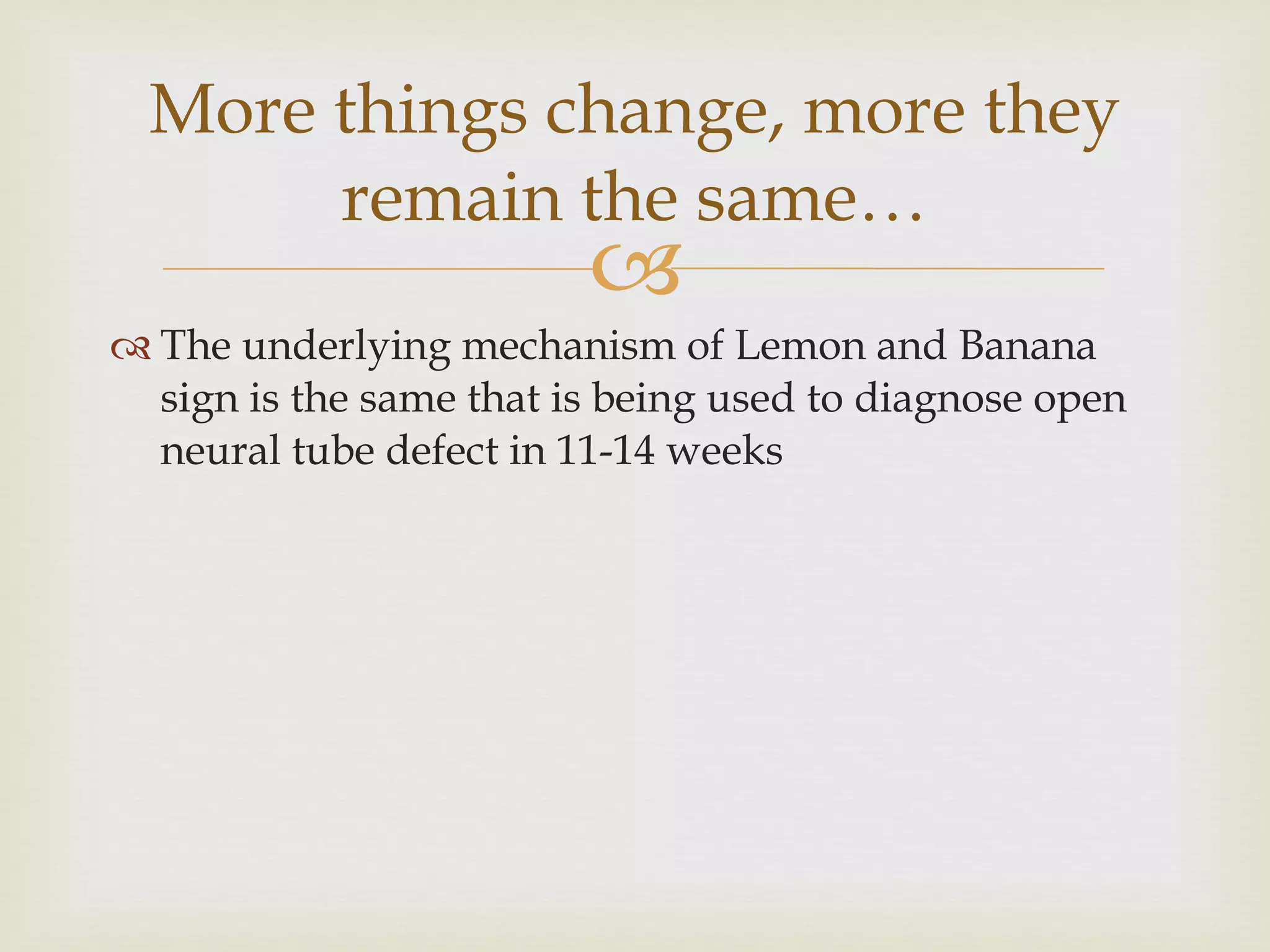
Dr.Sameer Dikshit MD, DGO,FCPS,FICOG Fetal Medicine Consultant, Mumbai
“ The eye only sees what the mind is prepared to comprehend” Henri Bergson, French Philosopher, 1927 Nobel prize in literature
 
The Lancet, Volume 328, Issue 8498, 1986 K.H.Nicolaides, S.G Gabbe, S. Cambell, R Guidetti 70 fetuses with Ultrasound diagnosis of spina bifida between 16-23 weeks Previous ultrasound records were reviewed
In 54 fetuses who had pictures taken at BPD plane showed scalloping of the frontal bones ( Lemon Sign )
In 12 of the 21 fetuses for which sub occipito bregnatic plane was taken, showed cerebellar hemispheres curved anteriorly with simultaneous obliteration of Cisterna Magna ( Banana Sign )
None of these changes were present in 100 normal  fetuses
 
 
Lemon sign 53% Ventriculomegaly 81% Small Cerebellum 96% Effaced Cisterna Magna 93% Small Posterior Fossa 96%
Spinal defect present but no CNS signs 0.04% 100% of the above were closed neural tube defects
Lemon sign & Banana sign present in 100% of cases diagnosed as open neural tube defect between 16-24 weeks Detected 1-2 weeks before noticing spinal defect Microcephaly in 69%
After 24 weeks, Lemon sign noted only in 25% Microcephaly and Banana sign noted in 100%
Evolution of Lemon sign and Banana sign
Open neural tube defect Leakage of CSF from the spinal defect Hypotension of Subarachnoid space Prolapse of cerebellum into the foramen magnum
Obstructive Hydrocephalus Abnormal Calvarial Development that is characteristic of the “Cranial Signs” Small posterior fossa
Arnold Chiari Malformation
Knowing earlier is always better than later !!!
Extrapolation of Arnold Chiari malformation in 1 st  Trimester
Acorn shaped head Small HC Parallel Cerebral Peduncles
 
The position of  parallel peduncles  is visible Visualisation of cerebellum  is difficult before 14 weeks  & may be confused with choroid plexus of the IV ventricle In sagittal view, the fetuses demonstrated a  flat occiput In axial view, the frontal bones are not convex but were retracted giving “acorn shape”
The contours of the metencephalon in spina bifida are straighter than normal This could be due to downward displacement of the brain
 
 
Normal Fetus Spina Bifida
1 st  Trimester Equivalent of Banana sign
Distance between posterior border of Aqueduct of Sylvius and anterior border of Occiput in Axial Plane AOS-to-Occiput Distance
 
In cases with spina bifida, juxtaposition of the midbrain to the occiput was a clue to the diagnosis In all the cases AOS-to-occiput distance was below the established normal range In contrast to subtle changes in the sagittal view, axial imaging of the midbrain reveals striking changes which can be quantified by AOS-to-occiput distance
 
First Trimester equivalent of Lemon sign
In fetuses with open spina bifida, at 11-13 weeks , the frontomaxillary facial angle is decreased and this measurement may be useful in early screening for this abnormality
Fronto Maxillo Facial Angle Spina Bifida, decreased angle Normal fetus, normal angle
This is due to caudal displacement of the fetal brain due to associated Arnold Chiarri malformation resulting in abnormal development of frontal bones This is thought to be the  underlying mechanism for producing “lemon sign”.
More technological refinement   3D USG
 
Brain Stem Diameter (BSOB) Brain Stem Occipital Bone Diameter
 
The underlying mechanism of Lemon and Banana sign is the same that is being used to diagnose open neural tube defect in 11-14 weeks More things change, more they remain the same…
Thank You

Lemon and Banana signs

  • 1.
    Dr.Sameer Dikshit MD,DGO,FCPS,FICOG Fetal Medicine Consultant, Mumbai
  • 2.
    “ The eyeonly sees what the mind is prepared to comprehend” Henri Bergson, French Philosopher, 1927 Nobel prize in literature
  • 3.
  • 4.
    The Lancet, Volume328, Issue 8498, 1986 K.H.Nicolaides, S.G Gabbe, S. Cambell, R Guidetti 70 fetuses with Ultrasound diagnosis of spina bifida between 16-23 weeks Previous ultrasound records were reviewed
  • 5.
    In 54 fetuseswho had pictures taken at BPD plane showed scalloping of the frontal bones ( Lemon Sign )
  • 6.
    In 12 ofthe 21 fetuses for which sub occipito bregnatic plane was taken, showed cerebellar hemispheres curved anteriorly with simultaneous obliteration of Cisterna Magna ( Banana Sign )
  • 7.
    None of thesechanges were present in 100 normal fetuses
  • 8.
  • 9.
  • 10.
    Lemon sign 53%Ventriculomegaly 81% Small Cerebellum 96% Effaced Cisterna Magna 93% Small Posterior Fossa 96%
  • 11.
    Spinal defect presentbut no CNS signs 0.04% 100% of the above were closed neural tube defects
  • 12.
    Lemon sign &Banana sign present in 100% of cases diagnosed as open neural tube defect between 16-24 weeks Detected 1-2 weeks before noticing spinal defect Microcephaly in 69%
  • 13.
    After 24 weeks,Lemon sign noted only in 25% Microcephaly and Banana sign noted in 100%
  • 14.
    Evolution of Lemonsign and Banana sign
  • 15.
    Open neural tubedefect Leakage of CSF from the spinal defect Hypotension of Subarachnoid space Prolapse of cerebellum into the foramen magnum
  • 16.
    Obstructive Hydrocephalus AbnormalCalvarial Development that is characteristic of the “Cranial Signs” Small posterior fossa
  • 17.
  • 18.
    Knowing earlier isalways better than later !!!
  • 19.
    Extrapolation of ArnoldChiari malformation in 1 st Trimester
  • 20.
    Acorn shaped headSmall HC Parallel Cerebral Peduncles
  • 21.
  • 22.
    The position of parallel peduncles is visible Visualisation of cerebellum is difficult before 14 weeks & may be confused with choroid plexus of the IV ventricle In sagittal view, the fetuses demonstrated a flat occiput In axial view, the frontal bones are not convex but were retracted giving “acorn shape”
  • 23.
    The contours ofthe metencephalon in spina bifida are straighter than normal This could be due to downward displacement of the brain
  • 24.
  • 25.
  • 26.
  • 27.
    1 st Trimester Equivalent of Banana sign
  • 28.
    Distance between posteriorborder of Aqueduct of Sylvius and anterior border of Occiput in Axial Plane AOS-to-Occiput Distance
  • 29.
  • 30.
    In cases withspina bifida, juxtaposition of the midbrain to the occiput was a clue to the diagnosis In all the cases AOS-to-occiput distance was below the established normal range In contrast to subtle changes in the sagittal view, axial imaging of the midbrain reveals striking changes which can be quantified by AOS-to-occiput distance
  • 31.
  • 32.
  • 33.
    In fetuses withopen spina bifida, at 11-13 weeks , the frontomaxillary facial angle is decreased and this measurement may be useful in early screening for this abnormality
  • 34.
    Fronto Maxillo FacialAngle Spina Bifida, decreased angle Normal fetus, normal angle
  • 35.
    This is dueto caudal displacement of the fetal brain due to associated Arnold Chiarri malformation resulting in abnormal development of frontal bones This is thought to be the underlying mechanism for producing “lemon sign”.
  • 36.
  • 37.
  • 38.
    Brain Stem Diameter(BSOB) Brain Stem Occipital Bone Diameter
  • 39.
  • 40.
    The underlying mechanismof Lemon and Banana sign is the same that is being used to diagnose open neural tube defect in 11-14 weeks More things change, more they remain the same…
  • 41.