![[object Object],[object Object],[object Object]](data:image/gif;base64,R0lGODlhAQABAIAAAAAAAP///yH5BAEAAAAALAAAAAABAAEAAAIBRAA7)
More Related Content
What's hot
What's hot (20)
Viewers also liked
Viewers also liked (20)
Similar to Pns New
Similar to Pns New (20)
Recently uploaded
Recently uploaded (20)
Pns New
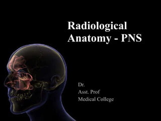
- 1. Radiological Anatomy - PNS Dr. Asst. Prof Medical College
- 5. WHERE ARE WE NOW…?
- 7. Changing Trends… The Gold Standard- CT Scan CT Scan MRI
- 23. BASAL LAMELLA
- 24. Pterygoid plates ppf Pt max fissure
- 26. Frontal Sinus
- 28. Frontal Recess
- 29. Uncinate Process
- 30. Terminal Recess
- 31. Frontal Cell I IV III II
- 32. Kero’s Classification I III II
- 33. Agger Nasi
- 34. Pneumatisation of crista galli
- 35. Pneumatisation of Uncinate Process
- 36. Ostiomeatal Complex ( OMC)
- 37. B/L CB, Lateralised UP, Narrow Infundibulum
- 38. Elongated UP
- 39. Uncinate Process
- 40. Haller Cell
- 43. Large bulla & concha bullosa narrowing infundibulum
- 45. Middle Turbinate
- 46. Middle Turbinate Part 1
- 47. Middle Turbinate Part 2
- 48. Middle Turbinate Part 3
- 49. Middle turbinate variants paradoxical interlamellar
- 50. Cocha Bullosa
- 51. Bulla Ethmoidalis Variants Absent bulla Bulla with multiple cells
- 52. Sinus Lateralis
- 53. Sinus Lateralis = Suprabullar recess and retrobullar recess
- 54. Maxillary Sinus
- 55. Height of Skullbase at Post.Ethmoid
- 57. Sphenoid Sinus
- 59. Pneumatisation
- 60. Anterior Clinoid Process Pneumatisn
- 61. Dehiscent Optic Nerve & ICA
- 62. Be careful…
- 72. Thank You
- 73. Haller cells multiple Large & flat
- 74. UP blocking EI