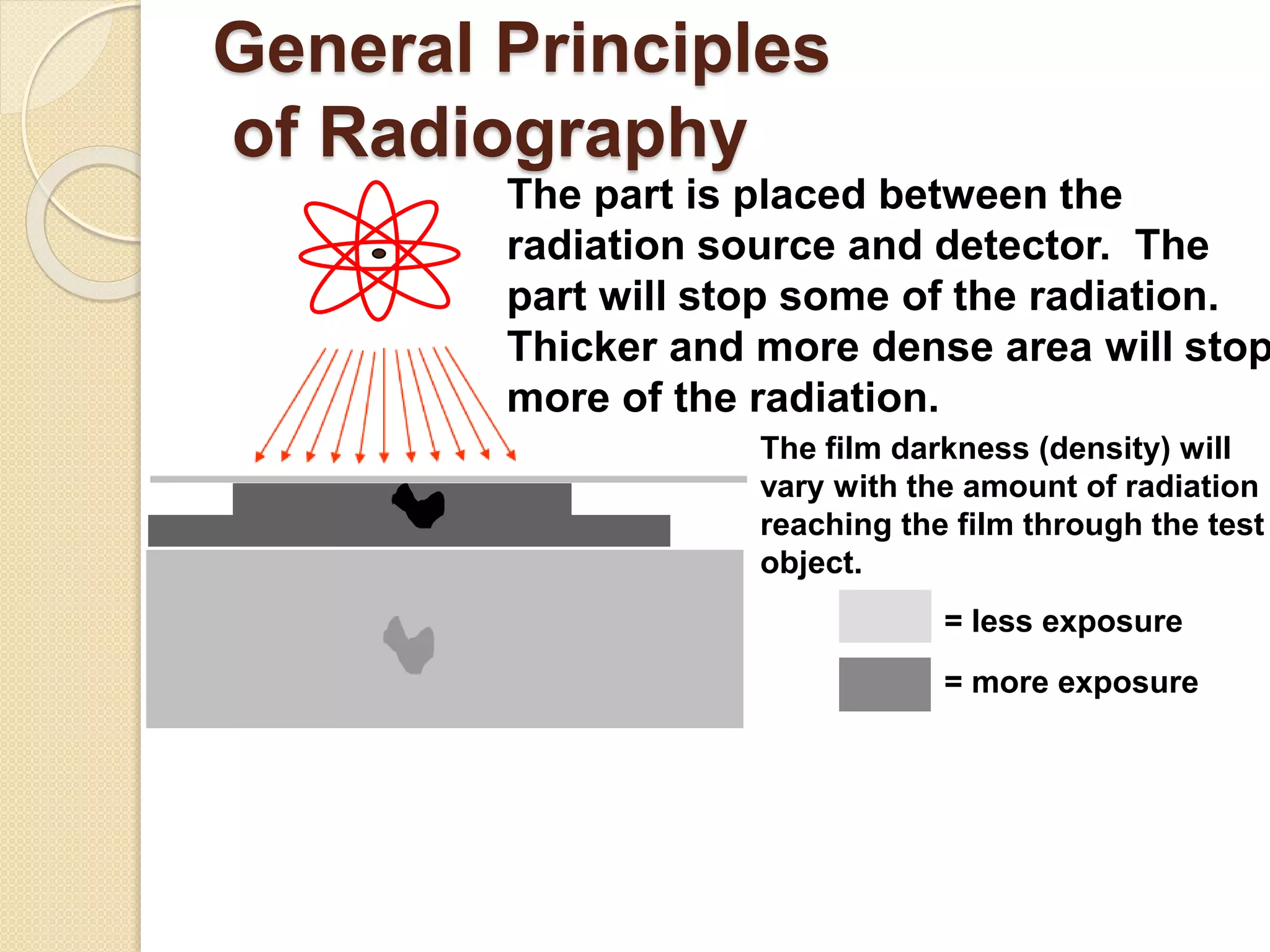
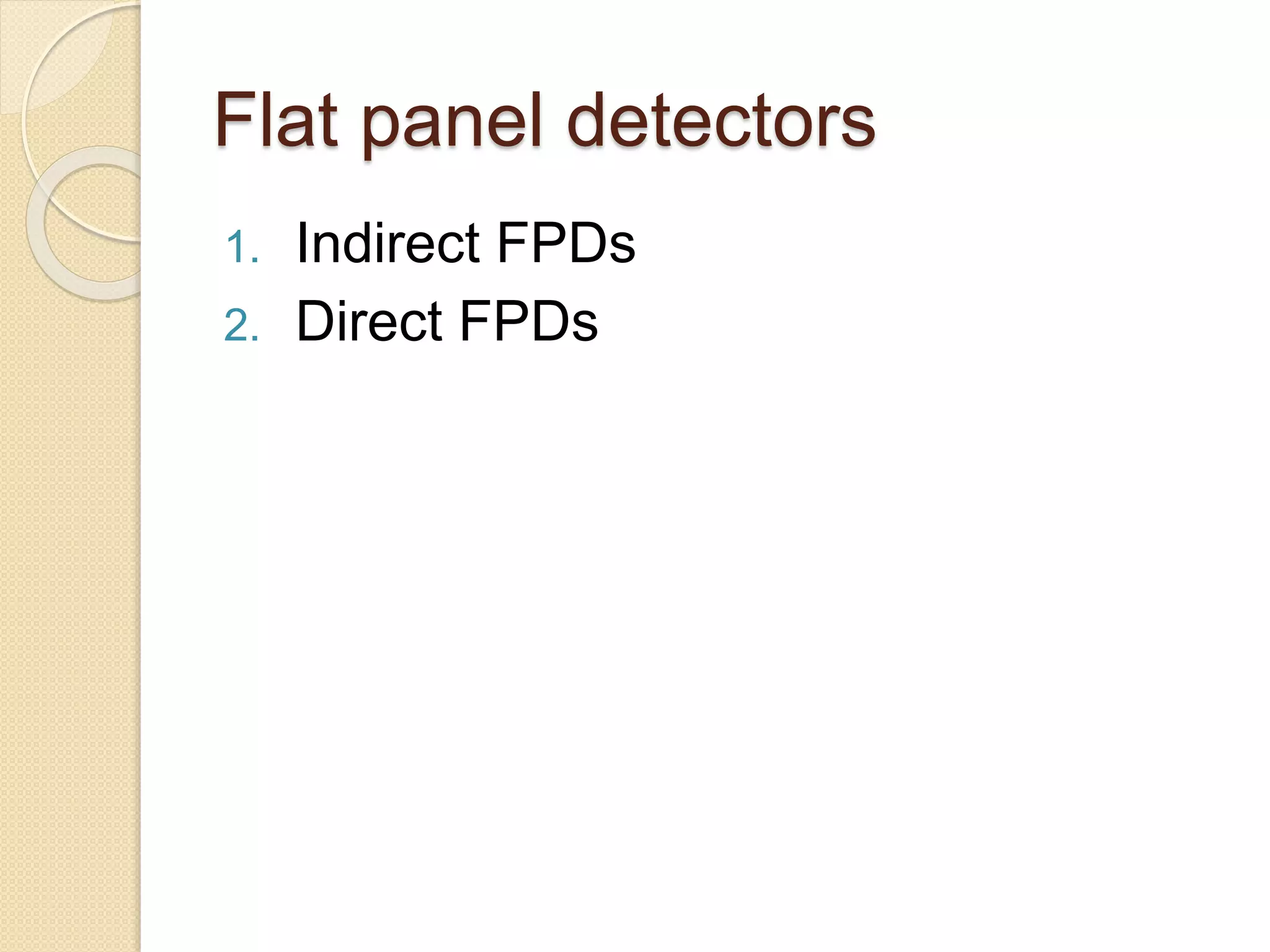
DIGITAL RADIOGRAPHY
PRESENTATED BY
AMBAR JYOTI HAZARIKA
BT/BME-13/03
OUTLINES
INTRODUCTION
HISTORY
GENERAL PRINCIPLE
DETECTORS
ADVANTAGE
DISADVANTAGE
CONCLUSION
What is radiography ?
 Radiography is an imaging technique
that’s uses electromagnetic radiation
other then visible light.
 Specially x-ray
 It is use to view internal structure of
human body
What Is Digital Radiography?
 Making radiographic images as
versatile as
pictures from digital camera
 Making radiographic images visible
and
storable on a personal computer or a
hospital-wide system
HISTORY
 The first digital radiography system
was developed by Fuji (Tokyo, Japan)
in 1980
General Principles
of Radiography
The part is placed between the
radiation source and detector. The
part will stop some of the radiation.
Thicker and more dense area will stop
more of the radiation.
The film darkness (density) will
vary with the amount of radiation
reaching the film through the test
object.
= less exposure
= more exposure
DR Overview
Detectors
1. Flat panel detectors(FPDs)
2. High density line-scan solid state
detectors.
Flat panel detectors
1. Indirect FPDs
2. Direct FPDs
Indirect FPDs
 Amorphous silicon(a-si) is most
common material of commercial
FPDs.
 Combining a-si detector with a
scintillator in the outer layer
 Which is made from caesium
iodide(Csl) or gadolinium
oxysulfide(Gd2o2s), converts x-ray to
light.
 Because of this conversion the a-si
detector considered an indirect image
 The light is channeled through the
amorphous silicon(a-si) photodiode
layer where it is converted to a digital
output signal.
 The digital signal read out by the
transistors
 Send to a computer for display.
Direct FPDs
 X-ray photon convert directly into the
charge.
 X-ray photon create electron hole
pairs in amorphous silicon(a-si) and
the transit of these electrons and
holes dependent on the potential of
the bias voltage charge
 As the holes are replaced with
electron
 The resultant charge pattern in the
High density line scan
detectors
 It is composed of a photostimulable
barium fluorobromide doped with
europium(BaFBr:Eu) or caesium
bromide(CsBr) phosphor.
 Phosphor detector records the x-ray
energy during exposure
 And scanned by laser diode to excite
the stored energy which is released
and read out by a digital image
Advantage
 Elimination of darkroom
 No chemical processing waste
 Elimination of radiography flim
 Instant viewing of image
 Less radiation dose to the patient
 Image can be send via email
 Save time
Disadvantage
 High cost
 Need a computer or network
Conclusion
 Digital radiography will change the
way technologists practice
radiography.
 However, it will not eliminate the need
for a quality education and an
understanding of radiology principles.
In fact, digital radiography will require
additional learning in order to
maximize its usefulness.
References
 https://www.aapm.org/meetings/04SS/
documents/yester.PDF
 http://iust.edu.sy/courses/DIGITAL%2
0RADIOGRAPHY.pdf
 Youtube videos
 http://radiopaedia.org/articles/flat-
panel-detectors
Digital Radiography

Digital Radiography

  • 1.
    DIGITAL RADIOGRAPHY PRESENTATED BY AMBARJYOTI HAZARIKA BT/BME-13/03
  • 2.
  • 3.
    What is radiography?  Radiography is an imaging technique that’s uses electromagnetic radiation other then visible light.  Specially x-ray  It is use to view internal structure of human body
  • 4.
    What Is DigitalRadiography?  Making radiographic images as versatile as pictures from digital camera  Making radiographic images visible and storable on a personal computer or a hospital-wide system
  • 5.
    HISTORY  The firstdigital radiography system was developed by Fuji (Tokyo, Japan) in 1980
  • 6.
    General Principles of Radiography Thepart is placed between the radiation source and detector. The part will stop some of the radiation. Thicker and more dense area will stop more of the radiation. The film darkness (density) will vary with the amount of radiation reaching the film through the test object. = less exposure = more exposure
  • 7.
  • 10.
    Detectors 1. Flat paneldetectors(FPDs) 2. High density line-scan solid state detectors.
  • 11.
    Flat panel detectors 1.Indirect FPDs 2. Direct FPDs
  • 12.
    Indirect FPDs  Amorphoussilicon(a-si) is most common material of commercial FPDs.  Combining a-si detector with a scintillator in the outer layer  Which is made from caesium iodide(Csl) or gadolinium oxysulfide(Gd2o2s), converts x-ray to light.  Because of this conversion the a-si detector considered an indirect image
  • 13.
     The lightis channeled through the amorphous silicon(a-si) photodiode layer where it is converted to a digital output signal.  The digital signal read out by the transistors  Send to a computer for display.
  • 14.
    Direct FPDs  X-rayphoton convert directly into the charge.  X-ray photon create electron hole pairs in amorphous silicon(a-si) and the transit of these electrons and holes dependent on the potential of the bias voltage charge  As the holes are replaced with electron  The resultant charge pattern in the
  • 15.
    High density linescan detectors  It is composed of a photostimulable barium fluorobromide doped with europium(BaFBr:Eu) or caesium bromide(CsBr) phosphor.  Phosphor detector records the x-ray energy during exposure  And scanned by laser diode to excite the stored energy which is released and read out by a digital image
  • 16.
    Advantage  Elimination ofdarkroom  No chemical processing waste  Elimination of radiography flim  Instant viewing of image  Less radiation dose to the patient  Image can be send via email  Save time
  • 17.
    Disadvantage  High cost Need a computer or network
  • 18.
    Conclusion  Digital radiographywill change the way technologists practice radiography.  However, it will not eliminate the need for a quality education and an understanding of radiology principles. In fact, digital radiography will require additional learning in order to maximize its usefulness.
  • 19.