Endodontics dr shabeel pn www.hi-dentfinishingschool.blogspot.com
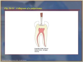
Endodontics  is the specialty of dentistry that manages the prevention, diagnosis, and treatment of the dental pulp and the  periradicular  tissues that surround the root of the tooth.   Introduction
Physical irritation   Most generally brought on by extensive decay. Trauma   Blow to a tooth or the jaw.  Causes of Pulpal Nerve Damage
Pain when biting down. Pain when chewing.  Sensitivity with hot or cold beverages.  Facial swelling.  Signs and Symptoms of Pulpal Nerve Damage
Subjective examination Chief complaint  Character and duration of pain  Painful stimuli  Sensitivity to biting and pressure  Endodontic Diagnosis
Objective examination Extent of decay   Periodontal conditions surrounding the tooth in question   Presence of an extensive restoration  Tooth mobility  Swelling or discoloration  Pulp exposure  Endodontic Diagnosis   cont’d
Percussion tests   Used to determine whether the inflammatory process has extended into the periapical tissues.   Completed by the dentist tapping on the incisal or occlusal surface of the tooth in question with the end of the mouth mirror handle held parallel to the long axis of the tooth.   Diagnostic Testing
Palpation tests Used to determine whether the inflammatory process has extended into the periapical tissues. The dentist applies firm pressure to the mucosa above the apex of the root.  Diagnostic Testing   cont’d
Thermal sensitivity Necrotic pulp will  not  respond to cold or hot. Cold test Ice, dry ice, or ethyl chloride used to determine the response of a tooth to cold. Heat test Piece of  gutta-percha  or instrument handle heated and applied to the facial surface of the tooth.   Diagnostic Testing   cont’d
Electric pulp testing Delivers a small electrical stimulus to the pulp. Factors that may influence readings: Teeth with extensive restorations. Teeth with more than  one canal.   Failing pulp can produce a variety of responses.   Control teeth may not respond as anticipated.   Moisture on the tooth during testing. Batteries in the tester may be weak.  Diagnostic Testing   cont’d
Fig. 54-4  Placement of a pulp tester.
Initial radiograph   Diagnosis.   Working length film   Used to determine the length of the canal.   Final instrumentation film   Taken with the final size files in all canals.   Root canal completion film   Taken after the tooth as been temporized.   Recall films   Taken at evaluations.  Radiographs in Endodontics
Show 4-5 mm beyond the apex of the tooth and the surrounding bone or pathologic condition.   Present an accurate image of the tooth without elongation or fore-shortening.   Exhibit good contrast so all pertinent structures are readily identifiable.   Requirements of Endodontic Films
Fig. 54-5  Quality radiograph in endodontics.
Normal pulp There are no subjective symptoms or objective signs. The tooth responds normally to sensory stimuli, and a healthy layer of dentin surrounds the pulp. Diagnostic Conclusions
Pulpitis  The pulp tissues have become inflamed. Reversible pulpitis The pulp is irritated, and the patient is experiencing pain to thermal stimuli.   Irreversible pulpitis The tooth will display symptoms of lingering pain.   Diagnostic Conclusions   cont’d
Periradicular abscess An inflammatory reaction to pulpal infection that can be chronic or have rapid onset with pain, tenderness of the tooth to pressure, pus formation, and swelling of the tissues.  Diagnostic Conclusions   cont’d
Periodontal abscess   An inflammatory reaction frequently caused by bacteria entrapped in the periodontal sulcus.  A patient will experience rapid onset, pain, tenderness of the tooth to pressure, pus formation, and swelling.   Diagnostic Conclusions   cont’d
Periradicular cyst A cyst that develops at or near the root of a necrotic tooth.  These types of cysts develop as an inflammatory response to pulpal infection and necrosis of the pulp.   Diagnostic Conclusions   cont’d
Pulp fibrosis The decrease of living cells within the pulp causing fibrous tissue to take over the pulpal canal.   Diagnostic Conclusions   cont’d
Necrotic tooth Also referred to as  nonvital .   Used to describe a tooth that does not respond to sensory stimulus.   Diagnostic Conclusions   cont’d
Pulp capping A covering of calcium hydroxide is placed over an exposed or nearly exposed pulp to encourage the formation of irritated dentin at the site of injury.  Indirect pulp cap  is indicated when a thin partition of dentin is still intact. Direct pulp cap  is indicated when the pulp has been slightly exposed.  Endodontic Procedures
Fig. 54-11  Spreader and plunger.   
Pulpotomy   Involves the removal of the coronal portion of an exposed vital pulp. Completed to preserve the vitality of the remaining portion of the pulp within the root of the tooth.  This procedure is commonly indicated for vital primary teeth, teeth with deep carious lesions, and emergency situations.  Endodontic Procedures   cont’d
Fig. 54-13  Example of a pulpotomy.
Pulpectomy   Also referred to as  root canal therapy ;  procedure involves the complete removal of the dental pulp.   Endodontic Procedures   cont’d
Fig. 54-14  A diagram of a pulpectomy.
Endodontic explorer   Endodontic spoon excavator   Broaches Endodontic files K-type Hedstrom Instruments and Accessories for Endodontic Procedures
Table 54‑1  Colors and Sizes of Endodontic Files
Rubber stops   Paper points   Spreaders  Pluggers   Glick No. 1 Millimeter ruler  Instruments and Accessories for Endodontic Procedures   cont’d
Rotary instruments  Gates-Glidden bur  Pesso reamer   Lentulo spiral  Instruments and Accessories for Endodontic Procedures   cont’d
Irrigation solution   Sodium hypochlorite   Hydrogen peroxide  Parachlorophenol (PCP) Medicaments and Dental Materials in Endodontics
Gutta-percha points Formocresol  Root canal sealer   Medicaments and Dental Materials in Endodontics   cont’d
Anesthesia and pain control Isolation and disinfection of the site Access preparation Debridement and shaping the canal  Obturation  Overview of Root Canal Therapy
Indications for surgical intervention Endodontic failure  caused by persistent infection, severely curved roots, perforation of the canal, fractured roots, extensive root resorption, pulp stones, or accessory canals that cannot be treated. Exploratory surgery  to determine why healing has not occurred.   Biopsy  Surgical Endodontics
To surgically remove the apical portion of the root with the use of a high‑speed handpiece and bur. To evaluate: Inadequate sealing of the canal.  Accessory canals.  Fractures of the root.  Pathological tissue around the root apex.  Apicoectomy and Apical Curettage
Completed when an apical seal is not adequate. A small class I preparation is made at the apex and sealed with filling materials such as gutta-percha, amalgam, or composite.   Retrograde Restoration
Root amputation A surgery performed to remove one or more roots of a multirooted tooth without removing the crown.   Hemisection A procedure in which the root and the crown are cut lengthwise and removed.   Root Amputation and Hemisection

endodontics

  • 1.
    Endodontics dr shabeelpn www.hi-dentfinishingschool.blogspot.com
  • 2.
    Endodontics isthe specialty of dentistry that manages the prevention, diagnosis, and treatment of the dental pulp and the periradicular tissues that surround the root of the tooth. Introduction
  • 3.
    Physical irritation Most generally brought on by extensive decay. Trauma Blow to a tooth or the jaw. Causes of Pulpal Nerve Damage
  • 4.
    Pain when bitingdown. Pain when chewing. Sensitivity with hot or cold beverages. Facial swelling. Signs and Symptoms of Pulpal Nerve Damage
  • 5.
    Subjective examination Chiefcomplaint Character and duration of pain Painful stimuli Sensitivity to biting and pressure Endodontic Diagnosis
  • 6.
    Objective examination Extentof decay Periodontal conditions surrounding the tooth in question Presence of an extensive restoration Tooth mobility Swelling or discoloration Pulp exposure Endodontic Diagnosis  cont’d
  • 7.
    Percussion tests Used to determine whether the inflammatory process has extended into the periapical tissues. Completed by the dentist tapping on the incisal or occlusal surface of the tooth in question with the end of the mouth mirror handle held parallel to the long axis of the tooth. Diagnostic Testing
  • 8.
    Palpation tests Usedto determine whether the inflammatory process has extended into the periapical tissues. The dentist applies firm pressure to the mucosa above the apex of the root. Diagnostic Testing  cont’d
  • 9.
    Thermal sensitivity Necroticpulp will not respond to cold or hot. Cold test Ice, dry ice, or ethyl chloride used to determine the response of a tooth to cold. Heat test Piece of gutta-percha or instrument handle heated and applied to the facial surface of the tooth. Diagnostic Testing  cont’d
  • 10.
    Electric pulp testingDelivers a small electrical stimulus to the pulp. Factors that may influence readings: Teeth with extensive restorations. Teeth with more than one canal. Failing pulp can produce a variety of responses. Control teeth may not respond as anticipated. Moisture on the tooth during testing. Batteries in the tester may be weak. Diagnostic Testing  cont’d
  • 11.
    Fig. 54-4 Placement of a pulp tester.
  • 12.
    Initial radiograph Diagnosis. Working length film Used to determine the length of the canal. Final instrumentation film Taken with the final size files in all canals. Root canal completion film Taken after the tooth as been temporized. Recall films Taken at evaluations. Radiographs in Endodontics
  • 13.
    Show 4-5 mmbeyond the apex of the tooth and the surrounding bone or pathologic condition. Present an accurate image of the tooth without elongation or fore-shortening. Exhibit good contrast so all pertinent structures are readily identifiable. Requirements of Endodontic Films
  • 14.
    Fig. 54-5 Quality radiograph in endodontics.
  • 15.
    Normal pulp Thereare no subjective symptoms or objective signs. The tooth responds normally to sensory stimuli, and a healthy layer of dentin surrounds the pulp. Diagnostic Conclusions
  • 16.
    Pulpitis Thepulp tissues have become inflamed. Reversible pulpitis The pulp is irritated, and the patient is experiencing pain to thermal stimuli. Irreversible pulpitis The tooth will display symptoms of lingering pain. Diagnostic Conclusions  cont’d
  • 17.
    Periradicular abscess Aninflammatory reaction to pulpal infection that can be chronic or have rapid onset with pain, tenderness of the tooth to pressure, pus formation, and swelling of the tissues. Diagnostic Conclusions  cont’d
  • 18.
    Periodontal abscess An inflammatory reaction frequently caused by bacteria entrapped in the periodontal sulcus. A patient will experience rapid onset, pain, tenderness of the tooth to pressure, pus formation, and swelling. Diagnostic Conclusions  cont’d
  • 19.
    Periradicular cyst Acyst that develops at or near the root of a necrotic tooth. These types of cysts develop as an inflammatory response to pulpal infection and necrosis of the pulp. Diagnostic Conclusions  cont’d
  • 20.
    Pulp fibrosis Thedecrease of living cells within the pulp causing fibrous tissue to take over the pulpal canal. Diagnostic Conclusions  cont’d
  • 21.
    Necrotic tooth Alsoreferred to as nonvital . Used to describe a tooth that does not respond to sensory stimulus. Diagnostic Conclusions  cont’d
  • 22.
    Pulp capping Acovering of calcium hydroxide is placed over an exposed or nearly exposed pulp to encourage the formation of irritated dentin at the site of injury. Indirect pulp cap is indicated when a thin partition of dentin is still intact. Direct pulp cap is indicated when the pulp has been slightly exposed. Endodontic Procedures
  • 23.
    Fig. 54-11 Spreader and plunger.  
  • 24.
    Pulpotomy Involves the removal of the coronal portion of an exposed vital pulp. Completed to preserve the vitality of the remaining portion of the pulp within the root of the tooth. This procedure is commonly indicated for vital primary teeth, teeth with deep carious lesions, and emergency situations. Endodontic Procedures  cont’d
  • 25.
    Fig. 54-13 Example of a pulpotomy.
  • 26.
    Pulpectomy Also referred to as root canal therapy ; procedure involves the complete removal of the dental pulp. Endodontic Procedures  cont’d
  • 27.
    Fig. 54-14 A diagram of a pulpectomy.
  • 28.
    Endodontic explorer Endodontic spoon excavator Broaches Endodontic files K-type Hedstrom Instruments and Accessories for Endodontic Procedures
  • 29.
    Table 54‑1 Colors and Sizes of Endodontic Files
  • 30.
    Rubber stops Paper points Spreaders Pluggers Glick No. 1 Millimeter ruler Instruments and Accessories for Endodontic Procedures  cont’d
  • 31.
    Rotary instruments Gates-Glidden bur Pesso reamer Lentulo spiral Instruments and Accessories for Endodontic Procedures  cont’d
  • 32.
    Irrigation solution Sodium hypochlorite Hydrogen peroxide Parachlorophenol (PCP) Medicaments and Dental Materials in Endodontics
  • 33.
    Gutta-percha points Formocresol Root canal sealer Medicaments and Dental Materials in Endodontics  cont’d
  • 34.
    Anesthesia and paincontrol Isolation and disinfection of the site Access preparation Debridement and shaping the canal Obturation Overview of Root Canal Therapy
  • 35.
    Indications for surgicalintervention Endodontic failure caused by persistent infection, severely curved roots, perforation of the canal, fractured roots, extensive root resorption, pulp stones, or accessory canals that cannot be treated. Exploratory surgery to determine why healing has not occurred. Biopsy Surgical Endodontics
  • 36.
    To surgically removethe apical portion of the root with the use of a high‑speed handpiece and bur. To evaluate: Inadequate sealing of the canal. Accessory canals. Fractures of the root. Pathological tissue around the root apex. Apicoectomy and Apical Curettage
  • 37.
    Completed when anapical seal is not adequate. A small class I preparation is made at the apex and sealed with filling materials such as gutta-percha, amalgam, or composite. Retrograde Restoration
  • 38.
    Root amputation Asurgery performed to remove one or more roots of a multirooted tooth without removing the crown. Hemisection A procedure in which the root and the crown are cut lengthwise and removed. Root Amputation and Hemisection