SlideShare a Scribd company logo
1 of 105
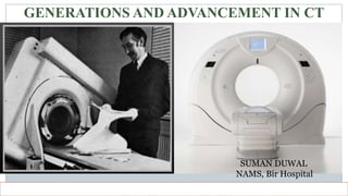
GENERATIONS AND ADVANCEMENT IN CT
SUMAN DUWAL
NAMS, Bir Hospital
overview
 Generations
 Advancement in CT
Advancement is focused more than generations in this presentation
Generations
 First generation
 Second generation
 Third generation
 Fourth generation
 Fifth generation
 Sixth generation
 Seventh generation
Among the generations, 5th generation is discussed more than other generations
First generation
 Pencil beam geometry
 Rotate/translate
 5 min required for a single
image.
Second generation
 Narrow fan beam geometry
 Translation/rotation
 Linear array of 30 detectors
 18 sec per slice
Third generation
 Rotate/rotate
 No of detectors increased to
more than 800
 Wide angle fan beam geometry
 Less than 5 sec to complete a
single slice
Fourth generation
•Rotate/stationary
•Incorporated with a large stationary
360 degrees ring of detectors with
the x-ray tube alone rotating around
the patient.
•Use of 4800 detectors
Fifth generation
 EBCT/EBT/UFCT/Cine CT
 Used for cardiac imaging which requires ultra fast scan time(<50ms)
 First introduced by the group of researchers in Douglas Boyd at the
university of San fransisco
 The company Imatron Inc. started commercial production and
distribution of EBCT system in 1984
 The sole manufacture of EBCT systems developed several scanner
generation( C-100, C-150, C-300)
Principle of EBCT
Fig: Imatron C 150 scanner
Fig: coronary angio performed in C 150
Fig: Imatron 300 scanner
 Imatron Inc. had a partnership with Siemens medical system which
results in several technical improvements of the scanner.
 In 2001, GEMS acquired Imatron Inc. and created daughter company
named ‘GE Imatron Inc’ for manufacturing, servicing and sales of
EBCT
 In Nov 2002 GEMS launched a new model EBCT called “e-speed” first
completely new EBCT scanner since company bought Imatron
e-speed
 Significant improvement over previous model C-300
 Has completely new power source(140 KW) for smoother and higher resolution.
 Enables scan time of 50 and even 33 ms(2-3 times faster then previous EBCT
scanners).
 e-speed is suitable for both cardiac and non cardiac applications.
 10 times faster than true scan speed of 256 slice CT(500 ms)
5 times faster than dual source CT(166ms)
 In terms of radiation dose 10 times lower radiation dose compared to 64 slice CT
 Due to its structural complexity and cost effectiveness and rapid advancement in
MDCT the role of EBCT is under the shadow.
Fig: e speed scanner and VRT image of heart demonstrating CAs
Sixth generation
 Spiral/helical CT
 Three technological development
 Slip ring technology
 High power x-ray tube
 interpolation
Fig: slip rings
Cylinder design Disc design
Fig: spiral/helical acquisition
Seventh generation
 MSCT/MDCT
 It is based on 3rd generation
geometry.
Advantage of MDCT
 The latest breakthrough in CT technology.
 The primary difference between single-slice CT (SSCT) and
MDCT hardware is in the design of the detector arrays.
 Faster Gantry rotation(sub second).
 Fast Data Acquisition System.
 High Speed image reconstruction system.
 The high rating x-ray tube i.e. 8 MHU or more.
 Multiple reconstruction technique.
Advancement
 Advancement in detectors
 Advancement in x-ray tube
 Gantry rotation time
 DECT
 Different techniques
 PET CT
 Portable CT
Advancement in detectors
 UFC
 Stellar detector
 Gemstone
Ultrafast ceramic
 Ultrafast ceramic is a hard yellow substance that resembles like
plastic and weighs as much as gold.
 UFC uses a crystal lattice of rare earth compounds like
gadolinium oxysulphide(GOS) and other compounds
Fig: UFC detector Fig: higher luminous efficiency
of UFC
Advantages of UFC
 High x-ray absorption efficiency
 Short afterglow
 Fast decay time
 Can be used with the fastest CT scanners, with rotational speeds well
under 0.3 seconds
 Resistance to air, humidity ,water , temperature and numerous
chemicals.
 The decay constant is nearly 3 ms and its optimized for
interrogation time of 20ms in order to maintain the image
quality.
 No permanent damage at all could be observed after 10 years of
operation(with 30kGray).
 UFC is a non-poisonous material that does not contain any toxic
elements as other solid state scintillation materials do.
Fig: afterglow behavior of UFC and other scintillator material
Stellar detectors
 The stellar detectors have combined all the analysis electronics in a
single chip
 The converted signals can be processed digitally with no loss which
makes possible to produce medical images with a noticeably higher
signal to electronic noise ratio(SNER) than before at the same radiation
dose.
 Reduces the image noise by 20 to 30% compared to conventional
detectors.
 Stellar detector consumes approximately 70% less power and dissipates
less heat than conventional detectors further reducing the image noise
Development of detectors
Fig : reduction in the image noise with the use of stellar detector
Gems stone detector
 It is the newly developed transparent polycrystalline scintillator
CT detector developed by GE health care.
 It has higher sensitivity to radiation and allow faster sampling
rate.
 It is made up of cerium activated rare earth based garnet material
 It is used in single source ultrafast dual energy switching ,
promising almost simultaneous spatial and temporal registration
and material decomposition.
 It has primary decay time only 30nsec i.e. 100 fold faster than
conventional scintillator.
 The most advantage of gemstone detector is the improved spatial
resolution.( high definition imaging up to 230mm resolution.)
 It has very low afterglow, extremely low radiation damage, very
good chemical durability and uniformity
Fig: decay time of gemstone and GOS
Fig: afterglow behavior of gemstone and GOS
Arrangement of the detector
 Matrix(uniform) array detector
 Adaptive(non- uniform) array detector
 Hybrid(mixed) array detector
Matrix array detector
 Also referred as uniform array detector.
 Contains detector elements that are equal in all dimensions.
 Used in GE scanners
Adaptive array detector
 Also called as non- uniform type of detector
 Detectors with variable thickness thinner rows centrally and
thicker rows peripherally.
 Used in Phillips and Siemens scanners
Hybrid array detector
 Detectors with two fixed thickness four thinner elements
centrally and thicker elements peripherally.
 Used in Toshiba scanners.
Advancement in CT tube
 Straton tube
 MRC( Maximus rotalix ceramic x-ray tube)
 LIMAX( liquid metal anode x-ray tube)
 Aquillion one x-ray tube
Straton tube
 One of the interesting development
is Siemens straton x-ray tube.
 The tube itself is a radical new
design where the entire tube body
rotates rather then just the anode
 This change allows all the bearings
to be located outside the evacuated
tube and enables the anode to be
cooled more efficiently.
 It has the low inherent heat capacity of 0.8 MHU but an extremely fast
cooling rate of 4.7 MHU/min ( Siemens claims the heat storage
capacity of 0 MHU)
 This compares with typical figures of 7-8MHU and up to 1.4MHU/min
for existing tubes.
 Tube cooled down within 20 sec
 Anode diameter: 120 mm
 Anode material : tungsten, zirconium and molybdenum
 Focal spot/track: alloy of tungsten and rhenium
 Enables gantry speed of 0.37 sec per rotation
 The electron beam in the tube is shaped and controlled by magnetic
deflection coil
Flying focal spot
• The no. of measurements channel can be doubled by rapid deflection of
X-ray tube focal spot for each projection increasing the image
resolution.
• This technology is achieved by electromagnetically deflecting the
electron beam within the X-ray Tube.
• For each focus position 2 – measured interlaced projection result, since
the detector continue to move continuously which double the sampling
frequency & enhance the spatial resolution.
(MRC) Maximus rotalix ceramic tube
 Introduced by Phillips in 1989
 Based on the technology of spiral groove bearing using liquid metal
alloy as the lubricant.
Fig: MRC x-ray tube
Fig: MRC model 600
Advantages of MRC
 Dissipation of the heat via the
liquid metal lubricant gave the
tube higher cooling capacity.
 Noiselessly rotating anode and
have a very long lifetime.
 Avoid waiting time during and
between examination.
 200 mm graphite backed anode
 Anode heat storage capacity- 8MHU
 Tube voltage- 90 to 140kv
 Tube current- 20 to 500 mA
 Anode angle- 7o
(LIMAX)Liquid metal anode x-ray tube
 The novel x-ray tube concept which predicts producing x-rays in a
turbulent flowing liquid metal which interacts with the electron beam
which is transmitted through a thin window.
 In this tubes an electron beam interacts with the liquid metal stream
flowing turbulently at high speed in a narrow channel
 Liquid metals eutectics, of SnPb, GaInSn, PbBi or PbBiInSn
Liquid metal
 It should contain element with high atomic number
 High thermal conductivity
 Chemically inert
 Liquid metal has to serve at least 2 purposes:
 It acts as the cooling medium
 It serves as the x-ray photon generating medium
 PbBi is the best choice as it got the higher x-ray output than other
liquid metals
Fig : photon emission spectra of three liquid metals covered with 5µm diamond
Electron beam window
 The x-ray photon will leave the anode in a reflection geometry , thus
the electron window has to have a low absorption coefficient
 Due to its extreme toxicity and relatively low melting point beryllium
cannot be used as the electron beam window material.
 Carbon in the form of diamond is the best choice due to high young’s
modulus , extremely good thermal conductivity, inertness to liquid
metals
Aquillion one
Aquillion one x-ray tube
 Heat storage capacity- 7.5 MHU
 Cooling rate- 1.7 MHU/min
 Anode is grounded
 Focal spot- 1.4mm* 1.4mm
 Air cooled tube
Gantry rotation time
 Rotation time is the time interval needed for a complete 360 ° rotation
of the tube and the detector system around the patient
 A short rotation time has following advantages
 Longer spiral length can be acquired in the same scan time
 Same volume and same slice thickness can be scanned in less time
 Increased temporal resolution
 Motions artefacts are reduced
Sub second gantry rotation
 In 16 slices- 0.5 sec
 In 64 slices- 0.3 sec
 In 320/640 slices- 0.27 sec
Post processing technique
 Multiplanar Reconstruction(MPR)
 MPR is the fast reconstruction method.
 Provide coronal or sagittal plane from axial scan.
 Depend on acquisition parameter- thin collimation
excellent results.
 Commonly used in orthopedic examination
 Shaded surface display(SSD)
 Also called surface rendering
 The marching cubes algorithm must be hallmark for surface
rendering.
 Commonly used for fracture, deformities of bone, CTA
 Volume Rendering Technique(VRT)
 Representation, visualization & manipulation of objects
represented as sampled data in 3 or more dimensions.
 Commonly used in CTA, CT Coronary angiography
 Maximum Intensity Projection(MIP)/ Minimum Intensity Projection(MinIP)
 MIP is a relatively simple method.
 for visualization permits easy viewing of vascular structures or air-filled
cavities.
 Virtual Endoscopy
 Used for 3D reconstruction of upper GI.
 Virtual Colonoscopy
 Used for 3D reconstruction of Colon.
 Virtual Bronchoscopy
 Used for 3D reconstruction of airways.
Fig: fly through technique
Dual energy MDCT
 DECT uses the principle that different material shows different attenuation at
varying energy level and this difference can be used for better tissue
characterization.
• With dual-energy CT, two image datasets are acquired in the same anatomic
location with two different x-ray spectra to allow the analysis of energy-dependent
changes in the attenuation of different materials.
• Each type of material demonstrates a relatively specific change in attenuation
between images obtained with a high-energy spectrum and those obtained with a
low-energy spectrum, and this attenuation difference allows a more distinct
characterization of the features depicted.
• Two different materials that show similar attenuation on images acquired with one
of the two energy spectra are often more easily differentiated on images acquired
with the other spectrum because of substantial differences in their attenuation
When dual-energy images reconstructed for 50 and 80 keV are compared, iodine
demonstrates a greater decrease in attenuation than calcium does at the higher energy,
whereas the attenuation of water remains more or less constant
Historical perspective
 In the early days, Sir Hounsfield proposed that ,two pictures are
taken of the same slice one at 100KV and other at 140 kv. tests
carried out have shown that iodine (z=53) can be readily
differentiated from calcium(z=20)
 Alvarez , Macovski and Calender also described the theoretical
basis of the dual energy scanning in the early 1980s.
 In 2006 DECT is introduced for the first time commercially
( somatom definition, Siemens)(DSDECT)
 First generation DSDECT have FOV limited to 26cm
 Whereas in second generation it was broaden up to 33cm
 Over the time new modifications and advancement on DECT
were made.
 Then later single source dual energy CT was introduced by GE
fig: 1st generation fig: 2nd generation
SSDECT
Types of DECT
 Source driven approach
 Dual source dual energy
 Single source single energy
 Detector driven approach
 Single source dual layer detectors
 Photon counting detectors
Clinical use of DECT
 Direct subtraction of bone.
 Differentiation between plaque and contrast agent
 Virtual unenhanced abdominal organ imaging
 Kidney stone characterization
 Visualization of cartilage, tendons, ligaments
 Evaluation of lung perfusion defects
 Heart perfusion blood volume
 Uric acid crystal visualization
 Lung vessel embolisation
 Brain hemorrhage differentiation.
Dose modulation techniques
 SURE DOSE used by Toshiba scanners
 DOSE RIGHT used by Phillips scanners
 CARE KV and CARE DOSE 4D by Siemens scanners
 AUTO MA or SMART MA by GEMS
Iterative reconstruction dose reduction
 AIDR-3D - Toshiba (adaptive iterative dose reconstruction)
 ASIR – GEMS(adaptive statistical iterative reconstruction)
 I DOSE IMR – Phillips(iterative model reconstruction)
 ADMIRE- Siemens(advanced modeled iterative reconstruction.
Metal artifact reduction techniques
 SEMAR- Toshiba
 MAR- GEMS
 iMAR- Siemens
 O-MAR- Phillips
O- MAR
matrix
80 x 80 512 x 512
PET CT
 PET and CT provide complementary information.
 PET provides functional information but little anatomic detail.
 CT provides anatomic and morphologic information (size,
shape, density of lesions ) but provides little physiologic
insight into tissues
Fig: PET CT
Portable CT
SUMMARY
bibliography
 Liquid-metal anode x-ray tube, DOI: 10.1117/12.504503
 https://www.spiedigitallibrary.org/conference-proceedings-of-
spie/5541/0000/Liquid-metal-anode-x-ray-tubes--interesting-but-
are/10.1117/12.561615.short?SSO=1
 http://spie.org/Publications/Proceedings/Paper/10.1117/12.561615
 Essential book of Physics - Busburg, third edition.
 Diagnostic radiology. Recent advances and applied and applied physics in
imaging.( Arun Kumar gupta , veena chowdhary , niranjan khandelwal)
 Christensen's Physics of Diagnostic Radiology Fourth edition
by Thomas S.curry,James Dowery
 Gemstone – The Ultimate Scintillator for Computed Tomography
(GE Healthcare)
 www.mahameru.com
 www.genewsroom.com
 Comparison of Three Generations of Electron Beam Tomography on Image Noise
and Reproducibility, a Phantom Study
Chau, Alex BS; Gopal, Ambarish MD; Mao, SongShou MD; Tseng, Philip H. BS;
Fischer, Hans MD, PhD; Budoff, Matthew J. MD
 matron's C-150 scanner as Evolution EBT (SCAN 4/12/95).
 Electron Beam Computed Tomography (EBCT)
Valentin E. Sinitsyn, Stephan Achenbach
 https://www.researchgate.net/figure/Virtual-endoscopy-showing-
concentric-narrowing-of-the-airway-a-View-of-the-proximal-
part_fig2_46289289
 www.radiopaedia.com
 https://www.jbsr.be/articles/10.5334/jbr-btr.1420/
 www.phillips.co.uk
Thank You!!!!

More Related Content

What's hot

MDCT Principles and Applications- Avinesh Shrestha
MDCT Principles and Applications- Avinesh ShresthaMDCT Principles and Applications- Avinesh Shrestha
MDCT Principles and Applications- Avinesh ShresthaAvinesh Shrestha
 
Mobile & portable radiography
Mobile & portable radiographyMobile & portable radiography
Mobile & portable radiographyPriyanka Parimala
 
Quality Assurance Programme in Computed Tomography
Quality Assurance Programme in Computed TomographyQuality Assurance Programme in Computed Tomography
Quality Assurance Programme in Computed TomographyRamzee Small
 
Multi slice ct ppt
Multi slice ct pptMulti slice ct ppt
Multi slice ct pptAnand Rk
 
Beam restricted device and filter used in x ray
Beam restricted device and filter used in x rayBeam restricted device and filter used in x ray
Beam restricted device and filter used in x raySushilPattar
 
Basic Pulse Sequences In MRI
Basic Pulse Sequences In MRIBasic Pulse Sequences In MRI
Basic Pulse Sequences In MRIUpakar Paudel
 
CT Physics
CT PhysicsCT Physics
CT PhysicsRMLIMS
 
Mri coils
Mri coilsMri coils
Mri coilsSelf
 
Ct tube and detectors
Ct tube and detectorsCt tube and detectors
Ct tube and detectorssandip suman
 
MACRORADIOGRAPHY.pptx
MACRORADIOGRAPHY.pptxMACRORADIOGRAPHY.pptx
MACRORADIOGRAPHY.pptxThejaTej6
 
Encoding and image formation
Encoding and image formationEncoding and image formation
Encoding and image formationAnkit Mishra
 
Computed radiography
Computed radiographyComputed radiography
Computed radiographyIllyasmk
 

What's hot (20)

Computed radiography
Computed radiographyComputed radiography
Computed radiography
 
MDCT Principles and Applications- Avinesh Shrestha
MDCT Principles and Applications- Avinesh ShresthaMDCT Principles and Applications- Avinesh Shrestha
MDCT Principles and Applications- Avinesh Shrestha
 
Mobile & portable radiography
Mobile & portable radiographyMobile & portable radiography
Mobile & portable radiography
 
radiation protection in ct
radiation protection in ctradiation protection in ct
radiation protection in ct
 
Mri artifacts
Mri artifactsMri artifacts
Mri artifacts
 
Quality Assurance Programme in Computed Tomography
Quality Assurance Programme in Computed TomographyQuality Assurance Programme in Computed Tomography
Quality Assurance Programme in Computed Tomography
 
CT artifact
CT artifact CT artifact
CT artifact
 
Multi slice ct ppt
Multi slice ct pptMulti slice ct ppt
Multi slice ct ppt
 
Beam restricted device and filter used in x ray
Beam restricted device and filter used in x rayBeam restricted device and filter used in x ray
Beam restricted device and filter used in x ray
 
Basic Pulse Sequences In MRI
Basic Pulse Sequences In MRIBasic Pulse Sequences In MRI
Basic Pulse Sequences In MRI
 
Ct Basics
Ct BasicsCt Basics
Ct Basics
 
CT Physics
CT PhysicsCT Physics
CT Physics
 
Mri coils
Mri coilsMri coils
Mri coils
 
Ct tube and detectors
Ct tube and detectorsCt tube and detectors
Ct tube and detectors
 
MACRORADIOGRAPHY.pptx
MACRORADIOGRAPHY.pptxMACRORADIOGRAPHY.pptx
MACRORADIOGRAPHY.pptx
 
Ultrasound contrast agents
Ultrasound contrast agentsUltrasound contrast agents
Ultrasound contrast agents
 
Encoding and image formation
Encoding and image formationEncoding and image formation
Encoding and image formation
 
CT Generations and Artefacts
CT Generations and ArtefactsCT Generations and Artefacts
CT Generations and Artefacts
 
Fluroscopy
FluroscopyFluroscopy
Fluroscopy
 
Computed radiography
Computed radiographyComputed radiography
Computed radiography
 

Similar to Generations and advancement of CT

Similar to Generations and advancement of CT (20)

CT SCAN equipment.pptx
CT SCAN equipment.pptxCT SCAN equipment.pptx
CT SCAN equipment.pptx
 
CT generations and peculiarities
CT generations and peculiaritiesCT generations and peculiarities
CT generations and peculiarities
 
Computed tomography
Computed tomographyComputed tomography
Computed tomography
 
Computed tomography
Computed tomographyComputed tomography
Computed tomography
 
CT scanner
CT scannerCT scanner
CT scanner
 
Basics in ct
Basics in ctBasics in ct
Basics in ct
 
Physics of ct mri
Physics of ct mriPhysics of ct mri
Physics of ct mri
 
Cttubeanddetectors
Cttubeanddetectors Cttubeanddetectors
Cttubeanddetectors
 
COMPUTED TOMOGRAPHY by Heena-1.pptx
COMPUTED TOMOGRAPHY by Heena-1.pptxCOMPUTED TOMOGRAPHY by Heena-1.pptx
COMPUTED TOMOGRAPHY by Heena-1.pptx
 
CT PHYSICS for first year radiology residents
CT PHYSICS for first year radiology residentsCT PHYSICS for first year radiology residents
CT PHYSICS for first year radiology residents
 
Nexray
NexrayNexray
Nexray
 
Basics of CT- Lecture 4.ppt
Basics of CT- Lecture 4.pptBasics of CT- Lecture 4.ppt
Basics of CT- Lecture 4.ppt
 
Computed Tomography
Computed TomographyComputed Tomography
Computed Tomography
 
computed Tomography
computed Tomographycomputed Tomography
computed Tomography
 
111
111111
111
 
Basics of CT & MRI
Basics of CT & MRIBasics of CT & MRI
Basics of CT & MRI
 
Computed tomography / dental implant courses
Computed tomography / dental implant coursesComputed tomography / dental implant courses
Computed tomography / dental implant courses
 
Basicsinct 110331052435-phpapp02
Basicsinct 110331052435-phpapp02Basicsinct 110331052435-phpapp02
Basicsinct 110331052435-phpapp02
 
basic of angiography physics and equipement.pdf
basic of angiography physics and equipement.pdfbasic of angiography physics and equipement.pdf
basic of angiography physics and equipement.pdf
 
Extended Abstract - Adam Moreau
Extended Abstract - Adam MoreauExtended Abstract - Adam Moreau
Extended Abstract - Adam Moreau
 

Recently uploaded

Russian Call Girls in Pune Tanvi 9907093804 Short 1500 Night 6000 Best call g...
Russian Call Girls in Pune Tanvi 9907093804 Short 1500 Night 6000 Best call g...Russian Call Girls in Pune Tanvi 9907093804 Short 1500 Night 6000 Best call g...
Russian Call Girls in Pune Tanvi 9907093804 Short 1500 Night 6000 Best call g...Miss joya
 
CALL ON ➥9907093804 🔝 Call Girls Baramati ( Pune) Girls Service
CALL ON ➥9907093804 🔝 Call Girls Baramati ( Pune)  Girls ServiceCALL ON ➥9907093804 🔝 Call Girls Baramati ( Pune)  Girls Service
CALL ON ➥9907093804 🔝 Call Girls Baramati ( Pune) Girls ServiceMiss joya
 
♛VVIP Hyderabad Call Girls Chintalkunta🖕7001035870🖕Riya Kappor Top Call Girl ...
♛VVIP Hyderabad Call Girls Chintalkunta🖕7001035870🖕Riya Kappor Top Call Girl ...♛VVIP Hyderabad Call Girls Chintalkunta🖕7001035870🖕Riya Kappor Top Call Girl ...
♛VVIP Hyderabad Call Girls Chintalkunta🖕7001035870🖕Riya Kappor Top Call Girl ...astropune
 
Aspirin presentation slides by Dr. Rewas Ali
Aspirin presentation slides by Dr. Rewas AliAspirin presentation slides by Dr. Rewas Ali
Aspirin presentation slides by Dr. Rewas AliRewAs ALI
 
Artifacts in Nuclear Medicine with Identifying and resolving artifacts.
Artifacts in Nuclear Medicine with Identifying and resolving artifacts.Artifacts in Nuclear Medicine with Identifying and resolving artifacts.
Artifacts in Nuclear Medicine with Identifying and resolving artifacts.MiadAlsulami
 
Call Girl Number in Vashi Mumbai📲 9833363713 💞 Full Night Enjoy
Call Girl Number in Vashi Mumbai📲 9833363713 💞 Full Night EnjoyCall Girl Number in Vashi Mumbai📲 9833363713 💞 Full Night Enjoy
Call Girl Number in Vashi Mumbai📲 9833363713 💞 Full Night Enjoybabeytanya
 
(👑VVIP ISHAAN ) Russian Call Girls Service Navi Mumbai🖕9920874524🖕Independent...
(👑VVIP ISHAAN ) Russian Call Girls Service Navi Mumbai🖕9920874524🖕Independent...(👑VVIP ISHAAN ) Russian Call Girls Service Navi Mumbai🖕9920874524🖕Independent...
(👑VVIP ISHAAN ) Russian Call Girls Service Navi Mumbai🖕9920874524🖕Independent...Taniya Sharma
 
💎VVIP Kolkata Call Girls Parganas🩱7001035870🩱Independent Girl ( Ac Rooms Avai...
💎VVIP Kolkata Call Girls Parganas🩱7001035870🩱Independent Girl ( Ac Rooms Avai...💎VVIP Kolkata Call Girls Parganas🩱7001035870🩱Independent Girl ( Ac Rooms Avai...
💎VVIP Kolkata Call Girls Parganas🩱7001035870🩱Independent Girl ( Ac Rooms Avai...Taniya Sharma
 
Bangalore Call Girls Nelamangala Number 7001035870 Meetin With Bangalore Esc...
Bangalore Call Girls Nelamangala Number 7001035870  Meetin With Bangalore Esc...Bangalore Call Girls Nelamangala Number 7001035870  Meetin With Bangalore Esc...
Bangalore Call Girls Nelamangala Number 7001035870 Meetin With Bangalore Esc...narwatsonia7
 
Call Girls Horamavu WhatsApp Number 7001035870 Meeting With Bangalore Escorts
Call Girls Horamavu WhatsApp Number 7001035870 Meeting With Bangalore EscortsCall Girls Horamavu WhatsApp Number 7001035870 Meeting With Bangalore Escorts
Call Girls Horamavu WhatsApp Number 7001035870 Meeting With Bangalore Escortsvidya singh
 
Lucknow Call girls - 8800925952 - 24x7 service with hotel room
Lucknow Call girls - 8800925952 - 24x7 service with hotel roomLucknow Call girls - 8800925952 - 24x7 service with hotel room
Lucknow Call girls - 8800925952 - 24x7 service with hotel roomdiscovermytutordmt
 
Call Girl Coimbatore Prisha☎️ 8250192130 Independent Escort Service Coimbatore
Call Girl Coimbatore Prisha☎️  8250192130 Independent Escort Service CoimbatoreCall Girl Coimbatore Prisha☎️  8250192130 Independent Escort Service Coimbatore
Call Girl Coimbatore Prisha☎️ 8250192130 Independent Escort Service Coimbatorenarwatsonia7
 
VIP Mumbai Call Girls Hiranandani Gardens Just Call 9920874524 with A/C Room ...
VIP Mumbai Call Girls Hiranandani Gardens Just Call 9920874524 with A/C Room ...VIP Mumbai Call Girls Hiranandani Gardens Just Call 9920874524 with A/C Room ...
VIP Mumbai Call Girls Hiranandani Gardens Just Call 9920874524 with A/C Room ...Garima Khatri
 
Premium Call Girls Cottonpet Whatsapp 7001035870 Independent Escort Service
Premium Call Girls Cottonpet Whatsapp 7001035870 Independent Escort ServicePremium Call Girls Cottonpet Whatsapp 7001035870 Independent Escort Service
Premium Call Girls Cottonpet Whatsapp 7001035870 Independent Escort Servicevidya singh
 
Vip Call Girls Anna Salai Chennai 👉 8250192130 ❣️💯 Top Class Girls Available
Vip Call Girls Anna Salai Chennai 👉 8250192130 ❣️💯 Top Class Girls AvailableVip Call Girls Anna Salai Chennai 👉 8250192130 ❣️💯 Top Class Girls Available
Vip Call Girls Anna Salai Chennai 👉 8250192130 ❣️💯 Top Class Girls AvailableNehru place Escorts
 
VIP Russian Call Girls in Varanasi Samaira 8250192130 Independent Escort Serv...
VIP Russian Call Girls in Varanasi Samaira 8250192130 Independent Escort Serv...VIP Russian Call Girls in Varanasi Samaira 8250192130 Independent Escort Serv...
VIP Russian Call Girls in Varanasi Samaira 8250192130 Independent Escort Serv...Neha Kaur
 
VIP Call Girls Pune Vrinda 9907093804 Short 1500 Night 6000 Best call girls S...
VIP Call Girls Pune Vrinda 9907093804 Short 1500 Night 6000 Best call girls S...VIP Call Girls Pune Vrinda 9907093804 Short 1500 Night 6000 Best call girls S...
VIP Call Girls Pune Vrinda 9907093804 Short 1500 Night 6000 Best call girls S...Miss joya
 
Bangalore Call Girls Hebbal Kempapura Number 7001035870 Meetin With Bangalor...
Bangalore Call Girls Hebbal Kempapura Number 7001035870  Meetin With Bangalor...Bangalore Call Girls Hebbal Kempapura Number 7001035870  Meetin With Bangalor...
Bangalore Call Girls Hebbal Kempapura Number 7001035870 Meetin With Bangalor...narwatsonia7
 
Call Girls Service In Shyam Nagar Whatsapp 8445551418 Independent Escort Service
Call Girls Service In Shyam Nagar Whatsapp 8445551418 Independent Escort ServiceCall Girls Service In Shyam Nagar Whatsapp 8445551418 Independent Escort Service
Call Girls Service In Shyam Nagar Whatsapp 8445551418 Independent Escort Serviceparulsinha
 

Recently uploaded (20)

Russian Call Girls in Pune Tanvi 9907093804 Short 1500 Night 6000 Best call g...
Russian Call Girls in Pune Tanvi 9907093804 Short 1500 Night 6000 Best call g...Russian Call Girls in Pune Tanvi 9907093804 Short 1500 Night 6000 Best call g...
Russian Call Girls in Pune Tanvi 9907093804 Short 1500 Night 6000 Best call g...
 
CALL ON ➥9907093804 🔝 Call Girls Baramati ( Pune) Girls Service
CALL ON ➥9907093804 🔝 Call Girls Baramati ( Pune)  Girls ServiceCALL ON ➥9907093804 🔝 Call Girls Baramati ( Pune)  Girls Service
CALL ON ➥9907093804 🔝 Call Girls Baramati ( Pune) Girls Service
 
♛VVIP Hyderabad Call Girls Chintalkunta🖕7001035870🖕Riya Kappor Top Call Girl ...
♛VVIP Hyderabad Call Girls Chintalkunta🖕7001035870🖕Riya Kappor Top Call Girl ...♛VVIP Hyderabad Call Girls Chintalkunta🖕7001035870🖕Riya Kappor Top Call Girl ...
♛VVIP Hyderabad Call Girls Chintalkunta🖕7001035870🖕Riya Kappor Top Call Girl ...
 
Aspirin presentation slides by Dr. Rewas Ali
Aspirin presentation slides by Dr. Rewas AliAspirin presentation slides by Dr. Rewas Ali
Aspirin presentation slides by Dr. Rewas Ali
 
Artifacts in Nuclear Medicine with Identifying and resolving artifacts.
Artifacts in Nuclear Medicine with Identifying and resolving artifacts.Artifacts in Nuclear Medicine with Identifying and resolving artifacts.
Artifacts in Nuclear Medicine with Identifying and resolving artifacts.
 
Call Girl Number in Vashi Mumbai📲 9833363713 💞 Full Night Enjoy
Call Girl Number in Vashi Mumbai📲 9833363713 💞 Full Night EnjoyCall Girl Number in Vashi Mumbai📲 9833363713 💞 Full Night Enjoy
Call Girl Number in Vashi Mumbai📲 9833363713 💞 Full Night Enjoy
 
(👑VVIP ISHAAN ) Russian Call Girls Service Navi Mumbai🖕9920874524🖕Independent...
(👑VVIP ISHAAN ) Russian Call Girls Service Navi Mumbai🖕9920874524🖕Independent...(👑VVIP ISHAAN ) Russian Call Girls Service Navi Mumbai🖕9920874524🖕Independent...
(👑VVIP ISHAAN ) Russian Call Girls Service Navi Mumbai🖕9920874524🖕Independent...
 
💎VVIP Kolkata Call Girls Parganas🩱7001035870🩱Independent Girl ( Ac Rooms Avai...
💎VVIP Kolkata Call Girls Parganas🩱7001035870🩱Independent Girl ( Ac Rooms Avai...💎VVIP Kolkata Call Girls Parganas🩱7001035870🩱Independent Girl ( Ac Rooms Avai...
💎VVIP Kolkata Call Girls Parganas🩱7001035870🩱Independent Girl ( Ac Rooms Avai...
 
Bangalore Call Girls Nelamangala Number 7001035870 Meetin With Bangalore Esc...
Bangalore Call Girls Nelamangala Number 7001035870  Meetin With Bangalore Esc...Bangalore Call Girls Nelamangala Number 7001035870  Meetin With Bangalore Esc...
Bangalore Call Girls Nelamangala Number 7001035870 Meetin With Bangalore Esc...
 
Call Girls Horamavu WhatsApp Number 7001035870 Meeting With Bangalore Escorts
Call Girls Horamavu WhatsApp Number 7001035870 Meeting With Bangalore EscortsCall Girls Horamavu WhatsApp Number 7001035870 Meeting With Bangalore Escorts
Call Girls Horamavu WhatsApp Number 7001035870 Meeting With Bangalore Escorts
 
Lucknow Call girls - 8800925952 - 24x7 service with hotel room
Lucknow Call girls - 8800925952 - 24x7 service with hotel roomLucknow Call girls - 8800925952 - 24x7 service with hotel room
Lucknow Call girls - 8800925952 - 24x7 service with hotel room
 
Call Girl Coimbatore Prisha☎️ 8250192130 Independent Escort Service Coimbatore
Call Girl Coimbatore Prisha☎️  8250192130 Independent Escort Service CoimbatoreCall Girl Coimbatore Prisha☎️  8250192130 Independent Escort Service Coimbatore
Call Girl Coimbatore Prisha☎️ 8250192130 Independent Escort Service Coimbatore
 
VIP Mumbai Call Girls Hiranandani Gardens Just Call 9920874524 with A/C Room ...
VIP Mumbai Call Girls Hiranandani Gardens Just Call 9920874524 with A/C Room ...VIP Mumbai Call Girls Hiranandani Gardens Just Call 9920874524 with A/C Room ...
VIP Mumbai Call Girls Hiranandani Gardens Just Call 9920874524 with A/C Room ...
 
Premium Call Girls Cottonpet Whatsapp 7001035870 Independent Escort Service
Premium Call Girls Cottonpet Whatsapp 7001035870 Independent Escort ServicePremium Call Girls Cottonpet Whatsapp 7001035870 Independent Escort Service
Premium Call Girls Cottonpet Whatsapp 7001035870 Independent Escort Service
 
Vip Call Girls Anna Salai Chennai 👉 8250192130 ❣️💯 Top Class Girls Available
Vip Call Girls Anna Salai Chennai 👉 8250192130 ❣️💯 Top Class Girls AvailableVip Call Girls Anna Salai Chennai 👉 8250192130 ❣️💯 Top Class Girls Available
Vip Call Girls Anna Salai Chennai 👉 8250192130 ❣️💯 Top Class Girls Available
 
sauth delhi call girls in Bhajanpura 🔝 9953056974 🔝 escort Service
sauth delhi call girls in Bhajanpura 🔝 9953056974 🔝 escort Servicesauth delhi call girls in Bhajanpura 🔝 9953056974 🔝 escort Service
sauth delhi call girls in Bhajanpura 🔝 9953056974 🔝 escort Service
 
VIP Russian Call Girls in Varanasi Samaira 8250192130 Independent Escort Serv...
VIP Russian Call Girls in Varanasi Samaira 8250192130 Independent Escort Serv...VIP Russian Call Girls in Varanasi Samaira 8250192130 Independent Escort Serv...
VIP Russian Call Girls in Varanasi Samaira 8250192130 Independent Escort Serv...
 
VIP Call Girls Pune Vrinda 9907093804 Short 1500 Night 6000 Best call girls S...
VIP Call Girls Pune Vrinda 9907093804 Short 1500 Night 6000 Best call girls S...VIP Call Girls Pune Vrinda 9907093804 Short 1500 Night 6000 Best call girls S...
VIP Call Girls Pune Vrinda 9907093804 Short 1500 Night 6000 Best call girls S...
 
Bangalore Call Girls Hebbal Kempapura Number 7001035870 Meetin With Bangalor...
Bangalore Call Girls Hebbal Kempapura Number 7001035870  Meetin With Bangalor...Bangalore Call Girls Hebbal Kempapura Number 7001035870  Meetin With Bangalor...
Bangalore Call Girls Hebbal Kempapura Number 7001035870 Meetin With Bangalor...
 
Call Girls Service In Shyam Nagar Whatsapp 8445551418 Independent Escort Service
Call Girls Service In Shyam Nagar Whatsapp 8445551418 Independent Escort ServiceCall Girls Service In Shyam Nagar Whatsapp 8445551418 Independent Escort Service
Call Girls Service In Shyam Nagar Whatsapp 8445551418 Independent Escort Service
 

Generations and advancement of CT

  • 1. GENERATIONS AND ADVANCEMENT IN CT SUMAN DUWAL NAMS, Bir Hospital
  • 2. overview  Generations  Advancement in CT Advancement is focused more than generations in this presentation
  • 3. Generations  First generation  Second generation  Third generation  Fourth generation  Fifth generation  Sixth generation  Seventh generation Among the generations, 5th generation is discussed more than other generations
  • 4. First generation  Pencil beam geometry  Rotate/translate  5 min required for a single image.
  • 5. Second generation  Narrow fan beam geometry  Translation/rotation  Linear array of 30 detectors  18 sec per slice
  • 6. Third generation  Rotate/rotate  No of detectors increased to more than 800  Wide angle fan beam geometry  Less than 5 sec to complete a single slice
  • 7. Fourth generation •Rotate/stationary •Incorporated with a large stationary 360 degrees ring of detectors with the x-ray tube alone rotating around the patient. •Use of 4800 detectors
  • 8. Fifth generation  EBCT/EBT/UFCT/Cine CT  Used for cardiac imaging which requires ultra fast scan time(<50ms)  First introduced by the group of researchers in Douglas Boyd at the university of San fransisco  The company Imatron Inc. started commercial production and distribution of EBCT system in 1984  The sole manufacture of EBCT systems developed several scanner generation( C-100, C-150, C-300)
  • 10. Fig: Imatron C 150 scanner
  • 11. Fig: coronary angio performed in C 150
  • 12. Fig: Imatron 300 scanner
  • 13.  Imatron Inc. had a partnership with Siemens medical system which results in several technical improvements of the scanner.  In 2001, GEMS acquired Imatron Inc. and created daughter company named ‘GE Imatron Inc’ for manufacturing, servicing and sales of EBCT  In Nov 2002 GEMS launched a new model EBCT called “e-speed” first completely new EBCT scanner since company bought Imatron
  • 14. e-speed  Significant improvement over previous model C-300  Has completely new power source(140 KW) for smoother and higher resolution.  Enables scan time of 50 and even 33 ms(2-3 times faster then previous EBCT scanners).  e-speed is suitable for both cardiac and non cardiac applications.  10 times faster than true scan speed of 256 slice CT(500 ms) 5 times faster than dual source CT(166ms)  In terms of radiation dose 10 times lower radiation dose compared to 64 slice CT  Due to its structural complexity and cost effectiveness and rapid advancement in MDCT the role of EBCT is under the shadow.
  • 15. Fig: e speed scanner and VRT image of heart demonstrating CAs
  • 16.
  • 17. Sixth generation  Spiral/helical CT  Three technological development  Slip ring technology  High power x-ray tube  interpolation
  • 18. Fig: slip rings Cylinder design Disc design
  • 20.
  • 21. Seventh generation  MSCT/MDCT  It is based on 3rd generation geometry.
  • 22. Advantage of MDCT  The latest breakthrough in CT technology.  The primary difference between single-slice CT (SSCT) and MDCT hardware is in the design of the detector arrays.  Faster Gantry rotation(sub second).  Fast Data Acquisition System.  High Speed image reconstruction system.  The high rating x-ray tube i.e. 8 MHU or more.  Multiple reconstruction technique.
  • 23. Advancement  Advancement in detectors  Advancement in x-ray tube  Gantry rotation time  DECT  Different techniques  PET CT  Portable CT
  • 24. Advancement in detectors  UFC  Stellar detector  Gemstone
  • 25. Ultrafast ceramic  Ultrafast ceramic is a hard yellow substance that resembles like plastic and weighs as much as gold.  UFC uses a crystal lattice of rare earth compounds like gadolinium oxysulphide(GOS) and other compounds
  • 26. Fig: UFC detector Fig: higher luminous efficiency of UFC
  • 27. Advantages of UFC  High x-ray absorption efficiency  Short afterglow  Fast decay time  Can be used with the fastest CT scanners, with rotational speeds well under 0.3 seconds  Resistance to air, humidity ,water , temperature and numerous chemicals.
  • 28.  The decay constant is nearly 3 ms and its optimized for interrogation time of 20ms in order to maintain the image quality.  No permanent damage at all could be observed after 10 years of operation(with 30kGray).  UFC is a non-poisonous material that does not contain any toxic elements as other solid state scintillation materials do.
  • 29.
  • 30. Fig: afterglow behavior of UFC and other scintillator material
  • 31. Stellar detectors  The stellar detectors have combined all the analysis electronics in a single chip  The converted signals can be processed digitally with no loss which makes possible to produce medical images with a noticeably higher signal to electronic noise ratio(SNER) than before at the same radiation dose.  Reduces the image noise by 20 to 30% compared to conventional detectors.  Stellar detector consumes approximately 70% less power and dissipates less heat than conventional detectors further reducing the image noise
  • 32.
  • 34.
  • 35.
  • 36.
  • 37.
  • 38. Fig : reduction in the image noise with the use of stellar detector
  • 39.
  • 40. Gems stone detector  It is the newly developed transparent polycrystalline scintillator CT detector developed by GE health care.  It has higher sensitivity to radiation and allow faster sampling rate.  It is made up of cerium activated rare earth based garnet material  It is used in single source ultrafast dual energy switching , promising almost simultaneous spatial and temporal registration and material decomposition.
  • 41.  It has primary decay time only 30nsec i.e. 100 fold faster than conventional scintillator.  The most advantage of gemstone detector is the improved spatial resolution.( high definition imaging up to 230mm resolution.)  It has very low afterglow, extremely low radiation damage, very good chemical durability and uniformity
  • 42.
  • 43. Fig: decay time of gemstone and GOS
  • 44. Fig: afterglow behavior of gemstone and GOS
  • 45.
  • 46. Arrangement of the detector  Matrix(uniform) array detector  Adaptive(non- uniform) array detector  Hybrid(mixed) array detector
  • 47. Matrix array detector  Also referred as uniform array detector.  Contains detector elements that are equal in all dimensions.  Used in GE scanners
  • 48. Adaptive array detector  Also called as non- uniform type of detector  Detectors with variable thickness thinner rows centrally and thicker rows peripherally.  Used in Phillips and Siemens scanners
  • 49. Hybrid array detector  Detectors with two fixed thickness four thinner elements centrally and thicker elements peripherally.  Used in Toshiba scanners.
  • 50. Advancement in CT tube  Straton tube  MRC( Maximus rotalix ceramic x-ray tube)  LIMAX( liquid metal anode x-ray tube)  Aquillion one x-ray tube
  • 51. Straton tube  One of the interesting development is Siemens straton x-ray tube.  The tube itself is a radical new design where the entire tube body rotates rather then just the anode  This change allows all the bearings to be located outside the evacuated tube and enables the anode to be cooled more efficiently.
  • 52.
  • 53.
  • 54.
  • 55.  It has the low inherent heat capacity of 0.8 MHU but an extremely fast cooling rate of 4.7 MHU/min ( Siemens claims the heat storage capacity of 0 MHU)  This compares with typical figures of 7-8MHU and up to 1.4MHU/min for existing tubes.  Tube cooled down within 20 sec  Anode diameter: 120 mm  Anode material : tungsten, zirconium and molybdenum  Focal spot/track: alloy of tungsten and rhenium
  • 56.  Enables gantry speed of 0.37 sec per rotation  The electron beam in the tube is shaped and controlled by magnetic deflection coil
  • 57. Flying focal spot • The no. of measurements channel can be doubled by rapid deflection of X-ray tube focal spot for each projection increasing the image resolution. • This technology is achieved by electromagnetically deflecting the electron beam within the X-ray Tube. • For each focus position 2 – measured interlaced projection result, since the detector continue to move continuously which double the sampling frequency & enhance the spatial resolution.
  • 58. (MRC) Maximus rotalix ceramic tube  Introduced by Phillips in 1989  Based on the technology of spiral groove bearing using liquid metal alloy as the lubricant. Fig: MRC x-ray tube Fig: MRC model 600
  • 59. Advantages of MRC  Dissipation of the heat via the liquid metal lubricant gave the tube higher cooling capacity.  Noiselessly rotating anode and have a very long lifetime.  Avoid waiting time during and between examination.
  • 60.  200 mm graphite backed anode  Anode heat storage capacity- 8MHU  Tube voltage- 90 to 140kv  Tube current- 20 to 500 mA  Anode angle- 7o
  • 61. (LIMAX)Liquid metal anode x-ray tube  The novel x-ray tube concept which predicts producing x-rays in a turbulent flowing liquid metal which interacts with the electron beam which is transmitted through a thin window.  In this tubes an electron beam interacts with the liquid metal stream flowing turbulently at high speed in a narrow channel  Liquid metals eutectics, of SnPb, GaInSn, PbBi or PbBiInSn
  • 62. Liquid metal  It should contain element with high atomic number  High thermal conductivity  Chemically inert  Liquid metal has to serve at least 2 purposes:  It acts as the cooling medium  It serves as the x-ray photon generating medium  PbBi is the best choice as it got the higher x-ray output than other liquid metals
  • 63. Fig : photon emission spectra of three liquid metals covered with 5µm diamond
  • 64. Electron beam window  The x-ray photon will leave the anode in a reflection geometry , thus the electron window has to have a low absorption coefficient  Due to its extreme toxicity and relatively low melting point beryllium cannot be used as the electron beam window material.  Carbon in the form of diamond is the best choice due to high young’s modulus , extremely good thermal conductivity, inertness to liquid metals
  • 65.
  • 67. Aquillion one x-ray tube  Heat storage capacity- 7.5 MHU  Cooling rate- 1.7 MHU/min  Anode is grounded  Focal spot- 1.4mm* 1.4mm  Air cooled tube
  • 68. Gantry rotation time  Rotation time is the time interval needed for a complete 360 ° rotation of the tube and the detector system around the patient  A short rotation time has following advantages  Longer spiral length can be acquired in the same scan time  Same volume and same slice thickness can be scanned in less time  Increased temporal resolution  Motions artefacts are reduced
  • 69. Sub second gantry rotation  In 16 slices- 0.5 sec  In 64 slices- 0.3 sec  In 320/640 slices- 0.27 sec
  • 70. Post processing technique  Multiplanar Reconstruction(MPR)  MPR is the fast reconstruction method.  Provide coronal or sagittal plane from axial scan.  Depend on acquisition parameter- thin collimation excellent results.  Commonly used in orthopedic examination
  • 71.  Shaded surface display(SSD)  Also called surface rendering  The marching cubes algorithm must be hallmark for surface rendering.  Commonly used for fracture, deformities of bone, CTA
  • 72.
  • 73.
  • 74.  Volume Rendering Technique(VRT)  Representation, visualization & manipulation of objects represented as sampled data in 3 or more dimensions.  Commonly used in CTA, CT Coronary angiography
  • 75.  Maximum Intensity Projection(MIP)/ Minimum Intensity Projection(MinIP)  MIP is a relatively simple method.  for visualization permits easy viewing of vascular structures or air-filled cavities.
  • 76.
  • 77.
  • 78.  Virtual Endoscopy  Used for 3D reconstruction of upper GI.  Virtual Colonoscopy  Used for 3D reconstruction of Colon.  Virtual Bronchoscopy  Used for 3D reconstruction of airways.
  • 79. Fig: fly through technique
  • 80. Dual energy MDCT  DECT uses the principle that different material shows different attenuation at varying energy level and this difference can be used for better tissue characterization. • With dual-energy CT, two image datasets are acquired in the same anatomic location with two different x-ray spectra to allow the analysis of energy-dependent changes in the attenuation of different materials. • Each type of material demonstrates a relatively specific change in attenuation between images obtained with a high-energy spectrum and those obtained with a low-energy spectrum, and this attenuation difference allows a more distinct characterization of the features depicted. • Two different materials that show similar attenuation on images acquired with one of the two energy spectra are often more easily differentiated on images acquired with the other spectrum because of substantial differences in their attenuation
  • 81. When dual-energy images reconstructed for 50 and 80 keV are compared, iodine demonstrates a greater decrease in attenuation than calcium does at the higher energy, whereas the attenuation of water remains more or less constant
  • 82. Historical perspective  In the early days, Sir Hounsfield proposed that ,two pictures are taken of the same slice one at 100KV and other at 140 kv. tests carried out have shown that iodine (z=53) can be readily differentiated from calcium(z=20)  Alvarez , Macovski and Calender also described the theoretical basis of the dual energy scanning in the early 1980s.
  • 83.  In 2006 DECT is introduced for the first time commercially ( somatom definition, Siemens)(DSDECT)  First generation DSDECT have FOV limited to 26cm  Whereas in second generation it was broaden up to 33cm  Over the time new modifications and advancement on DECT were made.  Then later single source dual energy CT was introduced by GE
  • 84. fig: 1st generation fig: 2nd generation
  • 86. Types of DECT  Source driven approach  Dual source dual energy  Single source single energy  Detector driven approach  Single source dual layer detectors  Photon counting detectors
  • 87.
  • 88.
  • 89. Clinical use of DECT  Direct subtraction of bone.  Differentiation between plaque and contrast agent  Virtual unenhanced abdominal organ imaging  Kidney stone characterization  Visualization of cartilage, tendons, ligaments  Evaluation of lung perfusion defects  Heart perfusion blood volume  Uric acid crystal visualization  Lung vessel embolisation  Brain hemorrhage differentiation.
  • 90. Dose modulation techniques  SURE DOSE used by Toshiba scanners  DOSE RIGHT used by Phillips scanners  CARE KV and CARE DOSE 4D by Siemens scanners  AUTO MA or SMART MA by GEMS
  • 91. Iterative reconstruction dose reduction  AIDR-3D - Toshiba (adaptive iterative dose reconstruction)  ASIR – GEMS(adaptive statistical iterative reconstruction)  I DOSE IMR – Phillips(iterative model reconstruction)  ADMIRE- Siemens(advanced modeled iterative reconstruction.
  • 92. Metal artifact reduction techniques  SEMAR- Toshiba  MAR- GEMS  iMAR- Siemens  O-MAR- Phillips
  • 93.
  • 94.
  • 95.
  • 97. matrix 80 x 80 512 x 512
  • 98. PET CT  PET and CT provide complementary information.  PET provides functional information but little anatomic detail.  CT provides anatomic and morphologic information (size, shape, density of lesions ) but provides little physiologic insight into tissues
  • 102. bibliography  Liquid-metal anode x-ray tube, DOI: 10.1117/12.504503  https://www.spiedigitallibrary.org/conference-proceedings-of- spie/5541/0000/Liquid-metal-anode-x-ray-tubes--interesting-but- are/10.1117/12.561615.short?SSO=1  http://spie.org/Publications/Proceedings/Paper/10.1117/12.561615  Essential book of Physics - Busburg, third edition.  Diagnostic radiology. Recent advances and applied and applied physics in imaging.( Arun Kumar gupta , veena chowdhary , niranjan khandelwal)  Christensen's Physics of Diagnostic Radiology Fourth edition by Thomas S.curry,James Dowery
  • 103.  Gemstone – The Ultimate Scintillator for Computed Tomography (GE Healthcare)  www.mahameru.com  www.genewsroom.com  Comparison of Three Generations of Electron Beam Tomography on Image Noise and Reproducibility, a Phantom Study Chau, Alex BS; Gopal, Ambarish MD; Mao, SongShou MD; Tseng, Philip H. BS; Fischer, Hans MD, PhD; Budoff, Matthew J. MD  matron's C-150 scanner as Evolution EBT (SCAN 4/12/95).  Electron Beam Computed Tomography (EBCT) Valentin E. Sinitsyn, Stephan Achenbach

Editor's Notes

  1. 15 times more faster than 1st generation
  2. Although the translation motion is eliminated in 3rd generation the gantry had to be stopped after each slice was accquired
  3. With the decay time of only 0.03microsec, gemstone is 100 times faster than GOS and is regarded as the fastest scintillator in CT industry.
  4. At 40ms the afterglow of gemstone is 0.001%, only 25% of that of GOS which results in higher spatial resolution and lower image noise.
  5. Gemstone is fully 20 times less sensitive to radiation damage or 20 times more stable than GOS