Downloaded 1,586 times

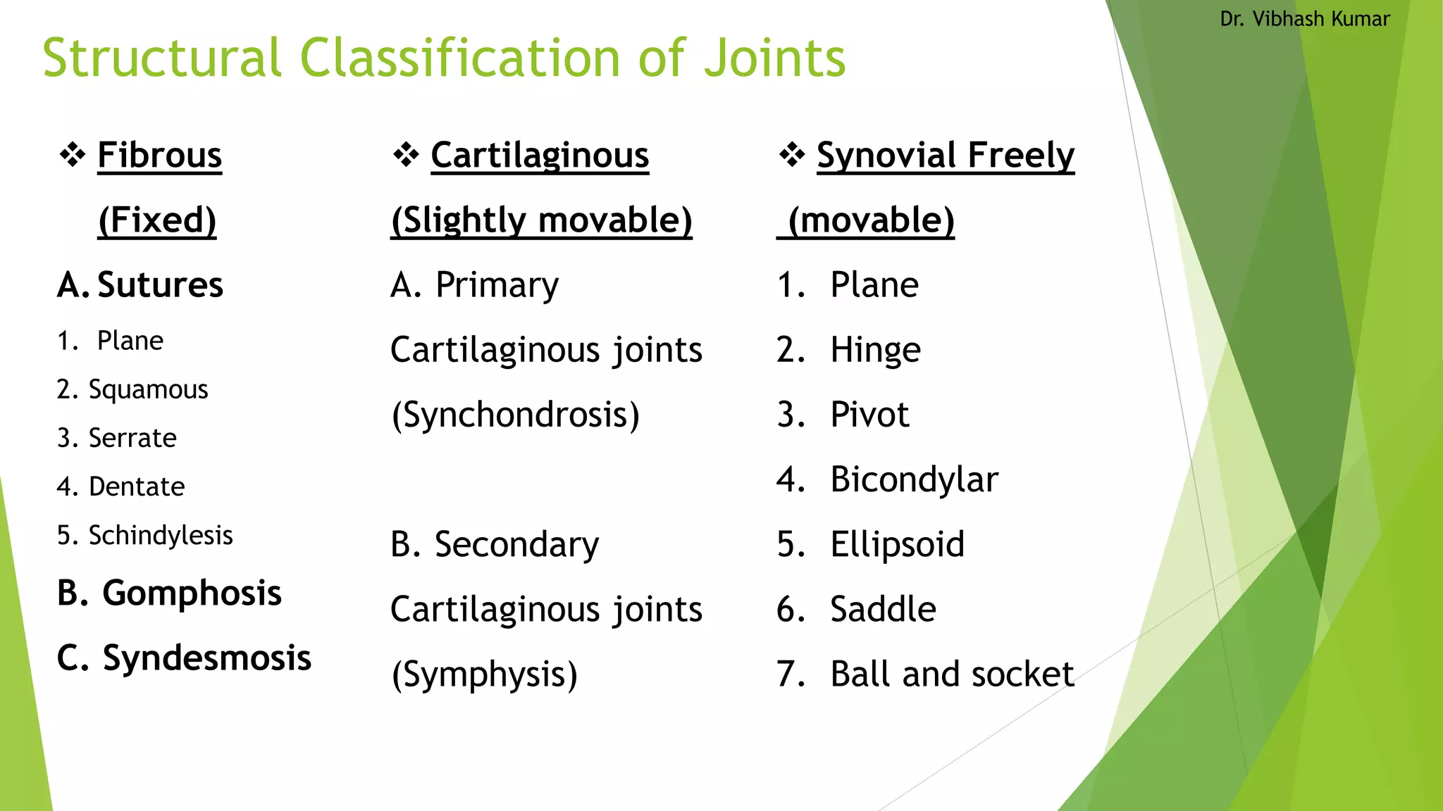

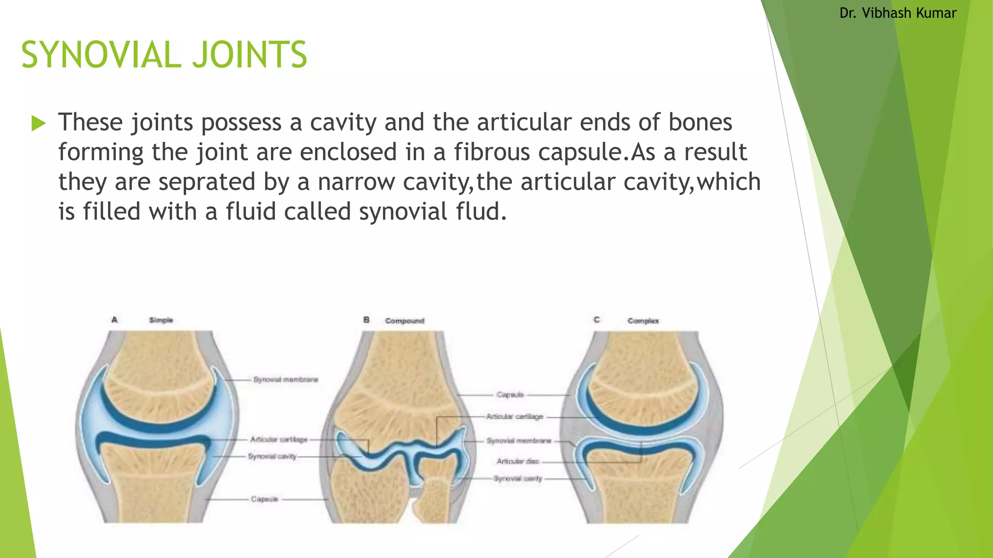
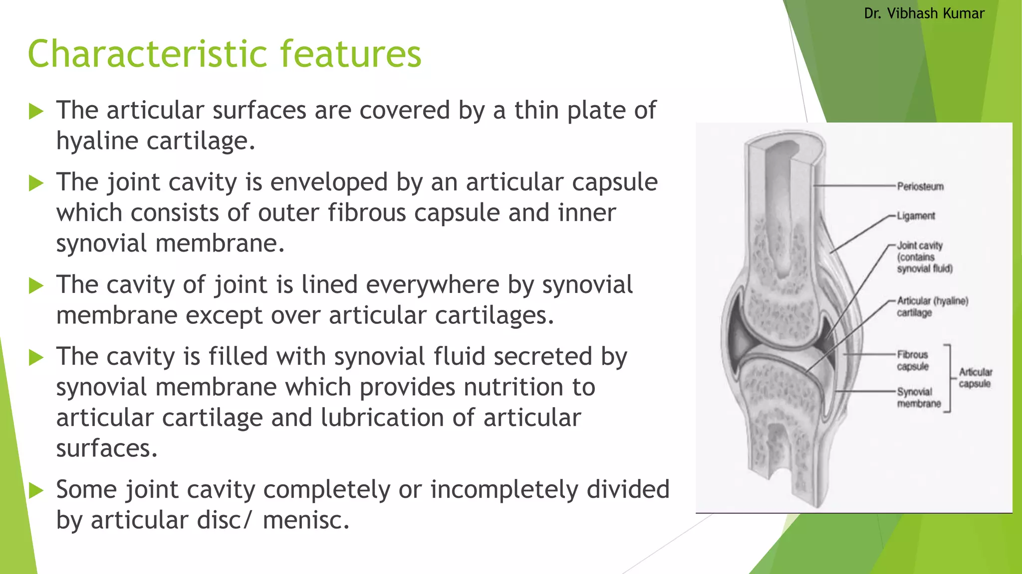


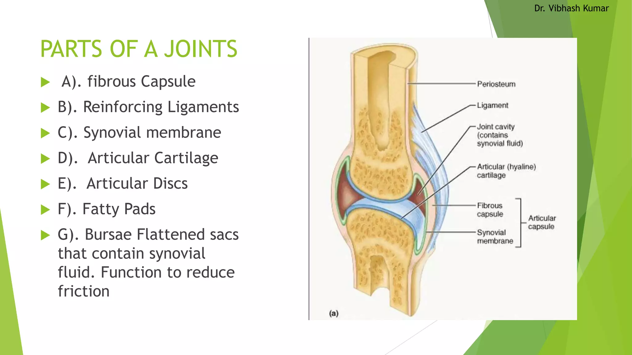


There are three main types of joints in the body - fibrous, cartilaginous, and synovial joints. Fibrous joints are immovable, cartilaginous joints allow slight movement, and synovial joints can move freely. Synovial joints are further classified by their shape and include hinge, ball-and-socket, and saddle joints. Each joint has a fibrous capsule, ligaments, synovial membrane, articular cartilage, and in some cases articular discs or bursae to facilitate movement and reduce friction between bones.


