Classification of Joints
•
31 likes•14,078 views

This document classifies and describes the main types of joints in the human body. It divides joints into four categories: head, trunk, upper limb, and lower limb. For each category it lists the specific joints and indicates whether they are fibrous, cartilaginous, or synovial joints. Synovial joints are further divided into typical and atypical types. Brief definitions are provided for the different joint types mentioned.

More Related Content
What's hot
What's hot (20)
More from Christiane Riedinger
More from Christiane Riedinger (20)
Classification of Joints
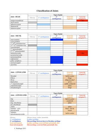
- 1. Classification of Joints Type of joint Joint - HEAD 2* Synovial Synovial Fibrous 1* cartilaginous cartilagenous (typical) (atypical*) temporomandibular + disc skull sutures sphenooccipital atlantoaxial atlantooccipital Type of joint Joint - TRUNK 2* Synovial Synovial Fibrous 1* cartilaginous cartilagenous (typical) (atypical*) intervertebral costovertebral 1st-10th costotransverse 11th 12th costotransverse costochondral 1st sternochondral 2nd-7th sternochondral sternoclavicular + disc acromioclavicular sternomanubrial xiphisternal pubic symphysis sacrococcygeal sacroiliac Type of joint Joint – UPPER LIMB 2* Synovial Synovial Fibrous 1* cartilaginous cartilagenous (typical) (atypical*) Shoulder ball & socket Elbow hinge Wrist ellipsoid, disc? Radioulnar pivot 1st carpometacarpal saddle condyloid carpometacarpal plane Interphalangeal hinge Type of joint Joint – LOWER LIMB 2* Synovial Synovial Fibrous 1* cartilaginous cartilagenous (typical) (atypical*) Hip Ball & socket Knee condyloid Sup. tibiofibular plane Inf. tibiofibular Ankle plane Subtalar – post Plane / ball & talocalcaneal socket Subtalar - ball & socket talocalcaneonavicular Fibrous fibrous tissue, rather immobile 1* cartilaginous hyaline cartilage 2* cartilaginous fibrocartilage between layers of hyaline cartilage Synovial (typical) hyaline cartilage, synovial fluid, potentially disc Synovial (atypical) fibrocartilage, synovial fluid, potentially disc C. Riedinger 2011