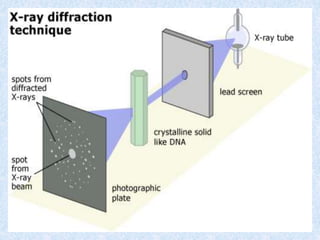
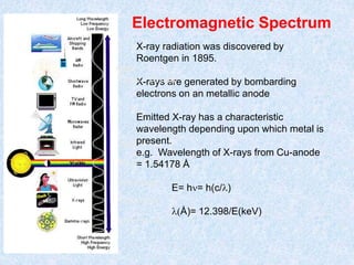
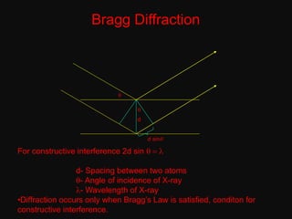
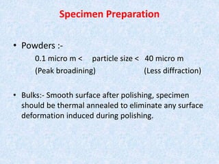
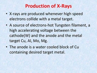
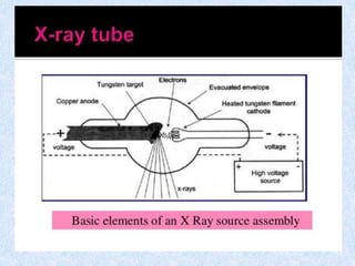
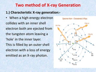
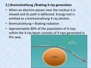
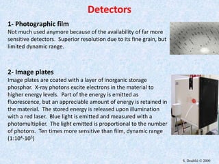
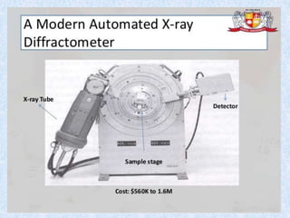
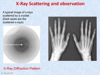
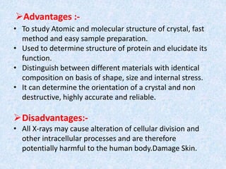
Artificial Intelligence In Microbiology by Dr. Prince C P
X ray diffraction
1. Presented by :- Faraz Ahmad Khan
(M.Sc. 3rd Semester)
“ X - Ray Diffraction ”Presented to :-
Dr. Dinesh K. Yadav
& Dr. Rajiv K. Yadav
2. Introduction
• X-Ray diffraction is a method of X-Ray
Crystallography, in which a beam of X-rays strikes
a sample (crystalline solid), land on a piece of a
film or other detector to produce scattered
beams.
• It is novel & non destructive method of chemical
analysis and a variety of X-Ray techniques in
practice.
• These are: 1.) X-Ray Absorption; 2.) X-Ray
Diffraction; 3.) X-Ray Fluorescence
3. • “Every crystalline substance gives a pattern;
the same substance always gives the same
pattern; and in a mixture of substance each
produces its pattern independently of the
others.”
• The X-Ray diffraction pattern of a pure
substance is, therefore, like a fingerprint of
the substance. It is based on the scattering of
x-rays by crystals.
• It shows the arrangement of atoms with in a
crystal.
4.
5. Electromagnetic Spectrum
10-1 to 10 nm
400 to 700 nm
10-4 to 10 -1 nm
10 to 400 nm
700 to 104 nm
X-ray radiation was discovered by
Roentgen in 1895.
X-rays are generated by bombarding
electrons on an metallic anode
Emitted X-ray has a characteristic
wavelength depending upon which metal is
present.
e.g. Wavelength of X-rays from Cu-anode
= 1.54178 Å
E= hn= h(c/l)
l(Å)= 12.398/E(keV)
NMR
10 um - 10 mm
6. Bragg Diffraction
d
q
q
d sinq
For constructive interference 2d sin q = l
d- Spacing between two atoms
q- Angle of incidence of X-ray
l- Wavelength of X-ray
•Diffraction occurs only when Bragg’s Law is satisfied, conditon for
constructive interference.
7. A protein crystal is placed in the x-ray beam
The x-rays are
diffracted by the
electron clouds
around atoms
The atomic structure
can be deduced from
the data
8. Specimen Preparation
• Powders :-
0.1 micro m < particle size < 40 micro m
(Peak broadining) (Less diffraction)
• Bulks:- Smooth surface after polishing, specimen
should be thermal annealed to eliminate any surface
deformation induced during polishing.
9. Production of X-Rays
• X-rays are produced whenever high speed
electrons collide with a metal target.
• A source of electrons-hot Tungsten filament, a
high accelerating voltage between the
cathode(W) and the anode and the metal
target Cu, Al, Mo, Mg.
• The anode is a water cooled block of Cu
containing desired target metal.
10.
11. Two method of X-ray Generation
1.) Characteristic X-ray generation:-
• When a high energy electron
collides with an inner shell
electron both are ejected from
the tungsten atom leaving a
'hole' in the inner layer.
This is filled by an outer shell
electron with a loss of energy
emitted as an X-ray photon.
12. 2.) Bremsstrahlung /Braking X-ray generation
• When an electron passes near the nucleus it is
slowed and its path is deflected. Energy lost is
emitted as a bremsstrahlung X-ray photon.
• Bremsstrahlung = Braking radiation
• Approximately 80% of the population of X-rays
within the X-ray beam consists of X-rays generated in
this way.
15. X-Ray Scattering and observation
A typical image of x-rays
scattered by a crystal:
(Dark spots are the
scattered x-rays)
X-Ray Diffraction Pattern
M. Rould ‘02
16. Advantages :-
• To study Atomic and molecular structure of crystal, fast
method and easy sample preparation.
• Used to determine structure of protein and elucidate its
function.
• Distinguish between different materials with identical
composition on basis of shape, size and internal stress.
• It can determine the orientation of a crystal and non
destructive, highly accurate and reliable.
Disadvantages:-
• All X-rays may cause alteration of cellular division and
other intracellular processes and are therefore
potentially harmful to the human body.Damage Skin.
17. Referances
• Principles and Techniques of Biochemistry and
Molecular Biology by Wilson and Walker.
• Fundamentals and Techniques of Biophysics
and Molecular Biology by Pranav Kumar.