


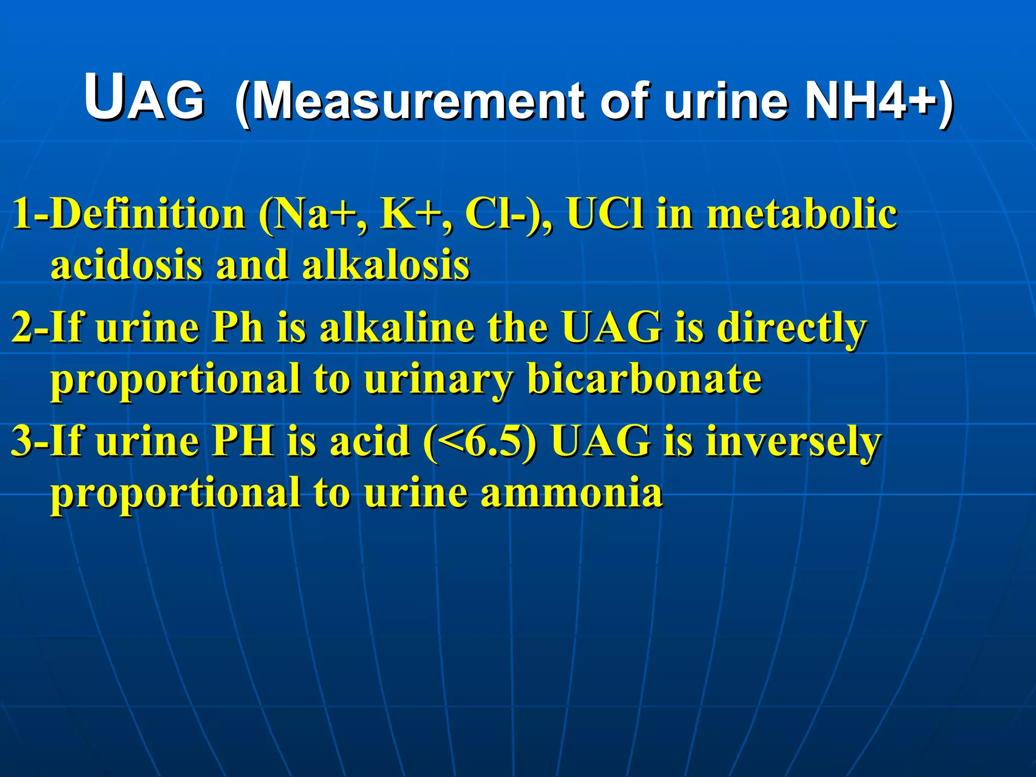
![AS a result, it is helpful to measure Ucl in a patient who appears to be hypovolemic but has a higher than expected UNa [like in m.Al due to vomiting that we have desire to excrete excess HCO3 (as Na-HCO3) to correct m.Al, this lead to a high UNa despite the presence of volume depletion and also UPH>6.5], in contrast we have Ucl in m.acidosis and hypovolemia (like in diarrhea) because of UNH4+ that excreted with chloride, but UNa is low.](https://image.slidesharecdn.com/urinalysis1-100811141145-phpapp01/75/Urinalysis-79-2048.jpg)


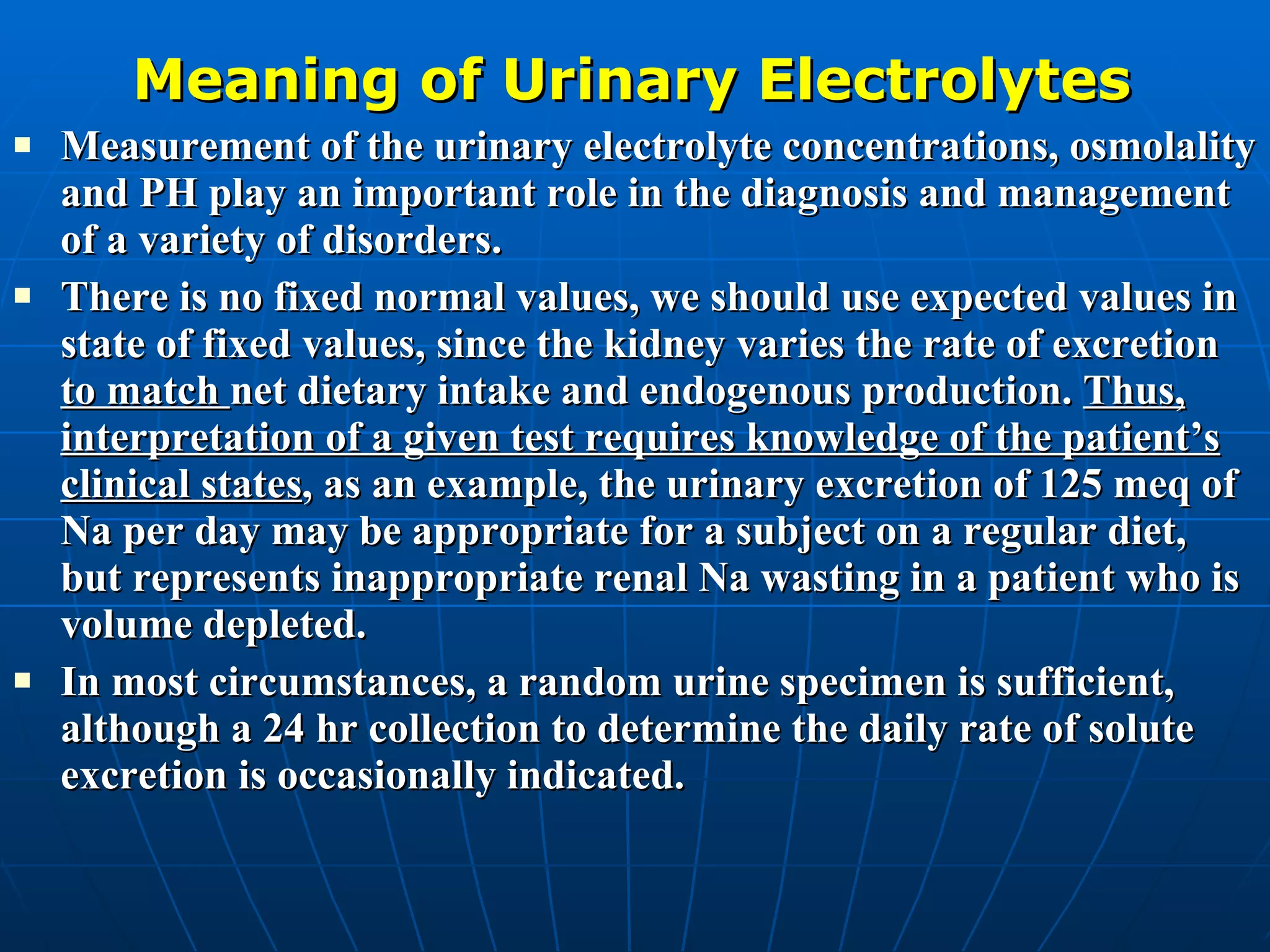
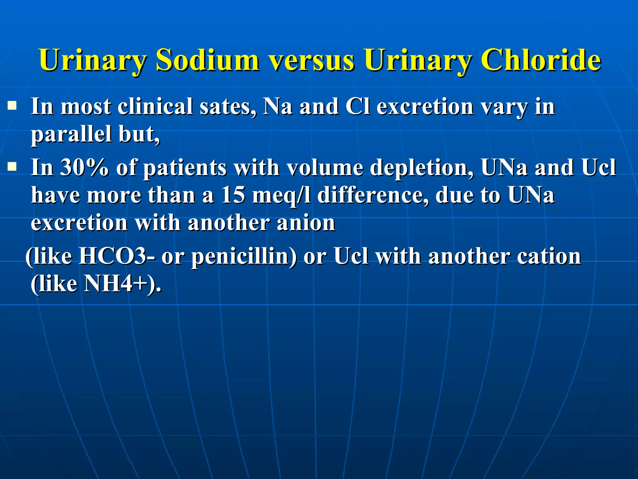
The document provides information on analyzing urinary electrolytes to evaluate extracellular volume status. It defines fractional excretion of sodium (FENa) and notes it is not dependent on urine volume and easy to calculate. While FENa and fractional excretion of chloride (FECl) generally vary in parallel, UNa and UCl can differ by more than 15 meq/L in 30% of volume depletion cases. Measuring both UNa and UCl is recommended in these situations to better evaluate volume status. Interpretation requires considering the clinical context as urinary electrolyte values considered normal may indicate inappropriate renal wasting in certain conditions.


