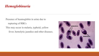
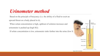
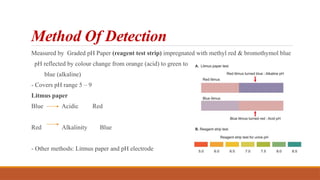
URINE EXAMINATION
Indications For Urinalysis
Suspected renal diseases like glomerulonephritis nephrotic syndrome, pyelonephritis, and renal
failure
Detection of urinary tract infection
Detection and management of metabolic disorders like diabetes mellitus
Differential diagnosis of jaundice
Detection and management of plasma cell dyscrasias
Diagnosis of pregnancy.
Composition of normal urine (24 hour)
in adults
Routine Urinalysis
Consists of two major components:
Physiochemical
examination
Microscopic
examination
Urine Collection
First morning, midstream: Routine urine examination.
Random, midstream: Routine urine examination.
First morning, midstream, clean catch: Bacteriological examination.
Postprandial: Estimation of glucose, urobilinogen
24-hour: Quantitative estimation of proteins or hormones.
Catheterised: Bacteriological examination in infants, bedridden patients, and in obstruction of
urinary tract.
Plastic bag (e.g. colostomy bag) tied around genitals: Infants; incontinent adults.
Time of analysis
Analyzed within 2h at room temp. or within 8hr at 2o
C- 8o
C
Changes which Occur in Standing Urine at Room Temperature
◦ Increase in pH due to production of ammonia from urea by urease-producing bacteria.
◦ Formation of crystals due to precipitation of phosphates and calcium (making the urine turbid)
◦ Loss of ketone bodies, since they are volatile.
◦ Decrease in glucose due to glycolysis and utilization of glucose by cells and bacteria.
◦ Oxidation of bilirubin to biliverdin causing false negative test for bilirubin
◦ Oxidation of urobilinogen to urobilin causing false negative test for urobilinogen
◦ Bacterial proliferation
Collection Of Urine Sample
For routine examination of urine, a wide-mouthed glass bottle of 20-30 ml capacity, which is dry,
chemically clean, leakproof, and with a tight fitting stopper is used.
Collection for bacterial culture
◦ Use sterile container
◦ Collect midstream, clean catch sample
◦ Must be plated within 2 hours of collection
◦ If refrigerated, must be plated within 24 hours of collection
◦ No preservative should be added.
Gross/Physical Examination
• Volume
• Odor
• Color
• Specific gravity
• Appearance
• pH
Urine Volume
The average adult produces from 600 to 2000 mL of urine per day, with night urine generally not
in excess of 400 mL.
Polyuria -Production of more than 2000 mL of urine in 24 hours
Nocturia- Excretion of more than 500 mL of urine at night with a specific gravity of less than
1.018
Oliguria- Excretion of less than 500 mL of urine per 24 hours
Anuria - Urinary output < 100 ml/24 hours or complete cessation of urine output. It occurs in
acute tubular necrosis (e.g. in shock, hemolytic transfusion reaction), acute glomerulonephritis,
and complete urinary tract obstruction..
Odor
Urine normally will have a faint, aromatic odor of undetermined source.
Amino acid disorders Odors
Cystinuria Rotten eggs
Tyrosinemia Rancid
Phenylketonuria Mousy,musty
Maple syrup urine disease (MSUD) Maple syrup
Isovaleric acidemia and glutaric acidemia Sweaty feet
Hawkinsinuria Swimming pool
Ketoacidosis Sweet, fruity
Appearance
Normal, freshly voided urine is clear in appearance.
Cause of cloudy or turbid urine-
Color
Normal urine color in a fresh state is pale yellow or amber due to the presence of
various pigments called urochrome.
Specific Gravity
Urea (20%), sodium chloride (25%), sulfate, and phosphate contribute most of the specific
gravity of normal urine.
Normal specific gravity 1.016 to 1.022
Several methods are available to measure specific gravity—
Reagent Strip,
Refractometer,
Urinometer, And
Falling Drop Method
Urinometer method
Based on the principle of buoyancy (i.e. the ability of a fluid to exert an
upward thrust on a body placed in it).
When solute concentration is high, upthrust of solution increases and
urinometer is pushed up (high SG).
If solute concentration is low, urinometer sinks further into the urine (low SG).
Reagent strip: Specific gravity
Reagent area has three main ingredients present: polyelectrolyte, indicator substance and buffer
Principle: pKa (acid dissociation constant) change of pre-treated polyelectrolytes in relation to the ionic
concentration of the urine
- Ionic concentration is high, the pKa is decreased, as is the pH
- Indicator substance then changes colour relative to ionic concentration, and this is translated to specific
gravity values
Urine pH
Healthy individuals: urine pH may vary from 4.6 to 8
Acidic –
◦ high protein intake, cranberries
◦ respiratory acidosis (CO2 retention)
◦ metabolic acidosis ( DKA, uremia, severe diarrhoea, starvation, UTI by E coli)
Alkaline –
◦ Respiratory alkalosis (hyperventilation)
◦ Metabolic alkalosis (vomiting, UTI by proteus, pseudomonas)
◦ Alkaline tide – post prandial reduction in acidity
Method Of Detection
Measured by Graded pH Paper (reagent test strip) impregnated with methyl red & bromothymol blue
pH reflected by colour change from orange (acid) to green to
blue (alkaline)
- Covers pH range 5 – 9
Litmus paper
Blue Acidic Red
Red Alkalinity Blue
- Other methods: Litmus paper and pH electrode
Chemical Screening
- Reagent strips :
- Primary method for chemical examination of urine
- Assess : pH, protein, sugar, ketones, blood, bilirubin,
urobilinogen, nitrites, leucocyte esterase,
ascorbic acids, porphyrins
Urine strip
Strip is filter paper or plastic which has chemical substance
(reagent) coated on it on different pads.
It gives color when react with substance in urine.
The produced color is compared with chart color visually or
mechanically assessed.
Glucose
Bilirubin
Ketones
Specific Gravity
Blood
pH
Protein
Urobilinogen
Nitrite
Leukocyte
Results are reported as:
In concentration (mg/dl)
As small, moderate, or large
Using the plus system (1+, 2+, 3+, 4+)
As positive, negative, or normal
Automated Urine Testing Machine
Urinalysis test strip
Strip include the tests:
Glucose
Bilirubin
Ketone
Specific Gravity
Blood
Protein
Urobilinogen
Nitrite
Leukocyte
pH
Proteinuria
Protein excretion in urine greater than 150 mg/24 hours in adults.
One third is albumin
Remaining plasma proteins include small globulins, such as α-, β-, and γ-globulins
Tamm Horsfall glycoprotein : secreted by distal tubular cells and cells of the ascending loop of Henle
constitutes one third or more of the total normal protein loss
Immunoglobulin A (IgA) in secretions of the urinary tract, enzymes, and proteins from tubular
epithelial cells, other desquamated cells and leukocytes
Urine Protein Electrophoresis
Selective proteinuria- passage of
comparatively lower molecular weight proteins
like albumin and transferrin
Nonselective proteinuria-larger molecular
weight proteins (γ globulins) are also excreted
along with albumin
Tubular proteinuria: low molecular weight
proteins like β2-microglobulin, retinol-binding
protein, lysozyme, α1-microglobulin, and free
immunoglobulin light chains(prominent α- and
βbands)
Heat precipitation test
Heat renders proteins insoluble & causes them to coagulate
Proteins are more susceptible to precipitation at pH near their isoelectric point
Fill 2/3 test tube with urine and hold bottom of test tube with holder
Boil upper portion of tube for 2 min
Cloudiness if appears: Protein/phosphate/carbonate
Add 3-5 drops 5-10% acetic acid + boil
If cloudiness disappears phosphate / carbonates
Methods:
Reagent Strip : - Proteins carry a charge at physiologic pH
- Presence will elicit a pH change
- Reagent strip is impregnated with tetrabromphenol blue buffered to an acid pH of 3 or
tetrachlorophenol - tetrabromosulfophthalein
- absence of protein, the strip is yellow
Sulfosalicylic Acid Method : - Specimen centrifuged and clear supernatant used
- To 3 mL of supernatant urine in clean test tube add equal amount of 3% SSA
- Invert to mix and let stand exactly 10 minutes
- Invert again twice and using ordinary room light (not a lamp) observe the degree of turbidity and/or
precipitation
- Interpretation:
Negative—no turbidity (≈5 mg/dL or less)
Trace—perceptible turbidity (≈20 mg/dL)
1+ —Distinct turbidity, but no discrete granulation (≈50 mg/dL)
2+ —Turbidity with granulation, but no flocculation (≈200 mg/dL)
3+ —Turbidity with granulation and flocculation (≈500 mg/dL)
4+ —Clumps of precipitated protein, or solid precipitate (≈1.0 g/dL or more)
Grading of proteinuria
Microalbuminuria
Presence of albumin in urine above the normal level but below the detectable range of
conventional urine dipstick methods.
urine albumin levels ranging from 20 to 200 mg/L
In diabetic patients, microalbuminuria is associated with a fourfold to sixfold increase in
cardiovascular mortality, and is an independent risk factor for renal mortality
Microalbuminuria cannot be detected by routine tests for proteinuria. Methods for detection
include:
Measurement of albumin-creatinine ratio in a random urine sample
Immunologic means using antibodies to the proteins, nephelometric methods,
radioimmunoassay, protein strip electrophoresis, high-performance liquid chromatography
(HPLC)
Bence Jones Proteinuria
Bence Jones proteins are monoclonal immunoglobulin light chains (either κ or λ) that are
synthesized by neoplastic plasma cells occurs in plasma cell dyscrasias like multiple myeloma and
primary amyloidosis.
Are excreted in urine because of low MW and
high concentration(overflow proteinuria).
When heated, Bence Jones proteins precipitate at
40°C to 60°C and precipitate disappears on
further heating at 85-100°C When cooled (60-85°C),
there is reappearance of precipitate of Bence
Jones proteins.
Glucose and Other Sugars in Urine :
Glucose - Presence of detectable amounts of glucose in urine is termed glycosuria
- Usually occurs when the blood level is greater than 180 to 200 mg/dL
Methods:
Reagent strip: Glucose oxidase and peroxidase method
- Reagent strips differ only in the chromogen used
- Method is specific for glucose
- Chemistry:
Copper Reduction Tests : Benedicts test and Clinitest
- Screening test glucose oxidase method will not detect increased levels of galactose or other
sugars in urine
- Benedict’s reagent: Copper sulphate 17.3 gm, sodium carbonate 100 gm, sodium citrate 173 gm,
d/w 1000 ml
- Copper reduction method be used especially for young paediatric patients
- Detects sufficient quantities of any reducing substances in the urine eg : lactose,fructose,
galactose, maltose and pentoses
- When urine is boiled in Benedict’s qualitative solution, blue alkaline copper sulphate is reduced
to red-brown cuprous oxide if a reducing agent is present
Interpretation
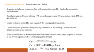
Blue solution – Negative
Green colour - Traces [<0.5gm/dL]
Green brown - + [0.5 - 1%]
yellow ppt - ++ [1 – 1.5%]
Orange ppt - +++ [1.5 - 2%]
Brick red ppt - ++++ [>2.0gm/dL]
Ketones in urine
3 ketone bodies present in the urine are acetoacetic (diacetic) acid (20%), acetone (2%), 3-hydroxybutyrate
(about 78%)
- Ketonuria: Diabetic and non-diabetic
- Methods: Nitroprusside strip and tablet tests based on the Rothera method
- Detect acetoacetic acid and acetone
- Ferric chloride (Gerhardt’s test) detects acetoacetic acid
Rothera Method
•Detects acetone/acetoacetic acid.
•Principle
- ketone bodies acetone and acetoacetic acid combine with
alkaline solution of sodium nitroprusside forming purple complex.
•Procedure:
•Take 5ml of urine in test tube
•Saturate with solid ammonium sulphate salt
•Add a few crystals of sodium nitroprusside and shake
•Add liquor ammonia from the side of the test tube
•Immediate formation of purple ring at junction indicates presence of
ketone bodies
•1-5mg/dl (acetoacetate), 10-25mg/dl (acetone)
Reagent Strip:
Nitroprusside (sodium nitroferricyanide) reaction for ketones
- Chemistrip reagent strips contain sodium nitroferricyanide and glycine which react with acetoacetic acid
and acetone in an alkaline medium to form a violet dye
- Positive result is indicated by a colour change from beige to violet, which is read at 60 seconds
- The method detects about 10mg/dL of acetoacetic acid and 70 mg/dL of acetone
Blood
The presence of abnormal number of intact red blood cells in urine is called as hematuria
Hemoglobinuria
Presence of heamoglobin in urine due to
rupturing of RBCs
This may occur in malaria, typhoid, yellow
fever, hemolytic jaundice and other diseases.
Chemical tests
Chemical tests are positive in hematuria, hemoglobinuria, and myoglobinuria.
Benzidine test: Make saturated solution of benzidine in glacial acetic acid. Mix 1 ml of this
solution with 1 ml of hydrogen peroxide in a test tube. Add 2 ml of urine. If green or blue color
develops within 5 minutes, the test is positive.
Orthotoluidine test: In this test, instead of benzidine, orthotoluidine is used. It is more sensitive
than benzidine test.
Reagent strip test: Various reagent strips are commercially available which use different
chromogens (o-toluidine, tetramethylbenzidine).
Reagent Strip for Heme Compounds (Hemoglobin, Myoglobin)
- Liberation of oxygen from peroxide in the reagent strip by the peroxidase-like activity of heme
in free hemoglobin, lysed erythrocytes, or myoglobin
- Reagent area is impregnated with buffered mixture of organic peroxide and the chromogen
tetramethylbenzidine
- Heme catalyzes the oxidation of tetramethylbenzidine to produce green colour
- Strip is read at 60 seconds following sample application
- Sensitivity is 0.05 to 0.3 mg Hb/dl
- 0.3 mg of hemoglobin per deciliter is equivalent to that from 10 lysed erythrocytes per microliter
Bilirubin in Urine
- Breakdown product of hemoglobin that is formed in the reticuloendothelial cells of the
spleen, liver and bone marrow
- Conjugated bilirubin is normally excreted in the bile into the duodenum
- Normal adult urine contains only 0.02 mg of bilirubin per deciliter
- Conjugated bilirubin appearing in urine : (1) obstruction to bile outflow from the liver
(intrahepatic or extrahepatic)
(2) hepatocellular disease with inability of hepatocytes to sufficiently excrete conjugated
bilirubin into bile
Methods:
Reagent Strip: Coupling reaction of bilirubin with a diazonium salt in acid medium
- Multistix uses diazotized 2,4-dichloroaniline with colour change from cream-buff to tan
after 20 seconds
- Detects even 0.8mg/dl of bilirubin
- Chemstrip uses 2,6-dichlorobenzene-diazonium tetrafluoroborate with colour change from
pink to violet at 30 to 60 seconds
- Detects even 0.5mg/dl of bilirubin
- Urine must be fresh
- Bilirubin glucuronide in urine quickly hydrolyzes to less reactive free bilirubin
- Large amounts of ascorbic acid and nitrite lower bilirubin results
Fouchet’s test
Take 5 ml of fresh urine in a test tube, add 2.5 ml of 10% of barium chloride, and mix well.
A precipitate of sulphates appears to which bilirubin is bound (barium sulphate-bilirubin
complex).
Filter to obtain the precipitate on a filter paper.
To the precipitate on the filter paper, add 1drop of Fouchet’s
reagent. (Fouchet’s reagent consists of 25 grams of
trichloroacetic acid, 10 ml of 10% ferric chloride, and
distilled water 100 ml).
Immediate development of blue-green color around
the drop indicates presence of bilirubin
Bile salts in urine
•Salts of cholic acid and chenodeoxycholoic acid. Normally not present in urine
Hay’s sulfur test:
•Bile salts lower the surface tension of the urine.
•Fill 50-100 ml beaker 2/3rd
to 3/4th
with urine.
•Sprinkle finely powdered sulfur powder which sinks if bile salts are present.
Cause: Obstructive jaundice and hepatocellular jaundice
Bile salts regurgitate into blood and excreted into urine
Urine leucocytes
This test detects any microbial infection in the body.
Depends on esterase method:
+ve result: means more than 5 leucocytes/hpf. (high power field)
If urine stand long time leucocytes lysis and more intense reaction occur.
False positives: occurs with vaginal contamination, presence of glucose, albumin, ascorbic
acid
Esterase + Ester 3-0H-5-phenyl pyrrole diazonium salt
pink -purple color
neutrophils reagent
strip
Microscopic Examination
Preparation of urine sediment for
microscopic examination
Urine Sample (12 ml) is centrifuged for 5
minutes at 1500 rpm and supernatant is poured
off
The tube is tapped at the bottom to resuspend
the sediment (in 0.5 ml of urine).
A drop of this is placed on a glass slide and
covered with a cover slip
The slide is examined under the microscope
using first the low power and then the high
power objective.
Erythrocytes
Fresh – normal, pale or yellowish, smooth biconcave discs without nuclei.
Hour glass appearance on side view
Dilute/hypotonic & alkaline urine – cells swell up – lyse – release Hb.
Lysed cells – ghost or shadow cells (faint circles – empty cell membranes).
Hypertonic urine – crenation of RBCS
Normal – 1-2 RBCs/HPF
If the RBCs have their typical shape &
color - hematuria associated with lower
urinary tract disease.
Dysmorphic RBCs indicate glomerular hematuria.
characterized by a great variation in the size of the cells
(anisocytosis) & many ghost cells
Leukocytes
Spherical, dull grey or greenish yellow.
Mostly neutrophils, identified by characteristic granules or nuclear lobulations
•Nuclei accentuated by 2% acetic acid
•Normal – 0-2 cells/ hpf
Renal tubular epithelial cells
Flat / cuboidal / columnar cells slightly larger
than leukocytes with large round nucleus
Increased no. suggests tubular damage –
pyelonephritis, ATN, salicylate toxicity,
transplant rejection
Transitional epithelial cell
Line the urinary tract from renal pelvis to upper portion of
the urethra
Round / pear shaped cells, 2 – 4 times larger than leukocytes
May have tail like projections ; Ocassionally 2 nuclei
Squamous epithelial cells
Most frequent; least significant
Large, flat, irregularly shaped cells with small
central nuclei & abundant cytoplasm
Cell edge often folded over & cell rolled up into
cylinder
Line the urethra & vagina
Urinary Casts
Formed in lumen of kidney tubules due to
◦ Precipitation of TammHorsfall protein
(mucoprotein secreted by renal tubules)
◦ Clumping of cells/ other material within a protein matrix
◦ Adherence of cells or material to matrix
◦ Conglutination of material within the lumen
◦ Some have serum proteins (as cast granules/ homogenous as in waxy casts)
indicator of intrinsic renal disease - glomerular/ tubular damage, renal inflammation/infection
Reported – no / LPF
Classified on basis of components into
MATRIX
CAST
Hyaline
Waxy
CELLULAR
CAST
Erythrocyte
Leukocyte
Renal tubular
epithelial cell cast
Mixed
INCLUSION
CAST
Granular
Fatty
Hemosiderin
Crystal
melanin
PIGMENT
CAST
Hb
Myoglobin
Bilirubin
drugs
TYPE OF
CAST
COMPOSITION ASSOCIATED CONDITIONS
Hyaline Mucoproteins Pyelonephritis, chronic renal disease
May be a normal finding
Erythrocyte Red blood
cells
Glomerulonephritis
Leukocyte White blood
cells
Pyelonephritis, glomerulonephritis,
interstitial nephritis, renal inflammatory
processes
Epithelial Renal tubule
cells
Acute tubular necrosis, interstitial
nephritis, eclampsia, nephritic syndrome,
allograft rejection, heavy metal ingestion,
renal disease
Type of
cast
Composition Associated conditions
Granular Various cell
types
Advanced renal disease
Waxy Various cell
types
Advanced renal disease
Fatty Lipid-laden
renal tubule cells
Nephrotic syndrome, renal disease,
hypothyroidism
Broad Various cell
types
End-stage renal disease
Hyaline casts
Most frequent
TammHorsfall protein
Very low refractive index
Colorless, homogenous, transparent
with rounded ends
Seen in renal disease; in normal urine
– following physical exercise/
physiological dehydration
Waxy casts
Very high refractive index
Yellow/grey/colorless, smooth
homogenous
Short broad casts with blunt or
broken ends with cracked/serrated
edges
Result of degeneration of granular
casts
Seen in CRF, malignant HT, renal
amyloidosis, Diabetic nephropathy,
acute renal disease, tubular
inflammation & degeneration, renal
allograft rejection
Red cell casts
Mean hematuria
Always pathologic
Brown to colorless
If RBCS intact –red cell cast
If degenerated – hemoglobin or blood
cast
Diagnostic of AGN, lupus nephritis,
Goodpasture’s syndrome, SABE, renal
trauma, infarction, severe
pyelonephritis, RVF, PAN
White cell casts
Majority – Polymorphonuclear neutrophils
Cells intact – nuclei clearly visible
Degenerated – cell membranes disappear
-- granular appearance
Renal infection & noninfectious
inflammation – acute pyelonephritis,
interstitial nephritis & lupus nephritis.
Also in glomerular disease
Epithelial cell casts
Result of stasis & desquamation of
renal tubular epithelial cells
After exposure to nephrotoxic
agents , viruses (CMV, hepatitis),
chronic renal disease (glomerular
injury + tubular damage), renal
allograft rejection
Cells arranged in parallel (come
from same segment of tubule)or
haphazard fashion in cast (come
from different portions of tubule)
Granular casts
Result of degeneration of cellular casts or
direct aggregation of serum proteins into
matrix of TammHorsfall mucoprotein
Large & coarse granules initially (black
due to density)
Fine granules due to prolonged stasis
(grey to pale yellow in color)
Occur in significant renal disease or after
strenous exercise
Fatty casts
Casts with incorporated free fat
droplets or oval fat bodies
Cholesterol - anisotropic droplets
which demonstrate characteristic
“maltese cross” formation under
polarized light
Triglyceride - stain with sudan III or
Oil Red O.
Seen in fatty degeneration of tubular
epithelium – degenerative tubular
disease.
Frequent in nephrotic syndrome,
diabetic glomerulosclerosis, CGN,
SLE, toxic renal poisoning
Crystals
Form when urine supersaturated leading to ppt of urinary salts due to alteration in factors
affecting their solubility – pH, temperature & concentration.
Precipitation in form of true crystals or amorphous material.
If precipitation occurs in urinary tract – urinary calculi
Formation is pH dependent hence knowledge of pH imp.
Identified by appearance, if necessary solubility characteristics
Alkaline pH Acidic pH
Triple phosphates Uric acid
Ammonium biurates Calcium oxalates
Calcium phosphates Cystine ,leucine, tyrosine
Amorphous phosphates Amorphous urates, Hippuric
acid, sodium urate, CaSO4
Calcium carbonates sulfa, cholesterol, bilirubin
Uric Acid Crystals
Diamond or rhombic prism, rosette
Yellow / red brown due to urinary
pigments
Soluble in NaOH, insoluble in HCl,
CH3COOH & alcohol
Occur in Normal urine/ pathological
states – gout, high purine
metabolism, acute febrile
conditions, chronic nephritis &
lesch nyhan syndrome
Calcium Oxalate Crystals
Colorless octahederal or “envelope”
shaped – small squares crossed by
intersecting diagonal lines
Rarely Oval spheres or biconcave discs –
look like dumb bells on side view
Soluble in HCl ; Insoluble in CH3COOH
Normal after ingestion of oxalate rich
foods – tomatoes, spinach, rhubarb,
garlic, oranges, asparagus & large dose
of vit C.
Pathologic – oxalate calculi, ethylene
glycol poisoning, DM, liver disease,
severe Chronic renal disease
Cystine Crystals
Colorless, refractile, hexagonal plates
Equal or unequal sides
Single/ on top of each other/ clusters
Soluble in HCl, alkali (NH3) – d/d uric acid crystal
Detected chemically with sodium cyanide-sodium nitroprusside test
Seen in congenital cystinosis, congenital cystinuria - calculi
Leucine
Oily, highly refractile, yellow or brown spheroids
with radial & concentric striations
Soluble in hot CH3COOH, hot alcohol, alkali
Insoluble in HCl
Found in maple syrup urine disease, terminal liver
cirrhosis, severe viral hepatitis
Tyrosine
Very fine, highly refractile needle in
sheaves or clusters
Black in centre
Yellow if bilirubin present
Soluble in NH4OH, HCl
Insoluble in CH3COOH
Occur in severe liver disease,
tyrosinosis
Amorphous urates
Urates of Na, K, Mg, Ca
yellow-red, granular
Soluble in alkali, at 600
C
Clinically insignificant
Hippuric acid crystals
Yellow brown or colorless
Elongated prisms/ plates
Resemble needles
Soluble in water, ether
No clinical significance
Ammonium Biurate
- yellow-brown color and appear as
spheres with radial or concentric
striations and irregular projections
or thorns (thorn apples)
- May also be seen in neutral and occasionally in
slightly acid urine
- They dissolve with heat at 60° C and with acetic
acid, reappearing as typical uric acid crystals
after about 20 minutes
Sodium urates
Amorphous / crystals
Colorless / yellowish needles
Soluble at 600
C, CH3COOH
No clinical significance
Calcium sulfate crystals
Long, thin, colorless needles or
prisms
d/d calcium phosphate crystals
(alkaline urine)
Soluble in CH3COOH
No clinical significance
Cholesterol
Large, flat, transparent plates
with notched corners
Variety of colors
Soluble in chloroform, ether,
hot alcohol
•Seen in nephritis, nephrotic conditions, chyluria
(due to thoracic or abdominal lymphatic drainage
– rupture of lymphatic vessels in renal pelvis or
urinary tract)
Sulfa drug crystals
Rare now due to soluble newer sulfa
drugs
Clear or brown Sheaves of needles
with eccentric binding
Soluble in acetone
Bilirubin
fine needles that regroup in a
clump or as red brown spheres
Triple Phosphates
Ammonium magnesium
phosphate
Colorless prisms with 3-5 sides
with oblique ends
Sometimes feathery or fern like
Soluble in CH3COOH
Occur in normal urine,
pathologic – chronic pyelitis,
chronic cystitis, enlarged
prostate, urinary retention
Calcium phosphate
- Long thin colorless prisms with one end pointed, as
rosettes or stars (stellate phosphate) or needles or
irregular granular plates
- May float on urine surface
- Soluble in CH3COOH
- Present normally or form calculi
Amorphous phosphates
Granular without any specific shape
Soluble in CH3COOH d/d amorphous
urates
No clinical significance
Calcium carbonate
Small, colorless dumb bell or spherical or
large granular
No clinical significance
Dissolve in CH3COOH with evolution of
CO2 gas
Parasites
Indigenous to urinary tract or vaginal / fecal contaminant
Trichomonas vaginalis – Most
frequent;
Flagellate, size of WBC.
Do not report in unstained mount
unless motile
Microfilariae - rare
Enterobius vermicularis ova – one
flat & one rounded side. Developing
larva may be seen through
transparent shell
Schistosoma hematobium
(Blood fluke in veins of urinary
bladder) eggs - Characteristic
terminal spine
Bacteria
Contaminant / denote UTI in
properly collected urine
Reported as no. / HPF
Yeast
• Smooth, colorless, ovoid cells
with doubly refractile walls
• Show budding ( v/s RBCs-
soluble in acid & alkali,
stain with eosin)
•UTI in DM
•Most common – candida albicans
Spermatozoa
oval bodies with long thin tail
Seen after epileptic convulsions /
nocturnal emissions /
spermatorrhea
Mucous threads
long, thin, wavy, ribbon like structures
with faint longitudinal striations
In normal urine / denote inflammation or
irritation of urinary tract
Thank You

URINE EXAMINATION and its analysis presentation.pptx

  • 1.
  • 2.
    Indications For Urinalysis Suspectedrenal diseases like glomerulonephritis nephrotic syndrome, pyelonephritis, and renal failure Detection of urinary tract infection Detection and management of metabolic disorders like diabetes mellitus Differential diagnosis of jaundice Detection and management of plasma cell dyscrasias Diagnosis of pregnancy.
  • 3.
    Composition of normalurine (24 hour) in adults
  • 4.
    Routine Urinalysis Consists oftwo major components: Physiochemical examination Microscopic examination
  • 5.
    Urine Collection First morning,midstream: Routine urine examination. Random, midstream: Routine urine examination. First morning, midstream, clean catch: Bacteriological examination. Postprandial: Estimation of glucose, urobilinogen 24-hour: Quantitative estimation of proteins or hormones. Catheterised: Bacteriological examination in infants, bedridden patients, and in obstruction of urinary tract. Plastic bag (e.g. colostomy bag) tied around genitals: Infants; incontinent adults.
  • 6.
    Time of analysis Analyzedwithin 2h at room temp. or within 8hr at 2o C- 8o C Changes which Occur in Standing Urine at Room Temperature ◦ Increase in pH due to production of ammonia from urea by urease-producing bacteria. ◦ Formation of crystals due to precipitation of phosphates and calcium (making the urine turbid) ◦ Loss of ketone bodies, since they are volatile. ◦ Decrease in glucose due to glycolysis and utilization of glucose by cells and bacteria. ◦ Oxidation of bilirubin to biliverdin causing false negative test for bilirubin ◦ Oxidation of urobilinogen to urobilin causing false negative test for urobilinogen ◦ Bacterial proliferation
  • 7.
    Collection Of UrineSample For routine examination of urine, a wide-mouthed glass bottle of 20-30 ml capacity, which is dry, chemically clean, leakproof, and with a tight fitting stopper is used. Collection for bacterial culture ◦ Use sterile container ◦ Collect midstream, clean catch sample ◦ Must be plated within 2 hours of collection ◦ If refrigerated, must be plated within 24 hours of collection ◦ No preservative should be added.
  • 8.
    Gross/Physical Examination • Volume •Odor • Color • Specific gravity • Appearance • pH
  • 9.
    Urine Volume The averageadult produces from 600 to 2000 mL of urine per day, with night urine generally not in excess of 400 mL. Polyuria -Production of more than 2000 mL of urine in 24 hours Nocturia- Excretion of more than 500 mL of urine at night with a specific gravity of less than 1.018 Oliguria- Excretion of less than 500 mL of urine per 24 hours Anuria - Urinary output < 100 ml/24 hours or complete cessation of urine output. It occurs in acute tubular necrosis (e.g. in shock, hemolytic transfusion reaction), acute glomerulonephritis, and complete urinary tract obstruction..
  • 10.
    Odor Urine normally willhave a faint, aromatic odor of undetermined source. Amino acid disorders Odors Cystinuria Rotten eggs Tyrosinemia Rancid Phenylketonuria Mousy,musty Maple syrup urine disease (MSUD) Maple syrup Isovaleric acidemia and glutaric acidemia Sweaty feet Hawkinsinuria Swimming pool Ketoacidosis Sweet, fruity
  • 11.
    Appearance Normal, freshly voidedurine is clear in appearance. Cause of cloudy or turbid urine-
  • 12.
    Color Normal urine colorin a fresh state is pale yellow or amber due to the presence of various pigments called urochrome.
  • 13.
    Specific Gravity Urea (20%),sodium chloride (25%), sulfate, and phosphate contribute most of the specific gravity of normal urine. Normal specific gravity 1.016 to 1.022 Several methods are available to measure specific gravity— Reagent Strip, Refractometer, Urinometer, And Falling Drop Method
  • 14.
    Urinometer method Based onthe principle of buoyancy (i.e. the ability of a fluid to exert an upward thrust on a body placed in it). When solute concentration is high, upthrust of solution increases and urinometer is pushed up (high SG). If solute concentration is low, urinometer sinks further into the urine (low SG).
  • 15.
    Reagent strip: Specificgravity Reagent area has three main ingredients present: polyelectrolyte, indicator substance and buffer Principle: pKa (acid dissociation constant) change of pre-treated polyelectrolytes in relation to the ionic concentration of the urine - Ionic concentration is high, the pKa is decreased, as is the pH - Indicator substance then changes colour relative to ionic concentration, and this is translated to specific gravity values
  • 16.
    Urine pH Healthy individuals:urine pH may vary from 4.6 to 8 Acidic – ◦ high protein intake, cranberries ◦ respiratory acidosis (CO2 retention) ◦ metabolic acidosis ( DKA, uremia, severe diarrhoea, starvation, UTI by E coli) Alkaline – ◦ Respiratory alkalosis (hyperventilation) ◦ Metabolic alkalosis (vomiting, UTI by proteus, pseudomonas) ◦ Alkaline tide – post prandial reduction in acidity
  • 17.
    Method Of Detection Measuredby Graded pH Paper (reagent test strip) impregnated with methyl red & bromothymol blue pH reflected by colour change from orange (acid) to green to blue (alkaline) - Covers pH range 5 – 9 Litmus paper Blue Acidic Red Red Alkalinity Blue - Other methods: Litmus paper and pH electrode
  • 18.
    Chemical Screening - Reagentstrips : - Primary method for chemical examination of urine - Assess : pH, protein, sugar, ketones, blood, bilirubin, urobilinogen, nitrites, leucocyte esterase, ascorbic acids, porphyrins
  • 19.
    Urine strip Strip isfilter paper or plastic which has chemical substance (reagent) coated on it on different pads. It gives color when react with substance in urine. The produced color is compared with chart color visually or mechanically assessed. Glucose Bilirubin Ketones Specific Gravity Blood pH Protein Urobilinogen Nitrite Leukocyte
  • 20.
    Results are reportedas: In concentration (mg/dl) As small, moderate, or large Using the plus system (1+, 2+, 3+, 4+) As positive, negative, or normal Automated Urine Testing Machine Urinalysis test strip
  • 21.
    Strip include thetests: Glucose Bilirubin Ketone Specific Gravity Blood Protein Urobilinogen Nitrite Leukocyte pH
  • 22.
    Proteinuria Protein excretion inurine greater than 150 mg/24 hours in adults. One third is albumin Remaining plasma proteins include small globulins, such as α-, β-, and γ-globulins Tamm Horsfall glycoprotein : secreted by distal tubular cells and cells of the ascending loop of Henle constitutes one third or more of the total normal protein loss Immunoglobulin A (IgA) in secretions of the urinary tract, enzymes, and proteins from tubular epithelial cells, other desquamated cells and leukocytes
  • 23.
    Urine Protein Electrophoresis Selectiveproteinuria- passage of comparatively lower molecular weight proteins like albumin and transferrin Nonselective proteinuria-larger molecular weight proteins (γ globulins) are also excreted along with albumin Tubular proteinuria: low molecular weight proteins like β2-microglobulin, retinol-binding protein, lysozyme, α1-microglobulin, and free immunoglobulin light chains(prominent α- and βbands)
  • 24.
    Heat precipitation test Heatrenders proteins insoluble & causes them to coagulate Proteins are more susceptible to precipitation at pH near their isoelectric point Fill 2/3 test tube with urine and hold bottom of test tube with holder Boil upper portion of tube for 2 min Cloudiness if appears: Protein/phosphate/carbonate Add 3-5 drops 5-10% acetic acid + boil If cloudiness disappears phosphate / carbonates
  • 25.
    Methods: Reagent Strip :- Proteins carry a charge at physiologic pH - Presence will elicit a pH change - Reagent strip is impregnated with tetrabromphenol blue buffered to an acid pH of 3 or tetrachlorophenol - tetrabromosulfophthalein - absence of protein, the strip is yellow
  • 26.
    Sulfosalicylic Acid Method: - Specimen centrifuged and clear supernatant used - To 3 mL of supernatant urine in clean test tube add equal amount of 3% SSA - Invert to mix and let stand exactly 10 minutes - Invert again twice and using ordinary room light (not a lamp) observe the degree of turbidity and/or precipitation - Interpretation: Negative—no turbidity (≈5 mg/dL or less) Trace—perceptible turbidity (≈20 mg/dL) 1+ —Distinct turbidity, but no discrete granulation (≈50 mg/dL) 2+ —Turbidity with granulation, but no flocculation (≈200 mg/dL) 3+ —Turbidity with granulation and flocculation (≈500 mg/dL) 4+ —Clumps of precipitated protein, or solid precipitate (≈1.0 g/dL or more)
  • 27.
  • 28.
    Microalbuminuria Presence of albuminin urine above the normal level but below the detectable range of conventional urine dipstick methods. urine albumin levels ranging from 20 to 200 mg/L In diabetic patients, microalbuminuria is associated with a fourfold to sixfold increase in cardiovascular mortality, and is an independent risk factor for renal mortality Microalbuminuria cannot be detected by routine tests for proteinuria. Methods for detection include: Measurement of albumin-creatinine ratio in a random urine sample Immunologic means using antibodies to the proteins, nephelometric methods, radioimmunoassay, protein strip electrophoresis, high-performance liquid chromatography (HPLC)
  • 29.
    Bence Jones Proteinuria BenceJones proteins are monoclonal immunoglobulin light chains (either κ or λ) that are synthesized by neoplastic plasma cells occurs in plasma cell dyscrasias like multiple myeloma and primary amyloidosis. Are excreted in urine because of low MW and high concentration(overflow proteinuria). When heated, Bence Jones proteins precipitate at 40°C to 60°C and precipitate disappears on further heating at 85-100°C When cooled (60-85°C), there is reappearance of precipitate of Bence Jones proteins.
  • 30.
    Glucose and OtherSugars in Urine : Glucose - Presence of detectable amounts of glucose in urine is termed glycosuria - Usually occurs when the blood level is greater than 180 to 200 mg/dL Methods: Reagent strip: Glucose oxidase and peroxidase method - Reagent strips differ only in the chromogen used - Method is specific for glucose - Chemistry:
  • 31.
    Copper Reduction Tests: Benedicts test and Clinitest - Screening test glucose oxidase method will not detect increased levels of galactose or other sugars in urine - Benedict’s reagent: Copper sulphate 17.3 gm, sodium carbonate 100 gm, sodium citrate 173 gm, d/w 1000 ml - Copper reduction method be used especially for young paediatric patients - Detects sufficient quantities of any reducing substances in the urine eg : lactose,fructose, galactose, maltose and pentoses - When urine is boiled in Benedict’s qualitative solution, blue alkaline copper sulphate is reduced to red-brown cuprous oxide if a reducing agent is present
  • 32.
    Interpretation Blue solution –Negative Green colour - Traces [<0.5gm/dL] Green brown - + [0.5 - 1%] yellow ppt - ++ [1 – 1.5%] Orange ppt - +++ [1.5 - 2%] Brick red ppt - ++++ [>2.0gm/dL]
  • 33.
    Ketones in urine 3ketone bodies present in the urine are acetoacetic (diacetic) acid (20%), acetone (2%), 3-hydroxybutyrate (about 78%) - Ketonuria: Diabetic and non-diabetic - Methods: Nitroprusside strip and tablet tests based on the Rothera method - Detect acetoacetic acid and acetone - Ferric chloride (Gerhardt’s test) detects acetoacetic acid
  • 34.
    Rothera Method •Detects acetone/acetoaceticacid. •Principle - ketone bodies acetone and acetoacetic acid combine with alkaline solution of sodium nitroprusside forming purple complex. •Procedure: •Take 5ml of urine in test tube •Saturate with solid ammonium sulphate salt •Add a few crystals of sodium nitroprusside and shake •Add liquor ammonia from the side of the test tube •Immediate formation of purple ring at junction indicates presence of ketone bodies •1-5mg/dl (acetoacetate), 10-25mg/dl (acetone)
  • 35.
    Reagent Strip: Nitroprusside (sodiumnitroferricyanide) reaction for ketones - Chemistrip reagent strips contain sodium nitroferricyanide and glycine which react with acetoacetic acid and acetone in an alkaline medium to form a violet dye - Positive result is indicated by a colour change from beige to violet, which is read at 60 seconds - The method detects about 10mg/dL of acetoacetic acid and 70 mg/dL of acetone
  • 36.
    Blood The presence ofabnormal number of intact red blood cells in urine is called as hematuria
  • 37.
    Hemoglobinuria Presence of heamoglobinin urine due to rupturing of RBCs This may occur in malaria, typhoid, yellow fever, hemolytic jaundice and other diseases.
  • 38.
    Chemical tests Chemical testsare positive in hematuria, hemoglobinuria, and myoglobinuria. Benzidine test: Make saturated solution of benzidine in glacial acetic acid. Mix 1 ml of this solution with 1 ml of hydrogen peroxide in a test tube. Add 2 ml of urine. If green or blue color develops within 5 minutes, the test is positive. Orthotoluidine test: In this test, instead of benzidine, orthotoluidine is used. It is more sensitive than benzidine test. Reagent strip test: Various reagent strips are commercially available which use different chromogens (o-toluidine, tetramethylbenzidine).
  • 39.
    Reagent Strip forHeme Compounds (Hemoglobin, Myoglobin) - Liberation of oxygen from peroxide in the reagent strip by the peroxidase-like activity of heme in free hemoglobin, lysed erythrocytes, or myoglobin - Reagent area is impregnated with buffered mixture of organic peroxide and the chromogen tetramethylbenzidine - Heme catalyzes the oxidation of tetramethylbenzidine to produce green colour - Strip is read at 60 seconds following sample application - Sensitivity is 0.05 to 0.3 mg Hb/dl - 0.3 mg of hemoglobin per deciliter is equivalent to that from 10 lysed erythrocytes per microliter
  • 40.
    Bilirubin in Urine -Breakdown product of hemoglobin that is formed in the reticuloendothelial cells of the spleen, liver and bone marrow - Conjugated bilirubin is normally excreted in the bile into the duodenum - Normal adult urine contains only 0.02 mg of bilirubin per deciliter - Conjugated bilirubin appearing in urine : (1) obstruction to bile outflow from the liver (intrahepatic or extrahepatic) (2) hepatocellular disease with inability of hepatocytes to sufficiently excrete conjugated bilirubin into bile
  • 41.
    Methods: Reagent Strip: Couplingreaction of bilirubin with a diazonium salt in acid medium - Multistix uses diazotized 2,4-dichloroaniline with colour change from cream-buff to tan after 20 seconds - Detects even 0.8mg/dl of bilirubin - Chemstrip uses 2,6-dichlorobenzene-diazonium tetrafluoroborate with colour change from pink to violet at 30 to 60 seconds - Detects even 0.5mg/dl of bilirubin - Urine must be fresh - Bilirubin glucuronide in urine quickly hydrolyzes to less reactive free bilirubin - Large amounts of ascorbic acid and nitrite lower bilirubin results
  • 42.
    Fouchet’s test Take 5ml of fresh urine in a test tube, add 2.5 ml of 10% of barium chloride, and mix well. A precipitate of sulphates appears to which bilirubin is bound (barium sulphate-bilirubin complex). Filter to obtain the precipitate on a filter paper. To the precipitate on the filter paper, add 1drop of Fouchet’s reagent. (Fouchet’s reagent consists of 25 grams of trichloroacetic acid, 10 ml of 10% ferric chloride, and distilled water 100 ml). Immediate development of blue-green color around the drop indicates presence of bilirubin
  • 43.
    Bile salts inurine •Salts of cholic acid and chenodeoxycholoic acid. Normally not present in urine Hay’s sulfur test: •Bile salts lower the surface tension of the urine. •Fill 50-100 ml beaker 2/3rd to 3/4th with urine. •Sprinkle finely powdered sulfur powder which sinks if bile salts are present. Cause: Obstructive jaundice and hepatocellular jaundice Bile salts regurgitate into blood and excreted into urine
  • 44.
    Urine leucocytes This testdetects any microbial infection in the body. Depends on esterase method: +ve result: means more than 5 leucocytes/hpf. (high power field) If urine stand long time leucocytes lysis and more intense reaction occur. False positives: occurs with vaginal contamination, presence of glucose, albumin, ascorbic acid Esterase + Ester 3-0H-5-phenyl pyrrole diazonium salt pink -purple color neutrophils reagent strip
  • 45.
  • 46.
    Preparation of urinesediment for microscopic examination Urine Sample (12 ml) is centrifuged for 5 minutes at 1500 rpm and supernatant is poured off The tube is tapped at the bottom to resuspend the sediment (in 0.5 ml of urine). A drop of this is placed on a glass slide and covered with a cover slip The slide is examined under the microscope using first the low power and then the high power objective.
  • 48.
    Erythrocytes Fresh – normal,pale or yellowish, smooth biconcave discs without nuclei. Hour glass appearance on side view Dilute/hypotonic & alkaline urine – cells swell up – lyse – release Hb. Lysed cells – ghost or shadow cells (faint circles – empty cell membranes). Hypertonic urine – crenation of RBCS Normal – 1-2 RBCs/HPF
  • 49.
    If the RBCshave their typical shape & color - hematuria associated with lower urinary tract disease. Dysmorphic RBCs indicate glomerular hematuria. characterized by a great variation in the size of the cells (anisocytosis) & many ghost cells
  • 50.
    Leukocytes Spherical, dull greyor greenish yellow. Mostly neutrophils, identified by characteristic granules or nuclear lobulations •Nuclei accentuated by 2% acetic acid •Normal – 0-2 cells/ hpf
  • 51.
    Renal tubular epithelialcells Flat / cuboidal / columnar cells slightly larger than leukocytes with large round nucleus Increased no. suggests tubular damage – pyelonephritis, ATN, salicylate toxicity, transplant rejection
  • 52.
    Transitional epithelial cell Linethe urinary tract from renal pelvis to upper portion of the urethra Round / pear shaped cells, 2 – 4 times larger than leukocytes May have tail like projections ; Ocassionally 2 nuclei
  • 53.
    Squamous epithelial cells Mostfrequent; least significant Large, flat, irregularly shaped cells with small central nuclei & abundant cytoplasm Cell edge often folded over & cell rolled up into cylinder Line the urethra & vagina
  • 54.
    Urinary Casts Formed inlumen of kidney tubules due to ◦ Precipitation of TammHorsfall protein (mucoprotein secreted by renal tubules) ◦ Clumping of cells/ other material within a protein matrix ◦ Adherence of cells or material to matrix ◦ Conglutination of material within the lumen ◦ Some have serum proteins (as cast granules/ homogenous as in waxy casts) indicator of intrinsic renal disease - glomerular/ tubular damage, renal inflammation/infection Reported – no / LPF
  • 55.
    Classified on basisof components into MATRIX CAST Hyaline Waxy CELLULAR CAST Erythrocyte Leukocyte Renal tubular epithelial cell cast Mixed INCLUSION CAST Granular Fatty Hemosiderin Crystal melanin PIGMENT CAST Hb Myoglobin Bilirubin drugs
  • 56.
    TYPE OF CAST COMPOSITION ASSOCIATEDCONDITIONS Hyaline Mucoproteins Pyelonephritis, chronic renal disease May be a normal finding Erythrocyte Red blood cells Glomerulonephritis Leukocyte White blood cells Pyelonephritis, glomerulonephritis, interstitial nephritis, renal inflammatory processes Epithelial Renal tubule cells Acute tubular necrosis, interstitial nephritis, eclampsia, nephritic syndrome, allograft rejection, heavy metal ingestion, renal disease
  • 57.
    Type of cast Composition Associatedconditions Granular Various cell types Advanced renal disease Waxy Various cell types Advanced renal disease Fatty Lipid-laden renal tubule cells Nephrotic syndrome, renal disease, hypothyroidism Broad Various cell types End-stage renal disease
  • 58.
    Hyaline casts Most frequent TammHorsfallprotein Very low refractive index Colorless, homogenous, transparent with rounded ends Seen in renal disease; in normal urine – following physical exercise/ physiological dehydration
  • 59.
    Waxy casts Very highrefractive index Yellow/grey/colorless, smooth homogenous Short broad casts with blunt or broken ends with cracked/serrated edges Result of degeneration of granular casts Seen in CRF, malignant HT, renal amyloidosis, Diabetic nephropathy, acute renal disease, tubular inflammation & degeneration, renal allograft rejection
  • 60.
    Red cell casts Meanhematuria Always pathologic Brown to colorless If RBCS intact –red cell cast If degenerated – hemoglobin or blood cast Diagnostic of AGN, lupus nephritis, Goodpasture’s syndrome, SABE, renal trauma, infarction, severe pyelonephritis, RVF, PAN
  • 61.
    White cell casts Majority– Polymorphonuclear neutrophils Cells intact – nuclei clearly visible Degenerated – cell membranes disappear -- granular appearance Renal infection & noninfectious inflammation – acute pyelonephritis, interstitial nephritis & lupus nephritis. Also in glomerular disease
  • 62.
    Epithelial cell casts Resultof stasis & desquamation of renal tubular epithelial cells After exposure to nephrotoxic agents , viruses (CMV, hepatitis), chronic renal disease (glomerular injury + tubular damage), renal allograft rejection Cells arranged in parallel (come from same segment of tubule)or haphazard fashion in cast (come from different portions of tubule)
  • 63.
    Granular casts Result ofdegeneration of cellular casts or direct aggregation of serum proteins into matrix of TammHorsfall mucoprotein Large & coarse granules initially (black due to density) Fine granules due to prolonged stasis (grey to pale yellow in color) Occur in significant renal disease or after strenous exercise
  • 64.
    Fatty casts Casts withincorporated free fat droplets or oval fat bodies Cholesterol - anisotropic droplets which demonstrate characteristic “maltese cross” formation under polarized light Triglyceride - stain with sudan III or Oil Red O. Seen in fatty degeneration of tubular epithelium – degenerative tubular disease. Frequent in nephrotic syndrome, diabetic glomerulosclerosis, CGN, SLE, toxic renal poisoning
  • 65.
    Crystals Form when urinesupersaturated leading to ppt of urinary salts due to alteration in factors affecting their solubility – pH, temperature & concentration. Precipitation in form of true crystals or amorphous material. If precipitation occurs in urinary tract – urinary calculi Formation is pH dependent hence knowledge of pH imp. Identified by appearance, if necessary solubility characteristics
  • 66.
    Alkaline pH AcidicpH Triple phosphates Uric acid Ammonium biurates Calcium oxalates Calcium phosphates Cystine ,leucine, tyrosine Amorphous phosphates Amorphous urates, Hippuric acid, sodium urate, CaSO4 Calcium carbonates sulfa, cholesterol, bilirubin
  • 67.
    Uric Acid Crystals Diamondor rhombic prism, rosette Yellow / red brown due to urinary pigments Soluble in NaOH, insoluble in HCl, CH3COOH & alcohol Occur in Normal urine/ pathological states – gout, high purine metabolism, acute febrile conditions, chronic nephritis & lesch nyhan syndrome
  • 68.
    Calcium Oxalate Crystals Colorlessoctahederal or “envelope” shaped – small squares crossed by intersecting diagonal lines Rarely Oval spheres or biconcave discs – look like dumb bells on side view Soluble in HCl ; Insoluble in CH3COOH Normal after ingestion of oxalate rich foods – tomatoes, spinach, rhubarb, garlic, oranges, asparagus & large dose of vit C. Pathologic – oxalate calculi, ethylene glycol poisoning, DM, liver disease, severe Chronic renal disease
  • 69.
    Cystine Crystals Colorless, refractile,hexagonal plates Equal or unequal sides Single/ on top of each other/ clusters Soluble in HCl, alkali (NH3) – d/d uric acid crystal Detected chemically with sodium cyanide-sodium nitroprusside test Seen in congenital cystinosis, congenital cystinuria - calculi
  • 70.
    Leucine Oily, highly refractile,yellow or brown spheroids with radial & concentric striations Soluble in hot CH3COOH, hot alcohol, alkali Insoluble in HCl Found in maple syrup urine disease, terminal liver cirrhosis, severe viral hepatitis
  • 71.
    Tyrosine Very fine, highlyrefractile needle in sheaves or clusters Black in centre Yellow if bilirubin present Soluble in NH4OH, HCl Insoluble in CH3COOH Occur in severe liver disease, tyrosinosis
  • 72.
    Amorphous urates Urates ofNa, K, Mg, Ca yellow-red, granular Soluble in alkali, at 600 C Clinically insignificant Hippuric acid crystals Yellow brown or colorless Elongated prisms/ plates Resemble needles Soluble in water, ether No clinical significance
  • 73.
    Ammonium Biurate - yellow-browncolor and appear as spheres with radial or concentric striations and irregular projections or thorns (thorn apples) - May also be seen in neutral and occasionally in slightly acid urine - They dissolve with heat at 60° C and with acetic acid, reappearing as typical uric acid crystals after about 20 minutes
  • 74.
    Sodium urates Amorphous /crystals Colorless / yellowish needles Soluble at 600 C, CH3COOH No clinical significance Calcium sulfate crystals Long, thin, colorless needles or prisms d/d calcium phosphate crystals (alkaline urine) Soluble in CH3COOH No clinical significance
  • 75.
    Cholesterol Large, flat, transparentplates with notched corners Variety of colors Soluble in chloroform, ether, hot alcohol •Seen in nephritis, nephrotic conditions, chyluria (due to thoracic or abdominal lymphatic drainage – rupture of lymphatic vessels in renal pelvis or urinary tract)
  • 76.
    Sulfa drug crystals Rarenow due to soluble newer sulfa drugs Clear or brown Sheaves of needles with eccentric binding Soluble in acetone Bilirubin fine needles that regroup in a clump or as red brown spheres
  • 77.
    Triple Phosphates Ammonium magnesium phosphate Colorlessprisms with 3-5 sides with oblique ends Sometimes feathery or fern like Soluble in CH3COOH Occur in normal urine, pathologic – chronic pyelitis, chronic cystitis, enlarged prostate, urinary retention
  • 78.
    Calcium phosphate - Longthin colorless prisms with one end pointed, as rosettes or stars (stellate phosphate) or needles or irregular granular plates - May float on urine surface - Soluble in CH3COOH - Present normally or form calculi
  • 79.
    Amorphous phosphates Granular withoutany specific shape Soluble in CH3COOH d/d amorphous urates No clinical significance Calcium carbonate Small, colorless dumb bell or spherical or large granular No clinical significance Dissolve in CH3COOH with evolution of CO2 gas
  • 80.
    Parasites Indigenous to urinarytract or vaginal / fecal contaminant Trichomonas vaginalis – Most frequent; Flagellate, size of WBC. Do not report in unstained mount unless motile Microfilariae - rare
  • 81.
    Enterobius vermicularis ova– one flat & one rounded side. Developing larva may be seen through transparent shell Schistosoma hematobium (Blood fluke in veins of urinary bladder) eggs - Characteristic terminal spine
  • 82.
    Bacteria Contaminant / denoteUTI in properly collected urine Reported as no. / HPF Yeast • Smooth, colorless, ovoid cells with doubly refractile walls • Show budding ( v/s RBCs- soluble in acid & alkali, stain with eosin) •UTI in DM •Most common – candida albicans
  • 83.
    Spermatozoa oval bodies withlong thin tail Seen after epileptic convulsions / nocturnal emissions / spermatorrhea Mucous threads long, thin, wavy, ribbon like structures with faint longitudinal striations In normal urine / denote inflammation or irritation of urinary tract
  • 84.