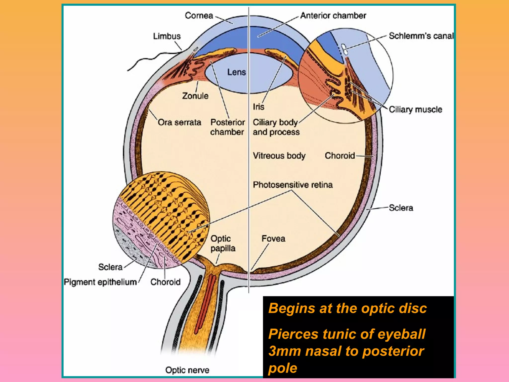
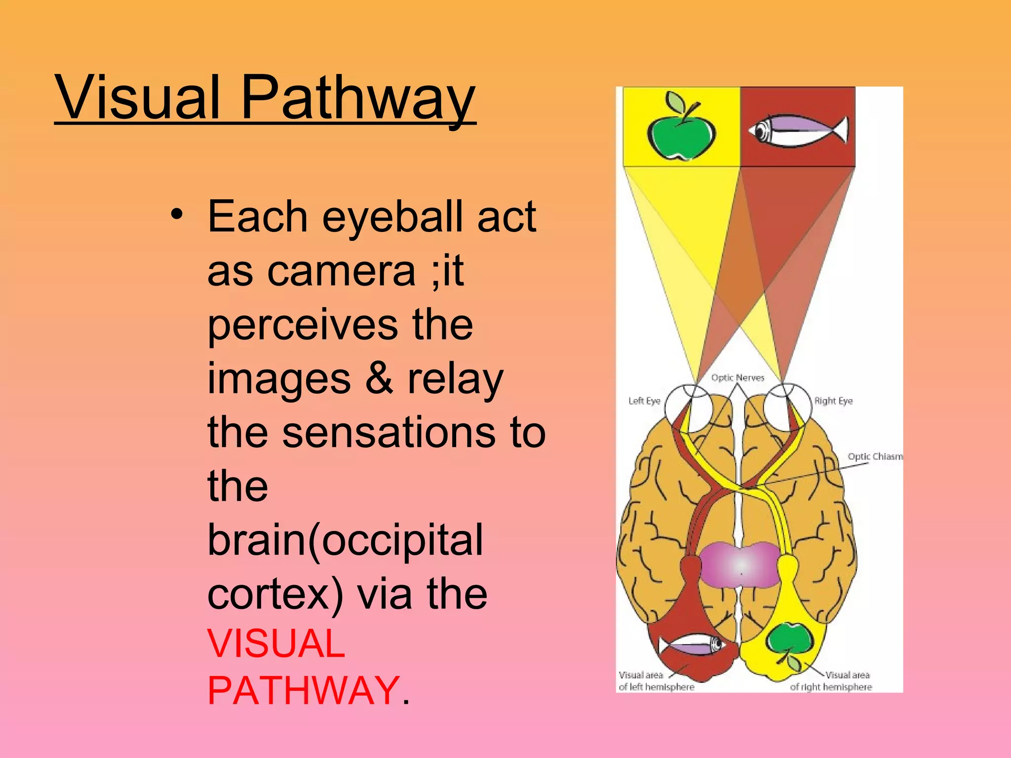
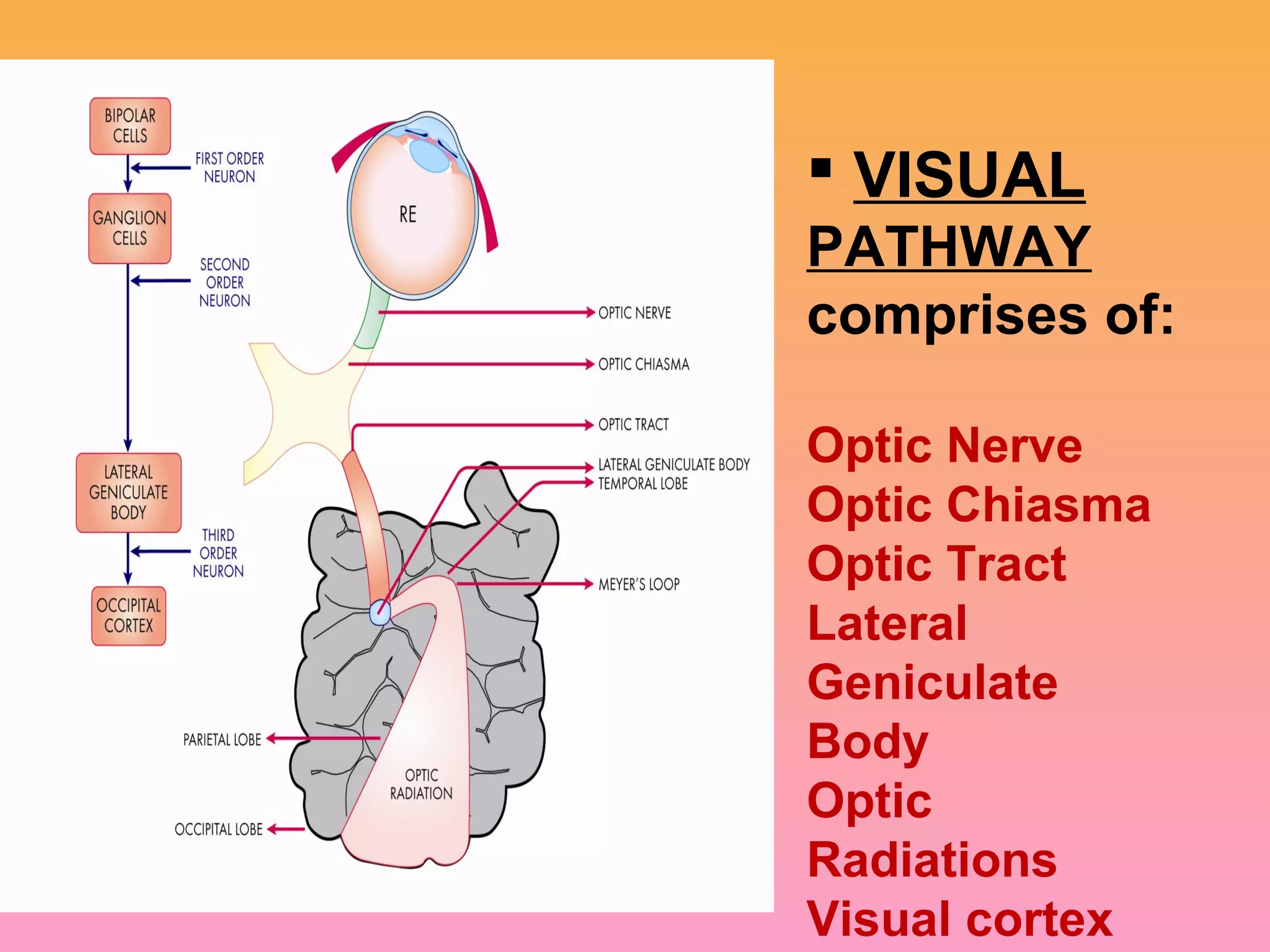
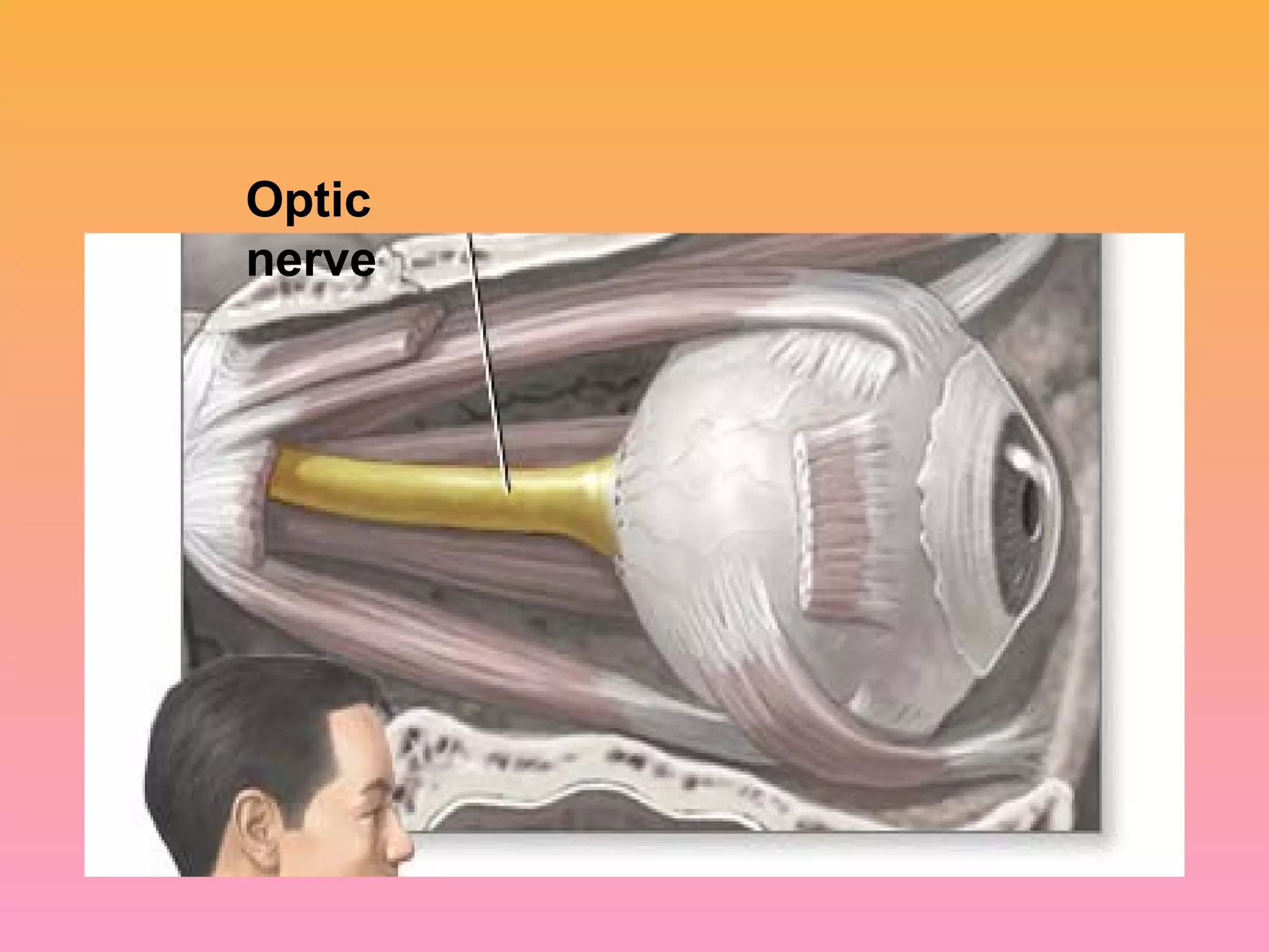
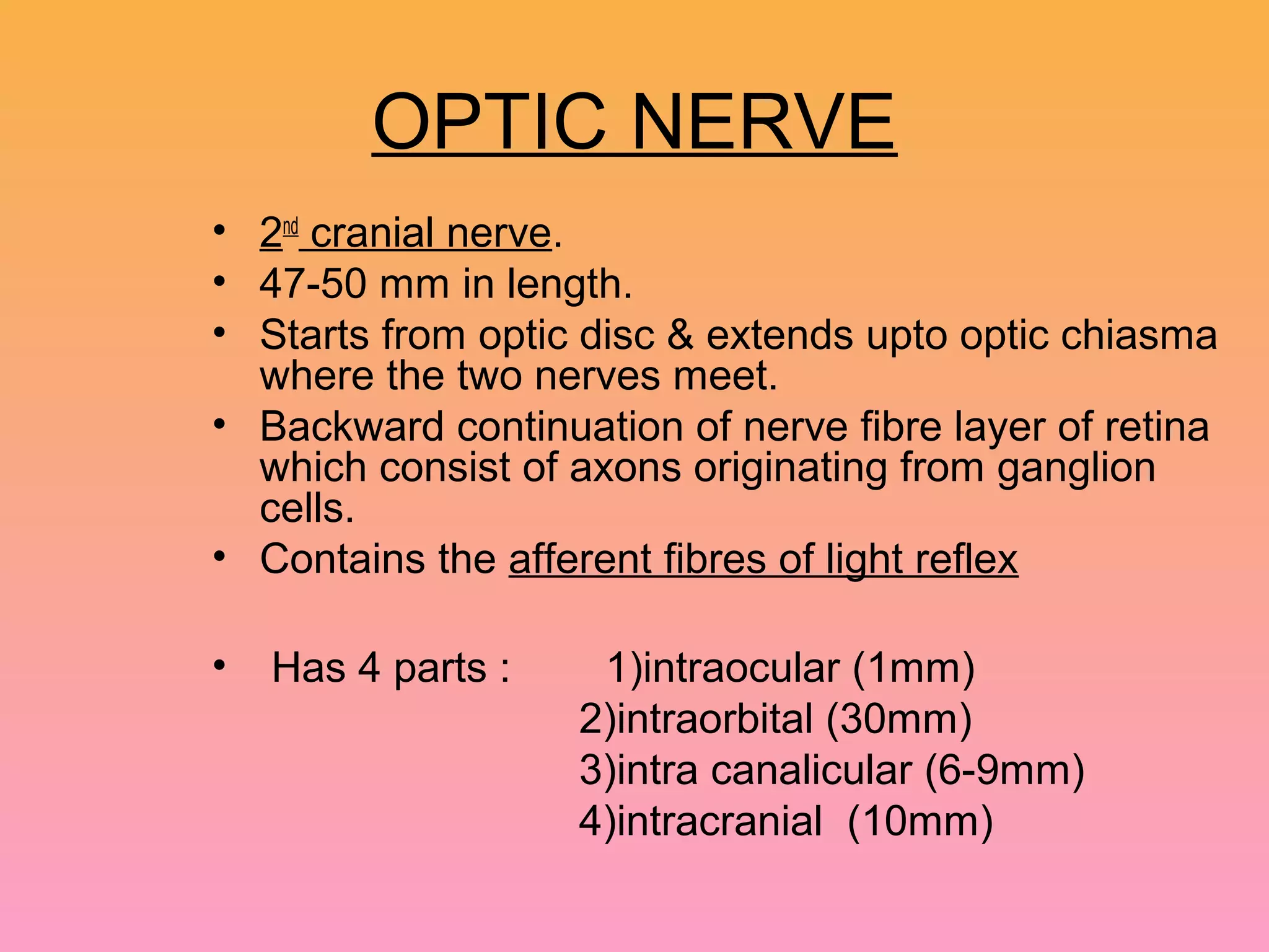
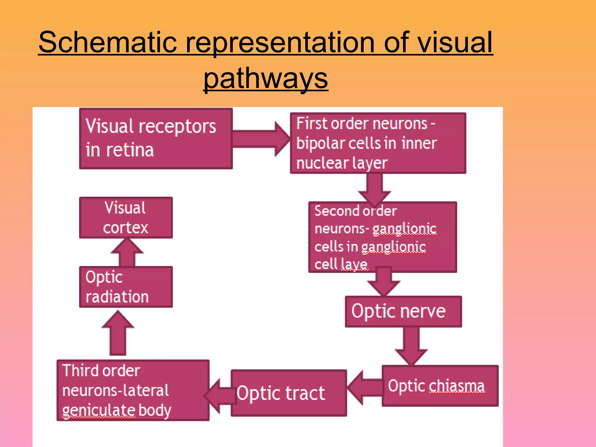
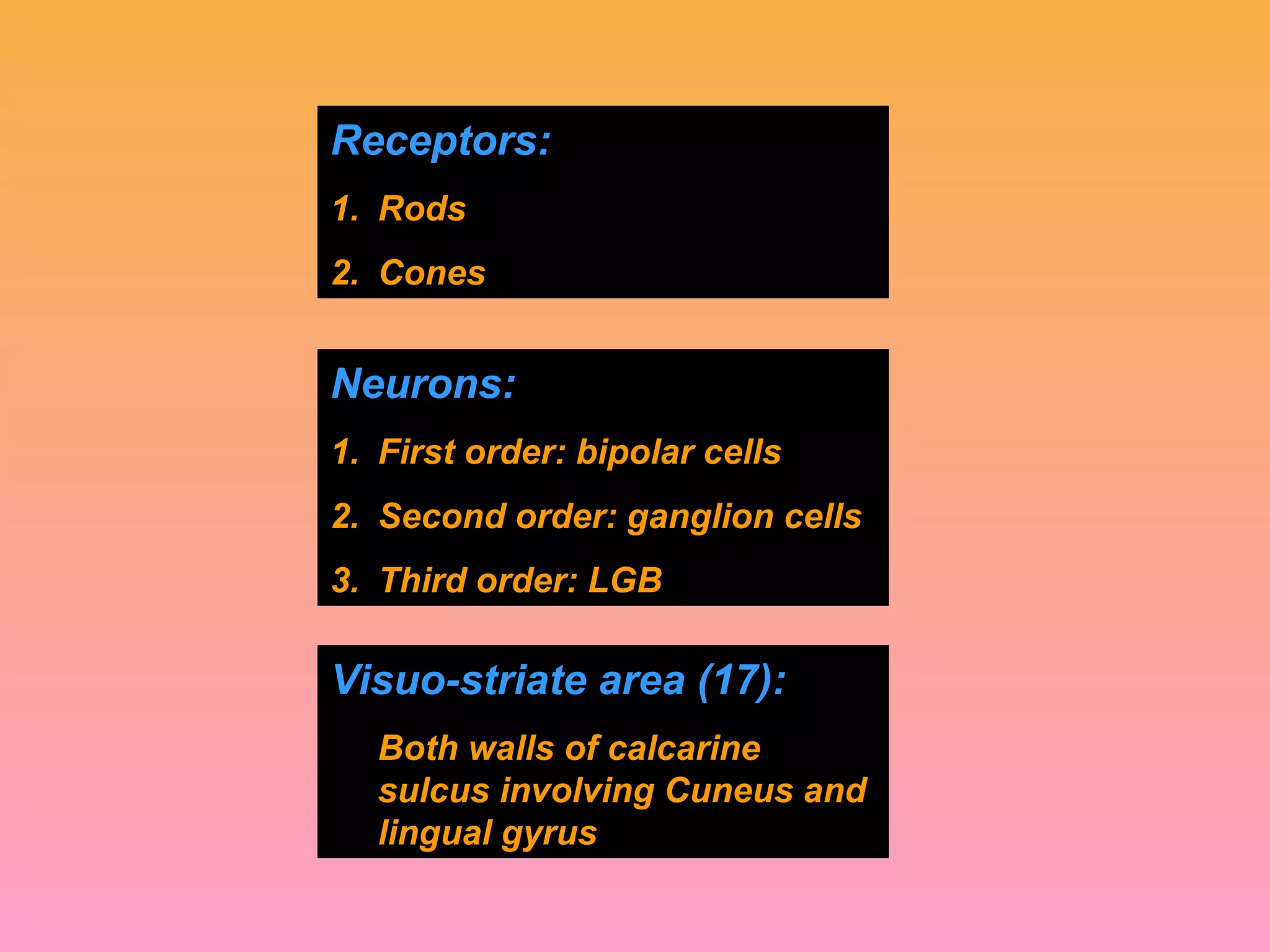
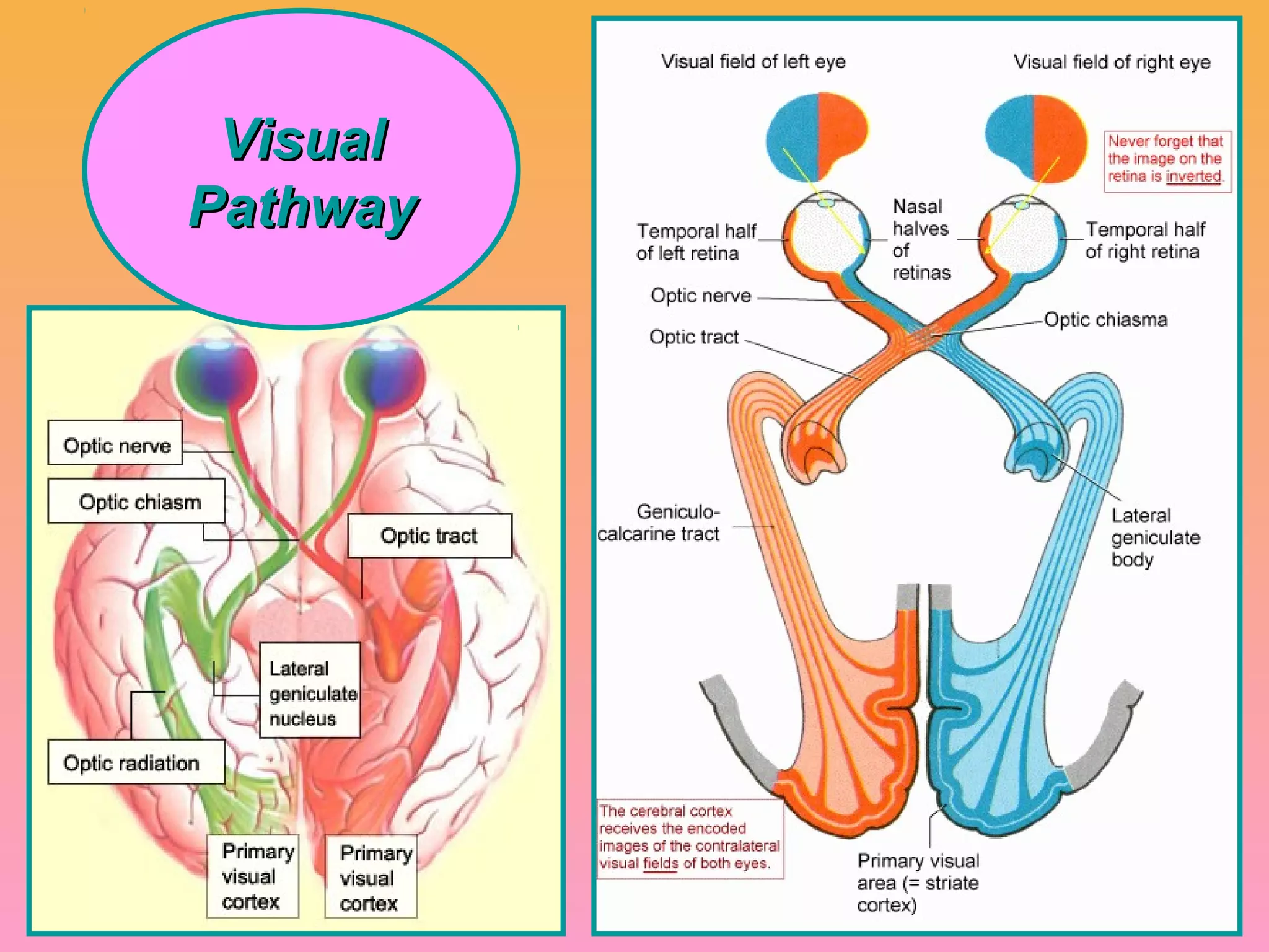
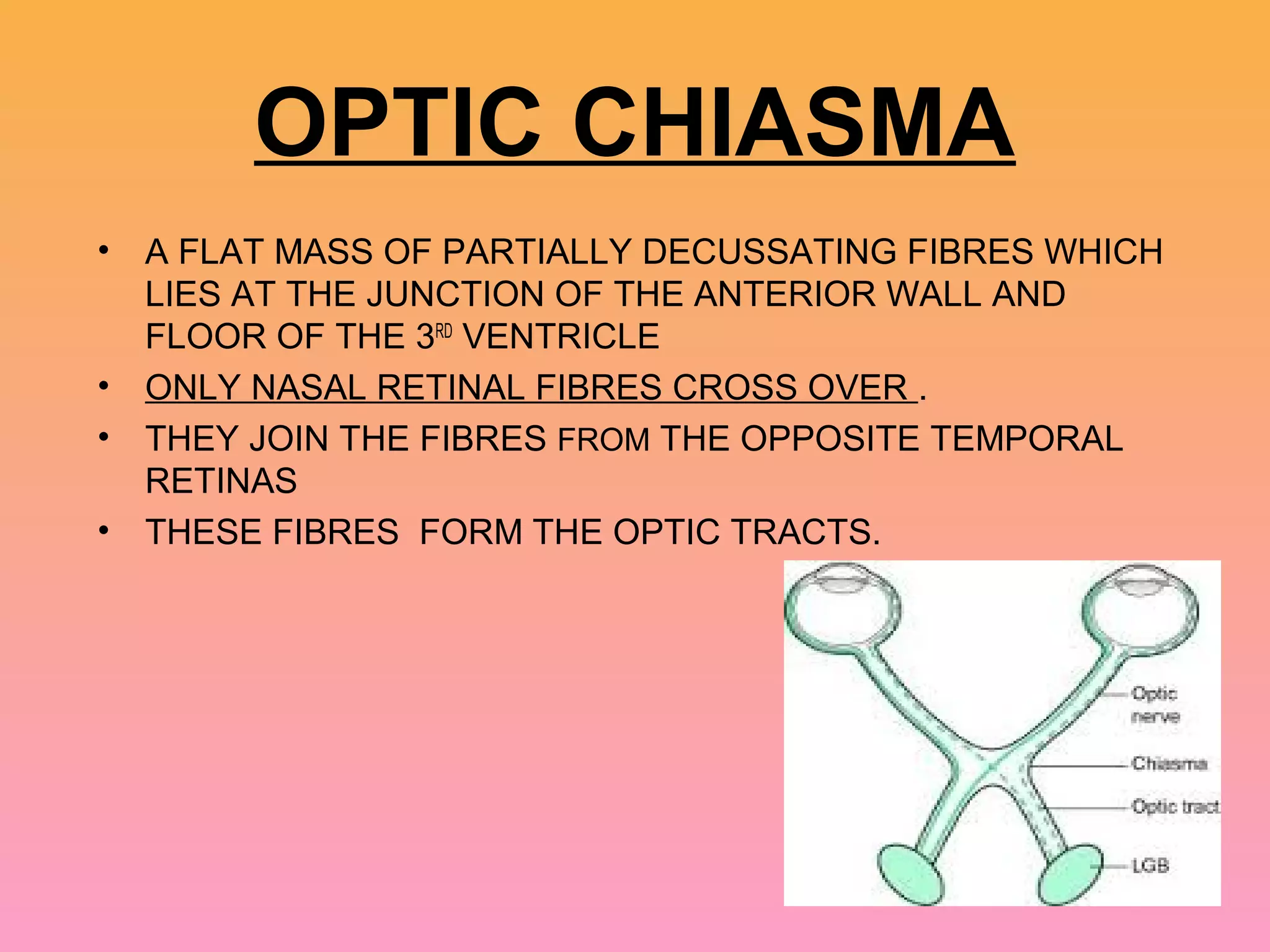
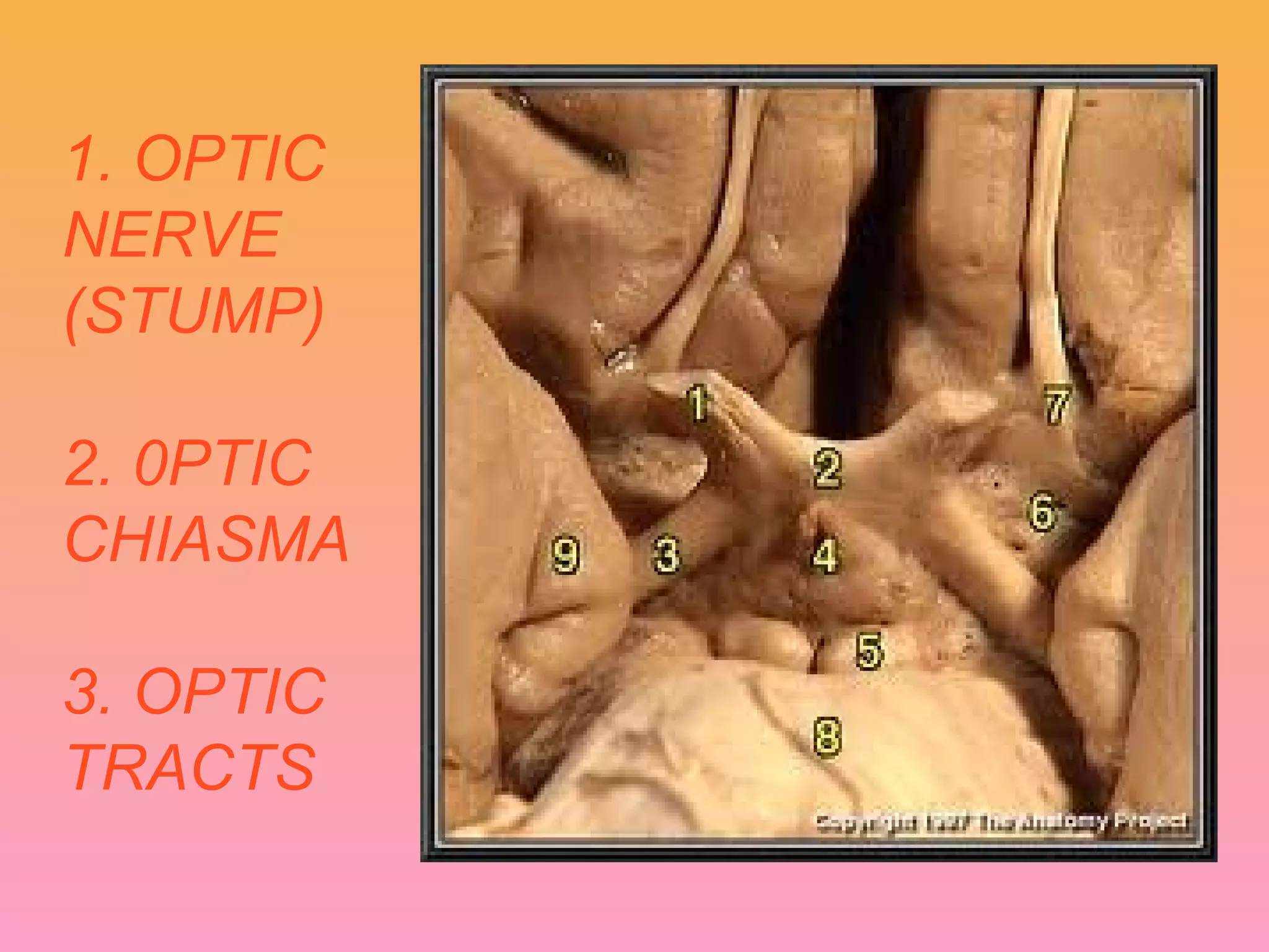
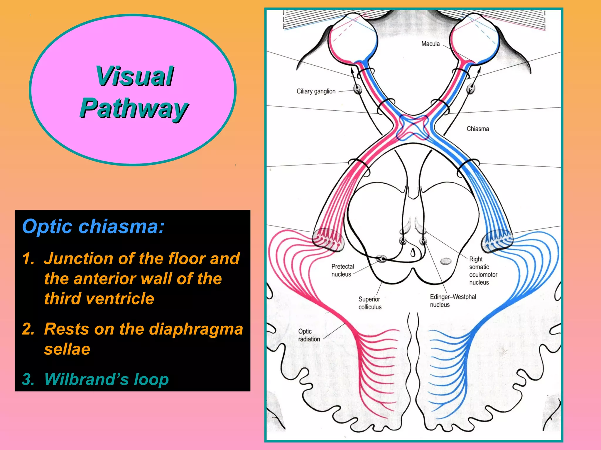
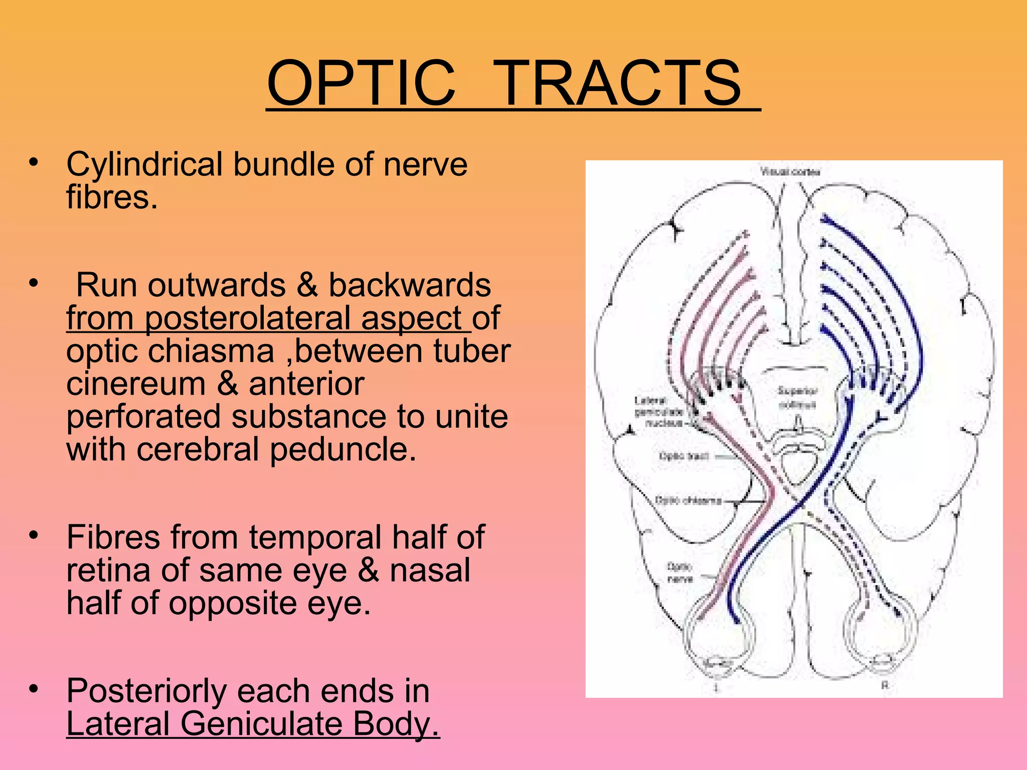
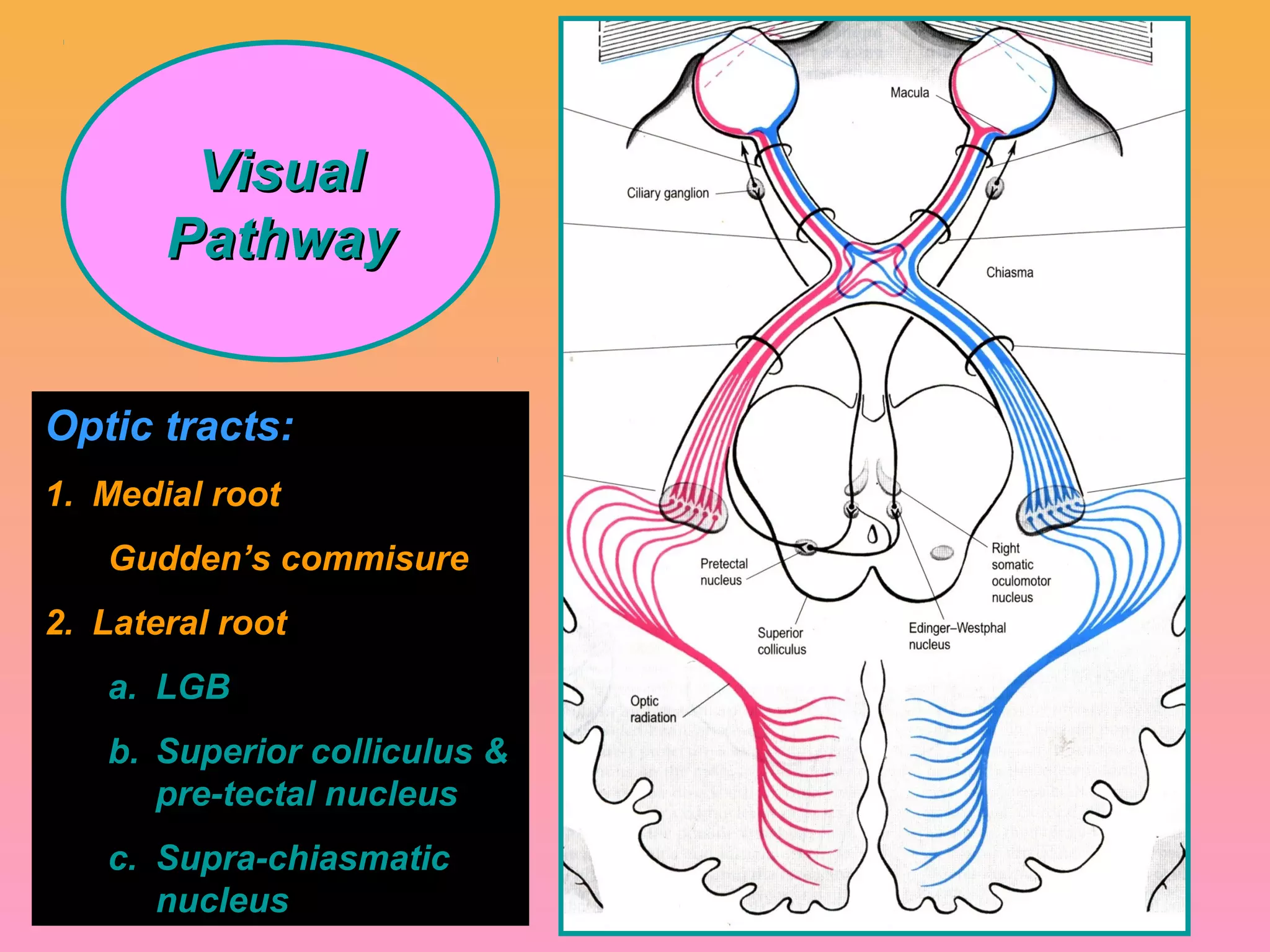
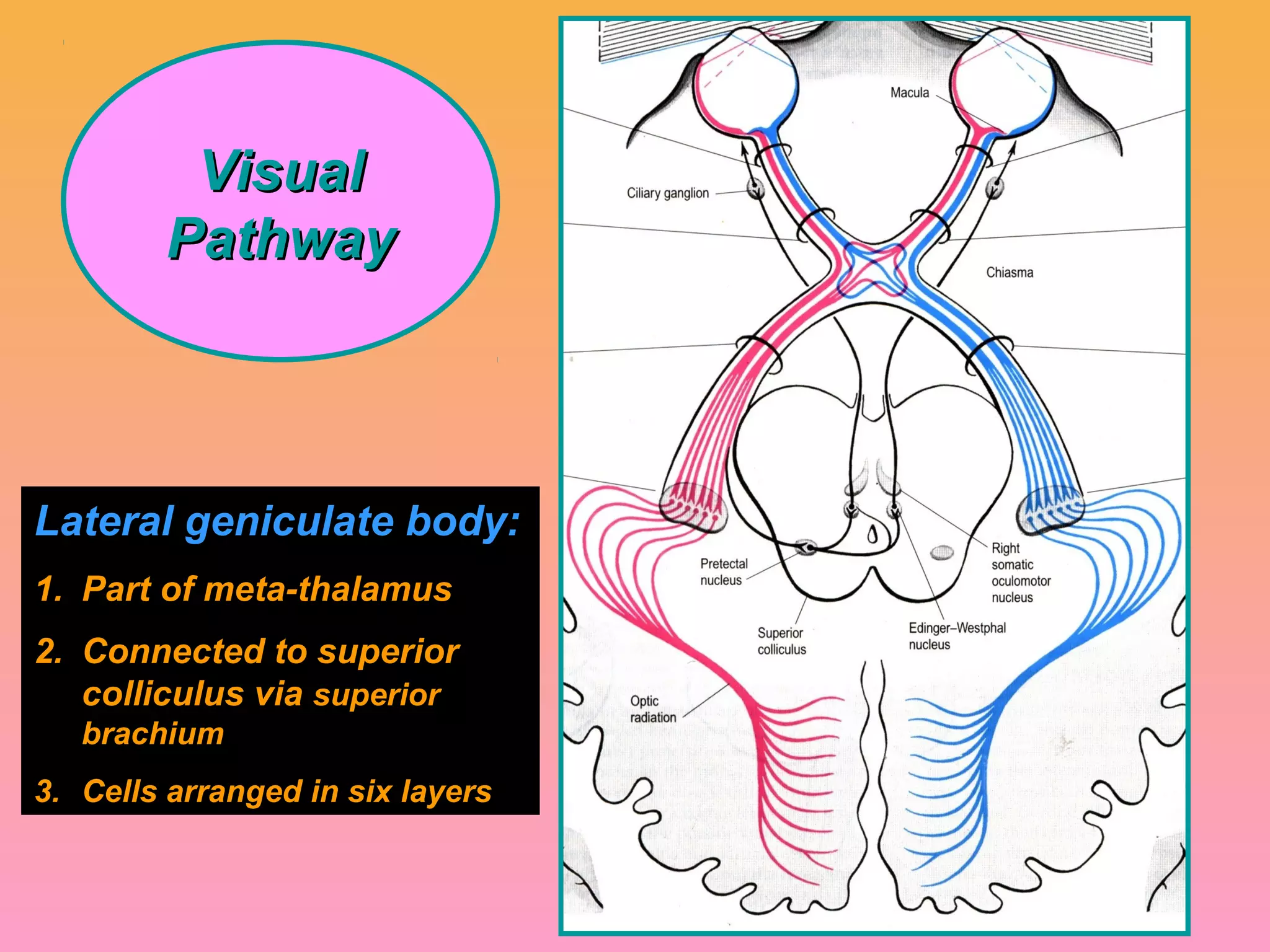
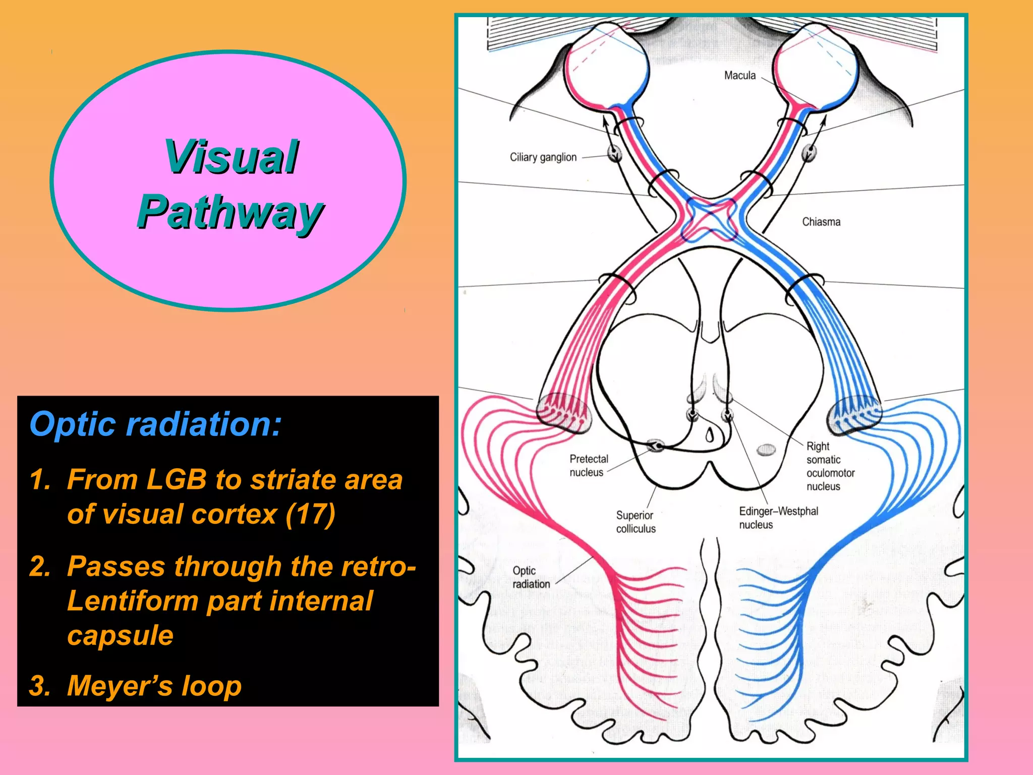
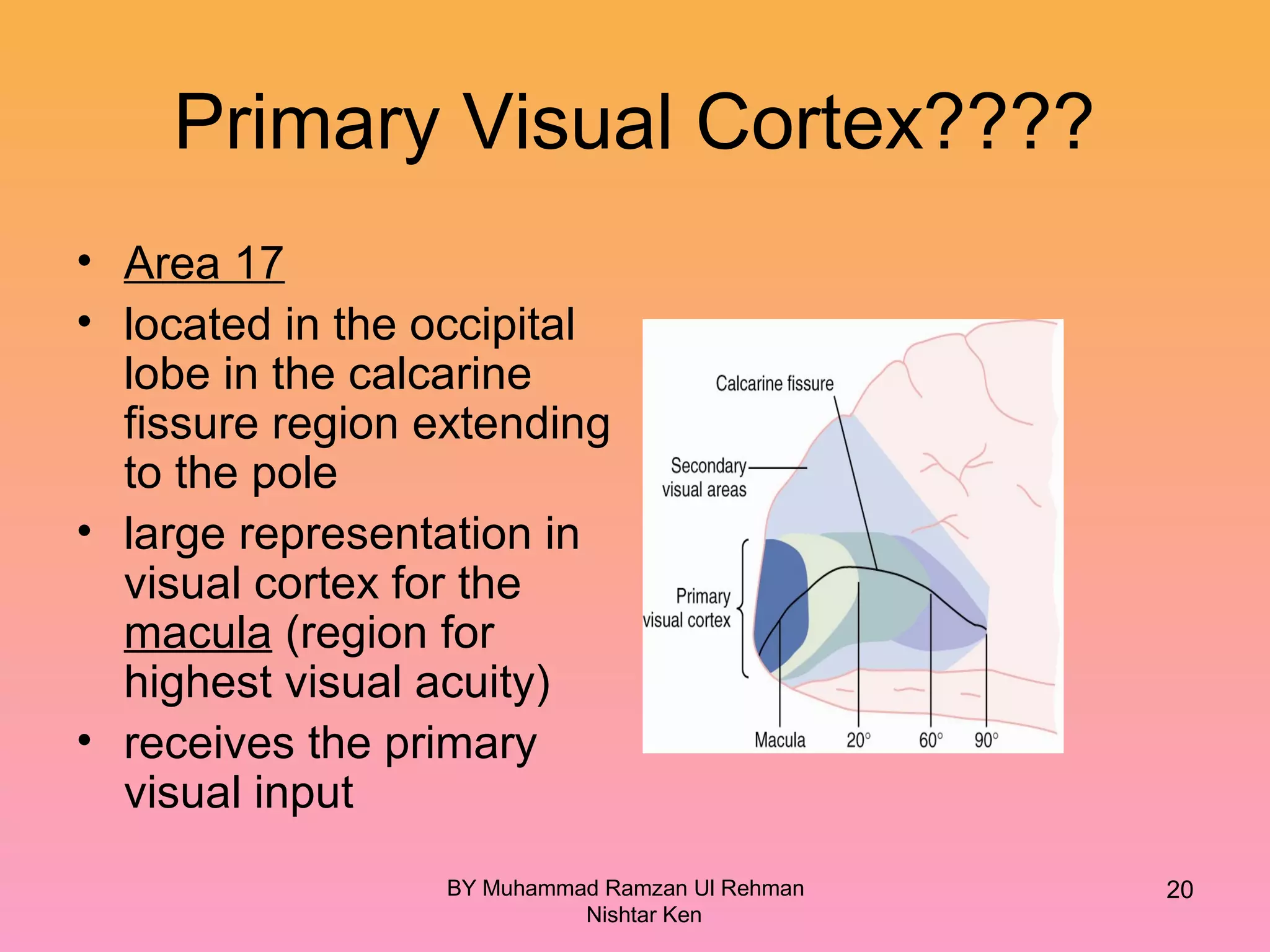
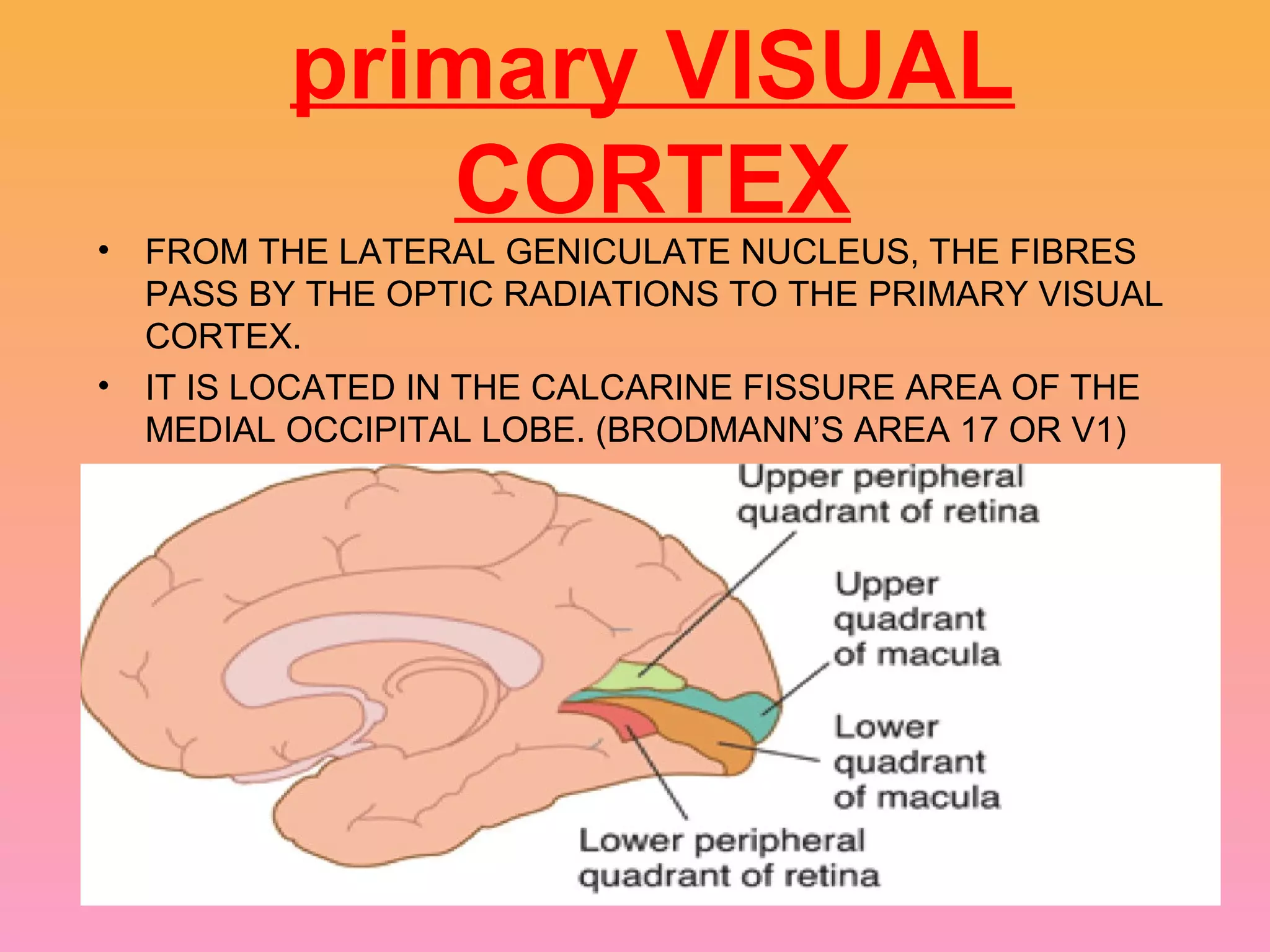
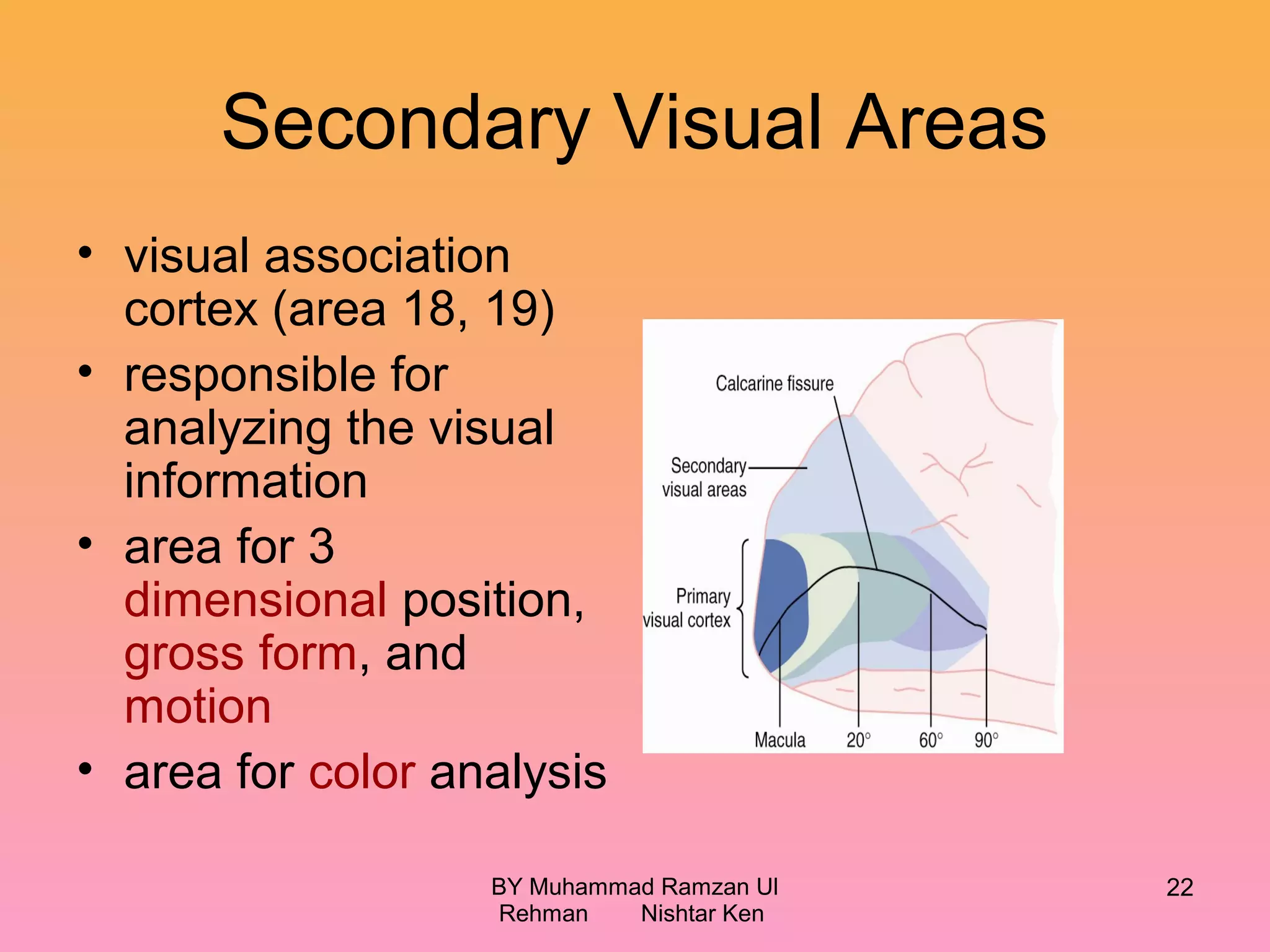
The visual pathway begins when light enters the eye and strikes the retina. Optic nerve fibers carry the visual information from the retina to the optic chiasm, where fibers from the nasal retina cross over. The fibers then continue along the optic tracts to the lateral geniculate bodies before projecting to the primary visual cortex via the optic radiations. The primary visual cortex is located in the occipital lobe.
6. OPTIC NERVE
• 2nd
cranial nerve.
• 47-50 mm in length.
• Starts from optic disc & extends upto optic chiasma
where the two nerves meet.
• Backward continuation of nerve fibre layer of retina
which consist of axons originating from ganglion
cells.
• Contains the afferent fibres of light reflex
• Has 4 parts : 1)intraocular (1mm)
2)intraorbital (30mm)
3)intra canalicular (6-9mm)
4)intracranial (10mm)
9. Receptors:
1. Rods
2. Cones
Neurons:
1. First order: bipolar cells
2. Second order: ganglion cells
3. Third order: LGB
Visuo-striate area (17):
Both walls of calcarine
sulcus involving Cuneus and
lingual gyrus
12. OPTIC CHIASMA
• A FLAT MASS OF PARTIALLY DECUSSATING FIBRES WHICH
LIES AT THE JUNCTION OF THE ANTERIOR WALL AND
FLOOR OF THE 3RD
VENTRICLE
• ONLY NASAL RETINAL FIBRES CROSS OVER .
• THEY JOIN THE FIBRES FROM THE OPPOSITE TEMPORAL
RETINAS
• THESE FIBRES FORM THE OPTIC TRACTS.
15. OPTIC TRACTS
• Cylindrical bundle of nerve
fibres.
• Run outwards & backwards
from posterolateral aspect of
optic chiasma ,between tuber
cinereum & anterior
perforated substance to unite
with cerebral peduncle.
• Fibres from temporal half of
retina of same eye & nasal
half of opposite eye.
• Posteriorly each ends in
Lateral Geniculate Body.
18. OPTIC RADIATIONS (Geniculo-
Calcarine Pathway)
• From LGB to the occipital cortex.
• Pass forwards then laterally through the area
of wernicke as optic peduncles.
• Anterior to lateral ventricle ,traversing the
retrolenticular part of internal capsule,medial
to auditory tract.
• Its fibres then spread out fanwise to form
medullary optic lamina.
• Inferior fibres subserve upper visual fields &
sweep anteroinferiorly in meyer’s loop &
temporal lobe to visual cortex.
• Superior fibres subserve inferior visual field
proceed posteriorly through parietal lobe to
visual cortex.
20. Primary Visual Cortex????
• Area 17
• located in the occipital
lobe in the calcarine
fissure region extending
to the pole
• large representation in
visual cortex for the
macula (region for
highest visual acuity)
• receives the primary
visual input
BY Muhammad Ramzan Ul Rehman
Nishtar Ken
20
21. primary VISUAL
CORTEX
• FROM THE LATERAL GENICULATE NUCLEUS, THE FIBRES
PASS BY THE OPTIC RADIATIONS TO THE PRIMARY VISUAL
CORTEX.
• IT IS LOCATED IN THE CALCARINE FISSURE AREA OF THE
MEDIAL OCCIPITAL LOBE. (BRODMANN’S AREA 17 OR V1)
22. Secondary Visual Areas
• visual association
cortex (area 18, 19)
• responsible for
analyzing the visual
information
• area for 3
dimensional position,
gross form, and
motion
• area for color analysis
BY Muhammad Ramzan Ul
Rehman Nishtar Ken
22
23. VISUAL CORTEX
• Located on the medial aspect of occipital lobe, in
& near calcarine fissure.
24. Retinal Projections to
Subcortical Regions
• suprachiasmatic nucleus of
the hypothalamus
– control of circadian rhythms??
• pretectal nuclei
– pupillary light reflex
– accommodation of the lens
• superior colliculus
– rapid directional movement of
both eyes
BY Muhammad Ramzan Ul Rehman
Nishtar Ken
24
26. The Autonomic Nerves to the
Eyes
• The eye is innervated by both parasympathetic and
sympathetic neurons.
• Parasympathetic fibers arise in the Edinger-
Westphal nucleus, pass in the 3rd cranial nerve to
the ciliary ganglion.
– Postganglionic fibers excite the ciliary muscle and sphincter
of the iris.
• Sympathetic fibers originate in the intermediolateral
horn cells of the superior cervical ganglion.
– Postganglionic fibers spread along the corotid
artery and eventually innervate the radial fibers of
the iris.
26
27. Control of Pupillary Diameter
• miosis: decreasing of pupillary
aperture due to stimulation of
parasympathetic nerves that
excite the pupillary sphincter
muscle
• mydriasis: dilation of pupillary
aperture due to stimulation of
sympathetic nerves that excite
the radial fibers of the iris
27
29. Visual
reflexes
• Light reflex or pupillary
reflex:
When light is shown to one
eye, normally the pupils of
both eyes constrict.
- Direct light reflex:
The constriction of pupils
upon which light is shown
is called direct light reflex.
- Indirect or consensual:
The constriction of pupil on
the other eye even though
no light is shown
30.
31. Pathways of direct and indirect light
reflexes (v.imp.)
BY Muhammad Ramzan Ul
Rehman Nishtar Ken
31
34. ACCCOMODATION REFLEX
• When the eyes are
focussed from a distant to
near object, three
reactions take place
• 1. Constriction of pupils
• 2. thickening of lens due to
contraction of ciliary
muscles
• 3. Convergence of both eye
balls
These three reactions
together constitute
Accommodation or near
reflex