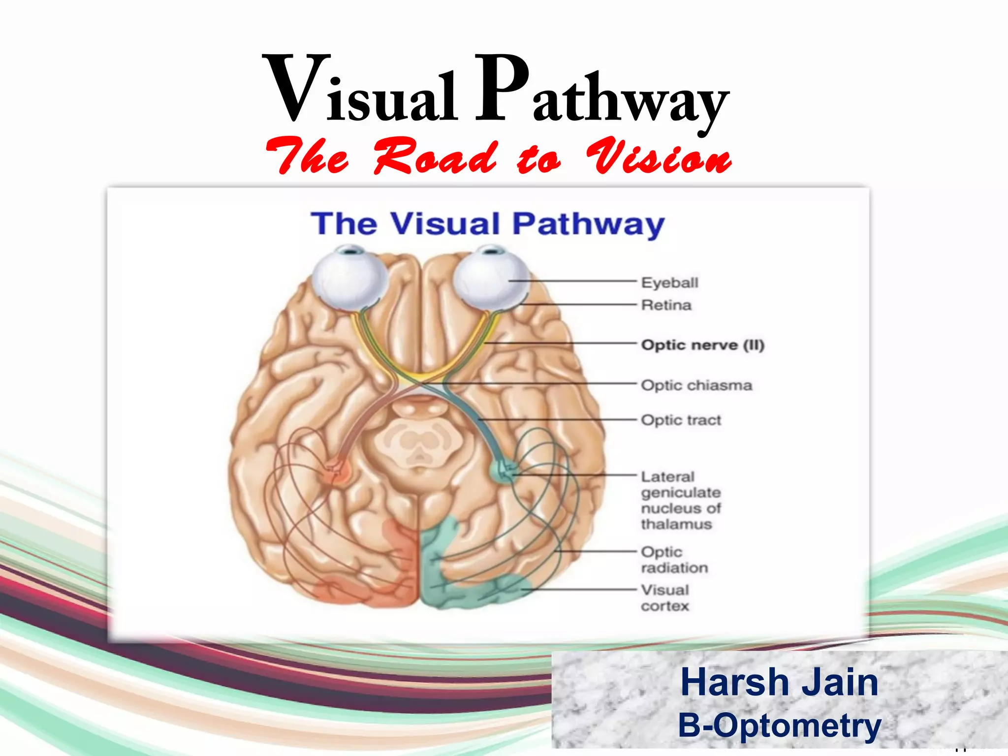
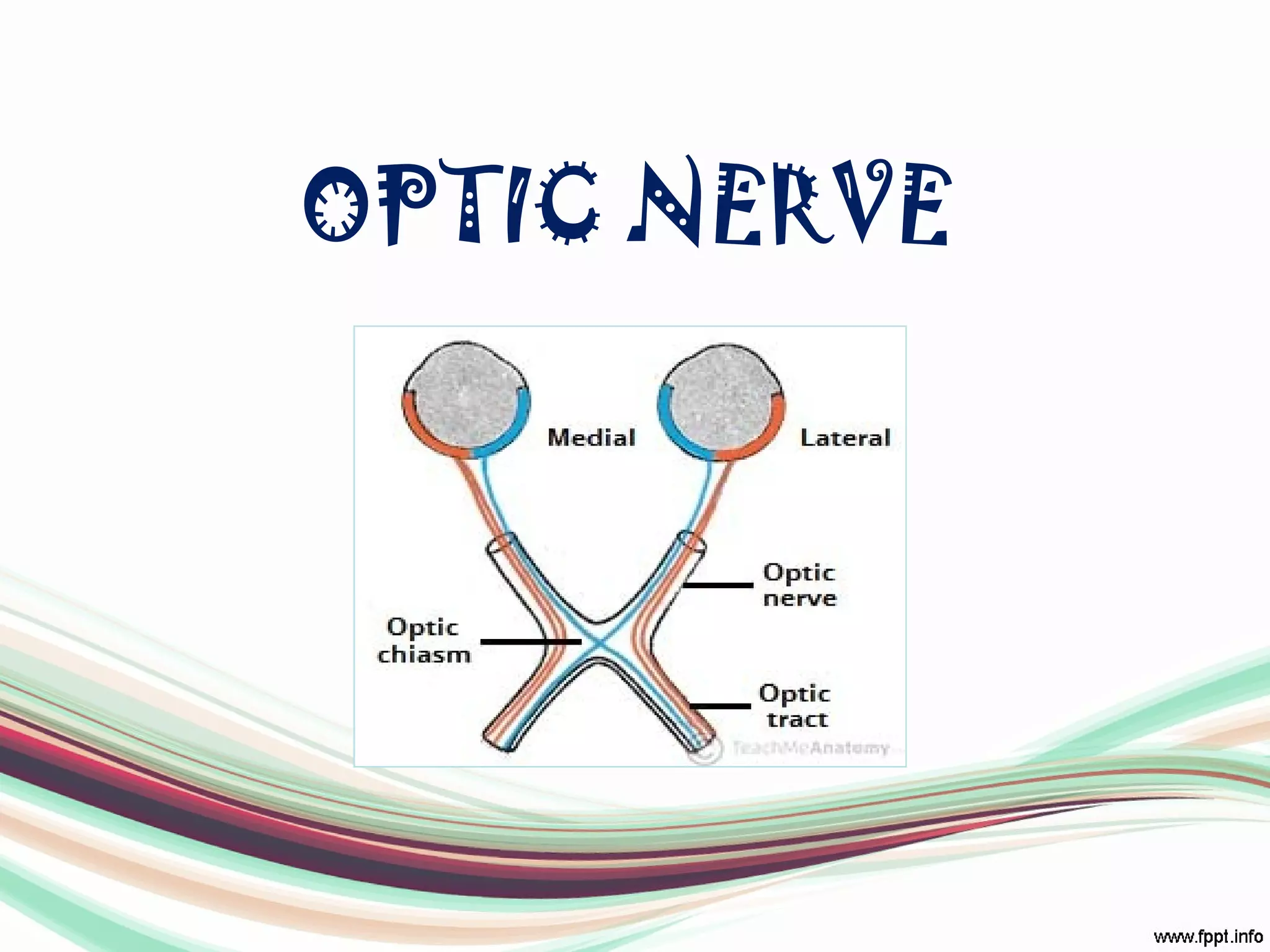
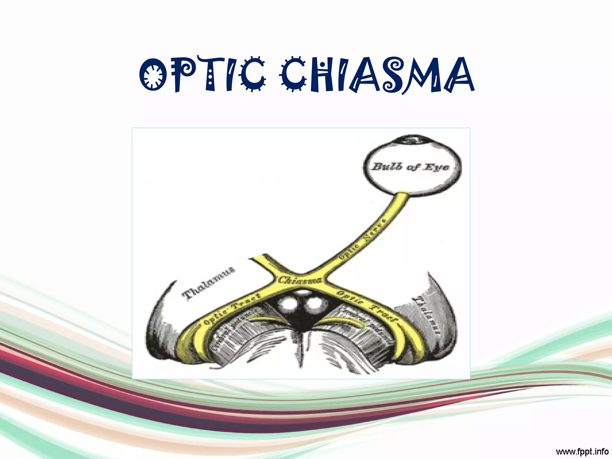
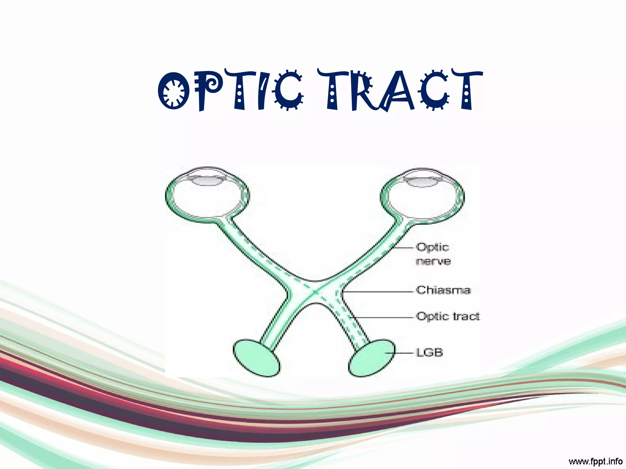
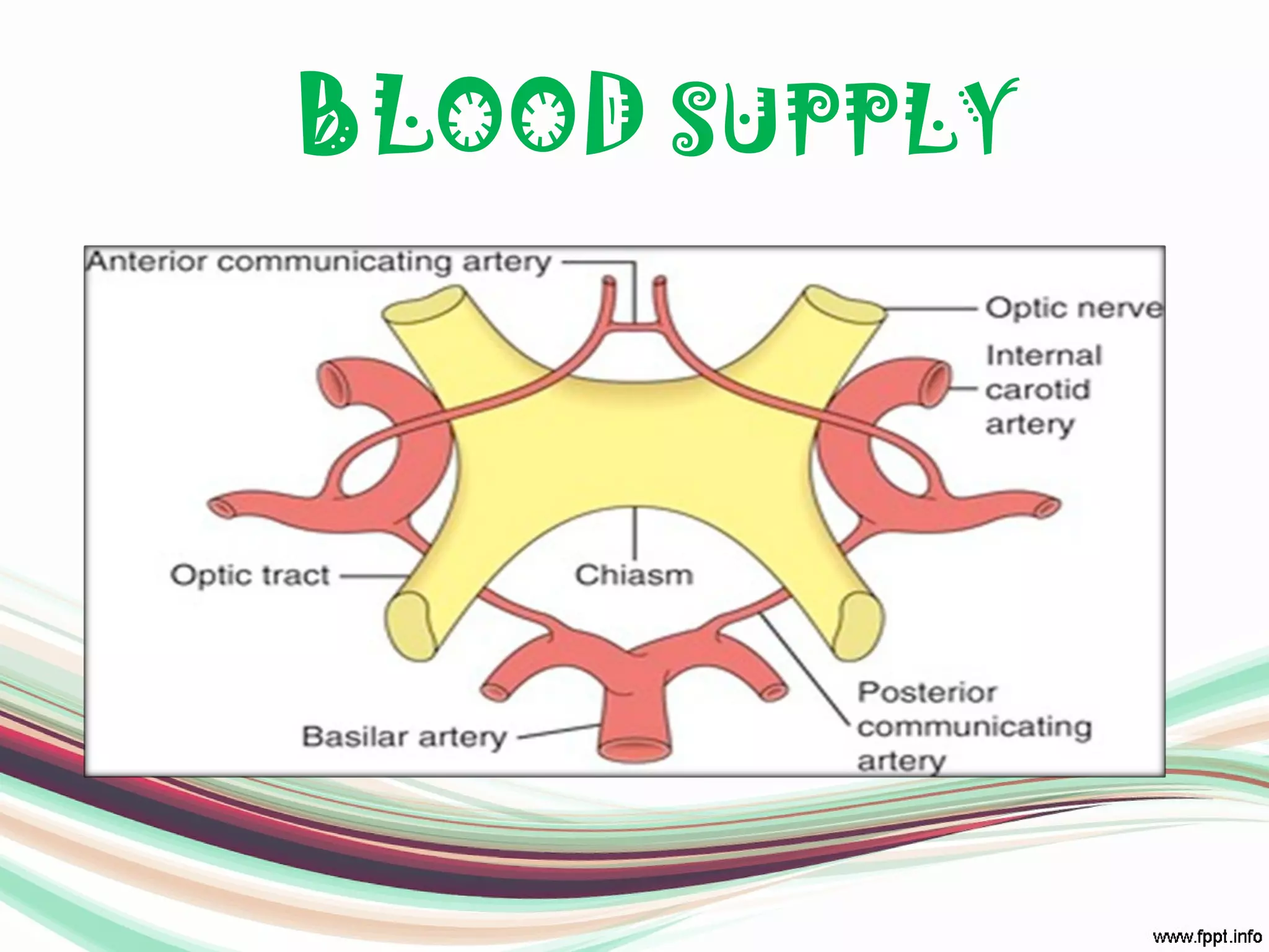
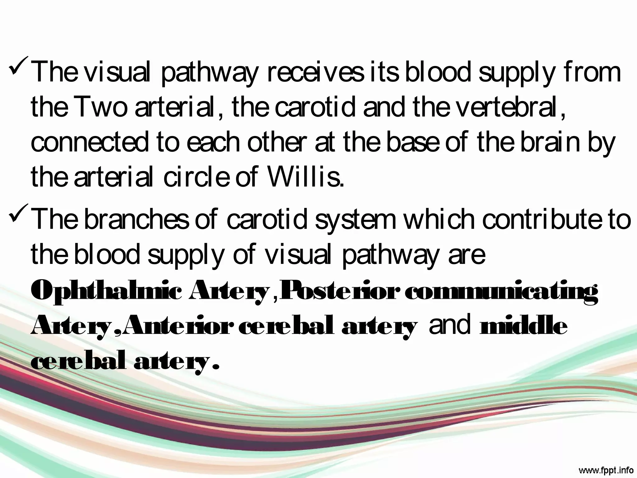
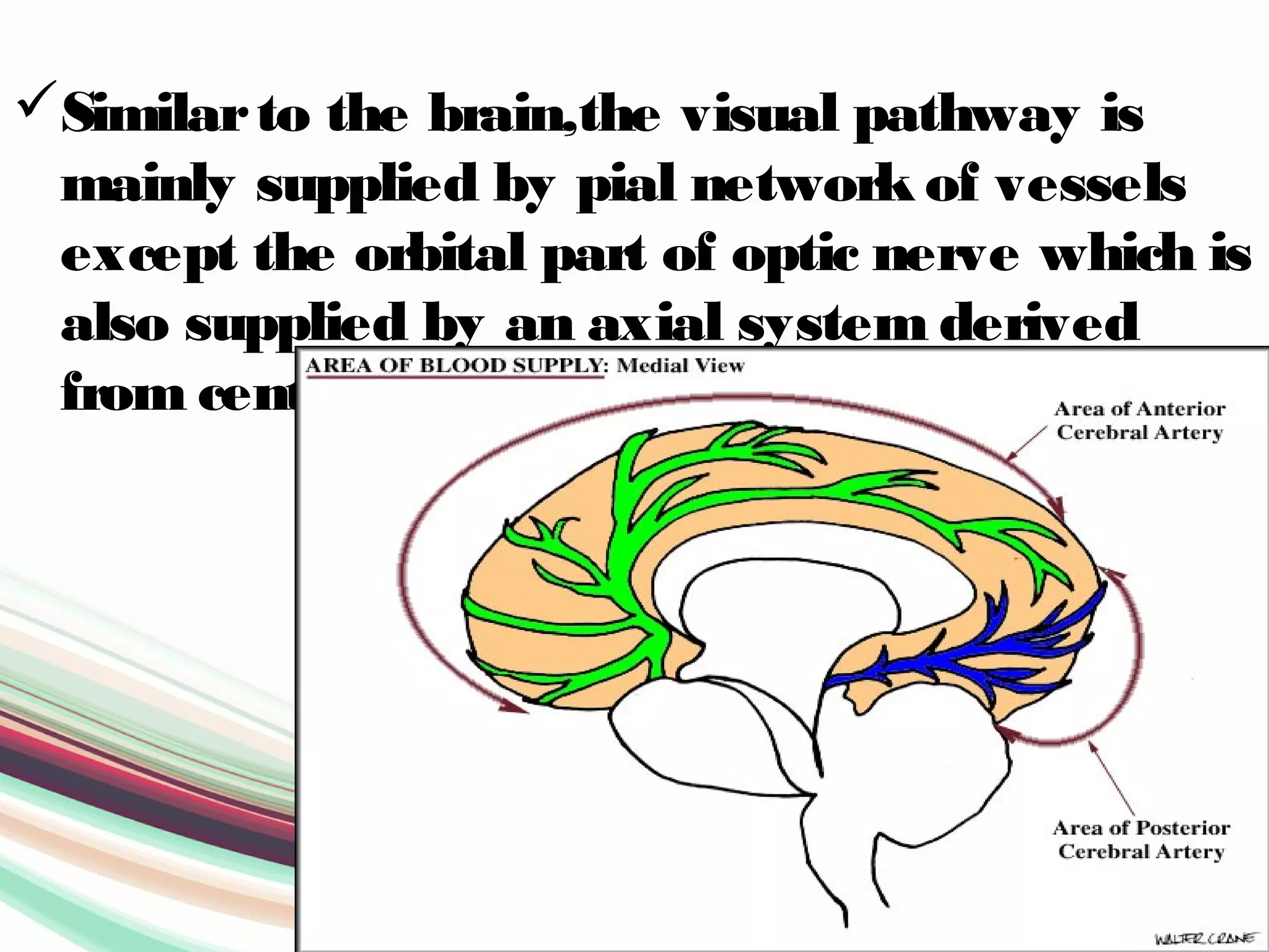
Visual Pathway
The Road to Vision
Harsh Jain
B-Optometry
Reference
Anatomy and physiology –A.K
Khurana.
Anatomy of the human eye –
Tripathi RC
Google wikipedia.
Visual Pathway
• Each eyeball acts as a camera.
• It perceives the image and relays the
sensations to the part of brain {Occipetal
Cortex} via the Visual pathway
Visual Pathway
It comprises of :-
o Optic Nerve.
o Optic Chiasma.
o Optic Tract.
o Lateral Geniculate body.
o Optic Radiations.
o Visual Cortex.
OPTIC NERVE
• Each optic nerve {CN IInd
} starts from the
optic disc and extends upto
chiasma,where the two nerve meets.
• It is backward continuation of NFLof
retina which consists of about 1-2 milion
axons originating from Ganglion cell
layer.
• It is afferent fibres of light reflex.
• Optic nerve is comparable to be sensory
tract .
• It is an outgrowth of the brain.
• Fibres of optic nerve, numbering about
million, are very fine 2-10μm in diameter
as compared to 20μm in sensory nerves.
• Optic nerve is surrounded by meninges.
• Unlike peripheral nerves it is not covered
by Neurilemma
OPTIC CHIASMA
• It is flattened structure measuring about
12mm horizontally and 8mm Antero-
posteriorly.
• It is En-sheathed by Pia and surrounded by
Cerebro-spinal fluid.
• The optic nerve fibres on the nasal sides of
each retina cross over(decussate) to the
opposite side of the brain via the optic
nerve at the optic chiasm (decussation of
medial fibers). The temporal hemiretina,
on the otherhand, stays on the same side.
Variation of location
Central Chiasma :-
• It liesdirectly abovethesella, so theexpanding
Pituitary tumoursinvolvethechiasmafirst.
Pre-fixed Chiasma :-
• It islocated moreanteriorly over thetuberculum
sellae.So thePituitary tumoursmay involvethe
Optic Tract first.
Post Fixed Chiasma :-
• It islocated moreposteriorly over the
Dorsum sellaeso that thepituitary tumours
damagetheOptic nervefirst
OPTIC TRACT
• Thesearecylinderical bundlesof nervefibres
running outwardsand backwardsfrom the
postero-lateral aspect of chiasma.
• Each optic nerveconsistsof fibresfrom the
temporal half of retinaof sameeyeand nasal
half of oppositeeye.
• Posterior each optic tract endsin LGB.
• Theoptic tract carriesretinal information relating to
thewholevisual field.
• Specifically, theleft optic tract correspondsto the
right visual field, whiletheright optic tract
correspondsto theleft visual field.
• To form theright visual field, temporal retinal fibers
from theleft eyeand nasal retinal fibersfrom the
right eyeform theleft optic tract, and to form the
left visual field, temporal retinal fibersfrom the
right eyeand nasal retinal fibersfrom theleft eye
form theright optic tract
LATERAL GENICULATE
BODY
• This are oval structures situated at the
optic tracts.
• Each LGBconsists of 6layers of Neurons
{grey matter} alternating with white
matterformed by optic fibres.
• Fibres of second orderneuron coming via
Optic tracts relay in these neurons.
OPTIC RADIATION
• It extends from the LGBto the visual
cortex.
• These are collection of axons from the
relay neurons in the LGBnucleus of the
thalamus.
• They carry visual information to the visual
cortex along the calcarine fissure.
VISUAL CORTEX
• It is located on the medial aspect of
occipetal lobe.
• The visual cortex is sub-divided into
Visual-sensory area {striate area no 17}
that receives the fibres of the Optic
Radiations and the surrounding visuao-
psychic {Peristriate area no 18 &
parastriate area no 19}.
1st
Visualarea{V1} in area 17
2nd
Visualarea{V2} occupying the greater
part of area no 18 but not whole
of it.
3rd
Visualarea{V3} occupying a narrow strip
overthe anteriorpart of area no 18.
4th
Visualarea{V4} within the area no 19.
5th
Visualarea{V5} at the posteriorend of
the superiortemporal gyrus.
BLOOD SUPPLY
Thevisual pathway receivesitsblood supply from
theTwo arterial, thecarotid and thevertebral,
connected to each other at thebaseof thebrain by
thearterial circleof Willis.
Thebranchesof carotid system which contributeto
theblood supply of visual pathway are
Ophthalmic Artery,Posteriorcommunicating
Artery,Anteriorcerebal artery and middle
cerebal artery.
Similarto the brain,the visual pathway is
mainly supplied by pial network of vessels
except the orbital part of optic nerve which is
also supplied by an axial system derived
from central retinal artery.
Reference
Anatomy and physiology –A.K
Khurana.
Anatomy of the human eye –
Tripathi RC
Google wikipedia.
Thank you in
Japanese

Visual pathway -"The road to Vision"

  • 1.
    Visual Pathway The Roadto Vision Harsh Jain B-Optometry
  • 2.
    Reference Anatomy and physiology–A.K Khurana. Anatomy of the human eye – Tripathi RC Google wikipedia.
  • 3.
    Visual Pathway • Eacheyeball acts as a camera. • It perceives the image and relays the sensations to the part of brain {Occipetal Cortex} via the Visual pathway
  • 4.
    Visual Pathway It comprisesof :- o Optic Nerve. o Optic Chiasma. o Optic Tract. o Lateral Geniculate body. o Optic Radiations. o Visual Cortex.
  • 5.
  • 6.
    • Each opticnerve {CN IInd } starts from the optic disc and extends upto chiasma,where the two nerve meets. • It is backward continuation of NFLof retina which consists of about 1-2 milion axons originating from Ganglion cell layer. • It is afferent fibres of light reflex.
  • 7.
    • Optic nerveis comparable to be sensory tract . • It is an outgrowth of the brain. • Fibres of optic nerve, numbering about million, are very fine 2-10μm in diameter as compared to 20μm in sensory nerves. • Optic nerve is surrounded by meninges. • Unlike peripheral nerves it is not covered by Neurilemma
  • 8.
  • 9.
    • It isflattened structure measuring about 12mm horizontally and 8mm Antero- posteriorly. • It is En-sheathed by Pia and surrounded by Cerebro-spinal fluid. • The optic nerve fibres on the nasal sides of each retina cross over(decussate) to the opposite side of the brain via the optic nerve at the optic chiasm (decussation of medial fibers). The temporal hemiretina, on the otherhand, stays on the same side.
  • 10.
    Variation of location CentralChiasma :- • It liesdirectly abovethesella, so theexpanding Pituitary tumoursinvolvethechiasmafirst. Pre-fixed Chiasma :- • It islocated moreanteriorly over thetuberculum sellae.So thePituitary tumoursmay involvethe Optic Tract first.
  • 11.
    Post Fixed Chiasma:- • It islocated moreposteriorly over the Dorsum sellaeso that thepituitary tumours damagetheOptic nervefirst
  • 12.
  • 13.
    • Thesearecylinderical bundlesofnervefibres running outwardsand backwardsfrom the postero-lateral aspect of chiasma. • Each optic nerveconsistsof fibresfrom the temporal half of retinaof sameeyeand nasal half of oppositeeye. • Posterior each optic tract endsin LGB.
  • 14.
    • Theoptic tractcarriesretinal information relating to thewholevisual field. • Specifically, theleft optic tract correspondsto the right visual field, whiletheright optic tract correspondsto theleft visual field. • To form theright visual field, temporal retinal fibers from theleft eyeand nasal retinal fibersfrom the right eyeform theleft optic tract, and to form the left visual field, temporal retinal fibersfrom the right eyeand nasal retinal fibersfrom theleft eye form theright optic tract
  • 15.
  • 16.
    • This areoval structures situated at the optic tracts. • Each LGBconsists of 6layers of Neurons {grey matter} alternating with white matterformed by optic fibres. • Fibres of second orderneuron coming via Optic tracts relay in these neurons.
  • 17.
  • 18.
    • It extendsfrom the LGBto the visual cortex. • These are collection of axons from the relay neurons in the LGBnucleus of the thalamus. • They carry visual information to the visual cortex along the calcarine fissure.
  • 19.
  • 20.
    • It islocated on the medial aspect of occipetal lobe. • The visual cortex is sub-divided into Visual-sensory area {striate area no 17} that receives the fibres of the Optic Radiations and the surrounding visuao- psychic {Peristriate area no 18 & parastriate area no 19}.
  • 21.
    1st Visualarea{V1} in area17 2nd Visualarea{V2} occupying the greater part of area no 18 but not whole of it. 3rd Visualarea{V3} occupying a narrow strip overthe anteriorpart of area no 18. 4th Visualarea{V4} within the area no 19. 5th Visualarea{V5} at the posteriorend of the superiortemporal gyrus.
  • 22.
  • 23.
    Thevisual pathway receivesitsbloodsupply from theTwo arterial, thecarotid and thevertebral, connected to each other at thebaseof thebrain by thearterial circleof Willis. Thebranchesof carotid system which contributeto theblood supply of visual pathway are Ophthalmic Artery,Posteriorcommunicating Artery,Anteriorcerebal artery and middle cerebal artery.
  • 24.
    Similarto the brain,thevisual pathway is mainly supplied by pial network of vessels except the orbital part of optic nerve which is also supplied by an axial system derived from central retinal artery.
  • 25.
    Reference Anatomy and physiology–A.K Khurana. Anatomy of the human eye – Tripathi RC Google wikipedia.
  • 26.