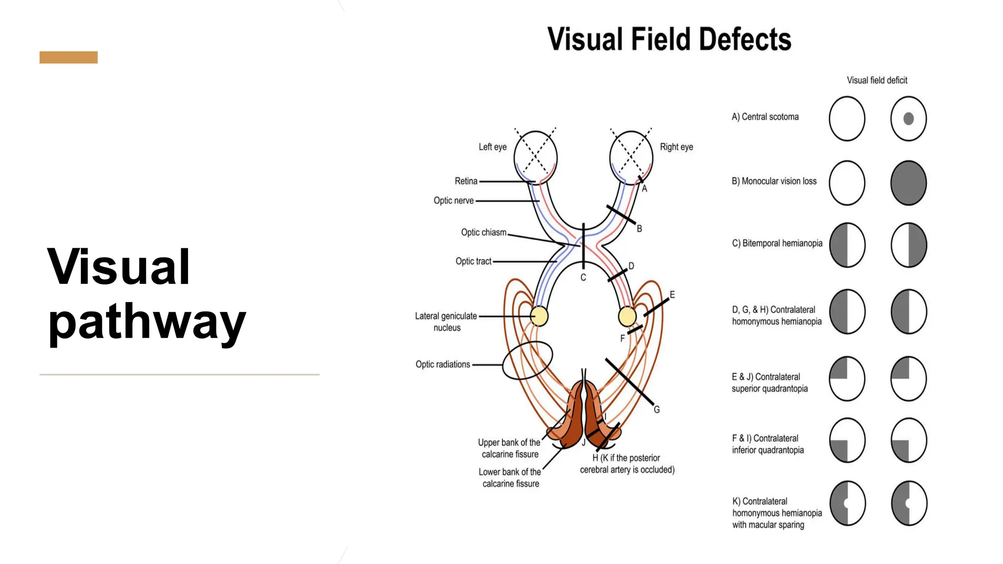
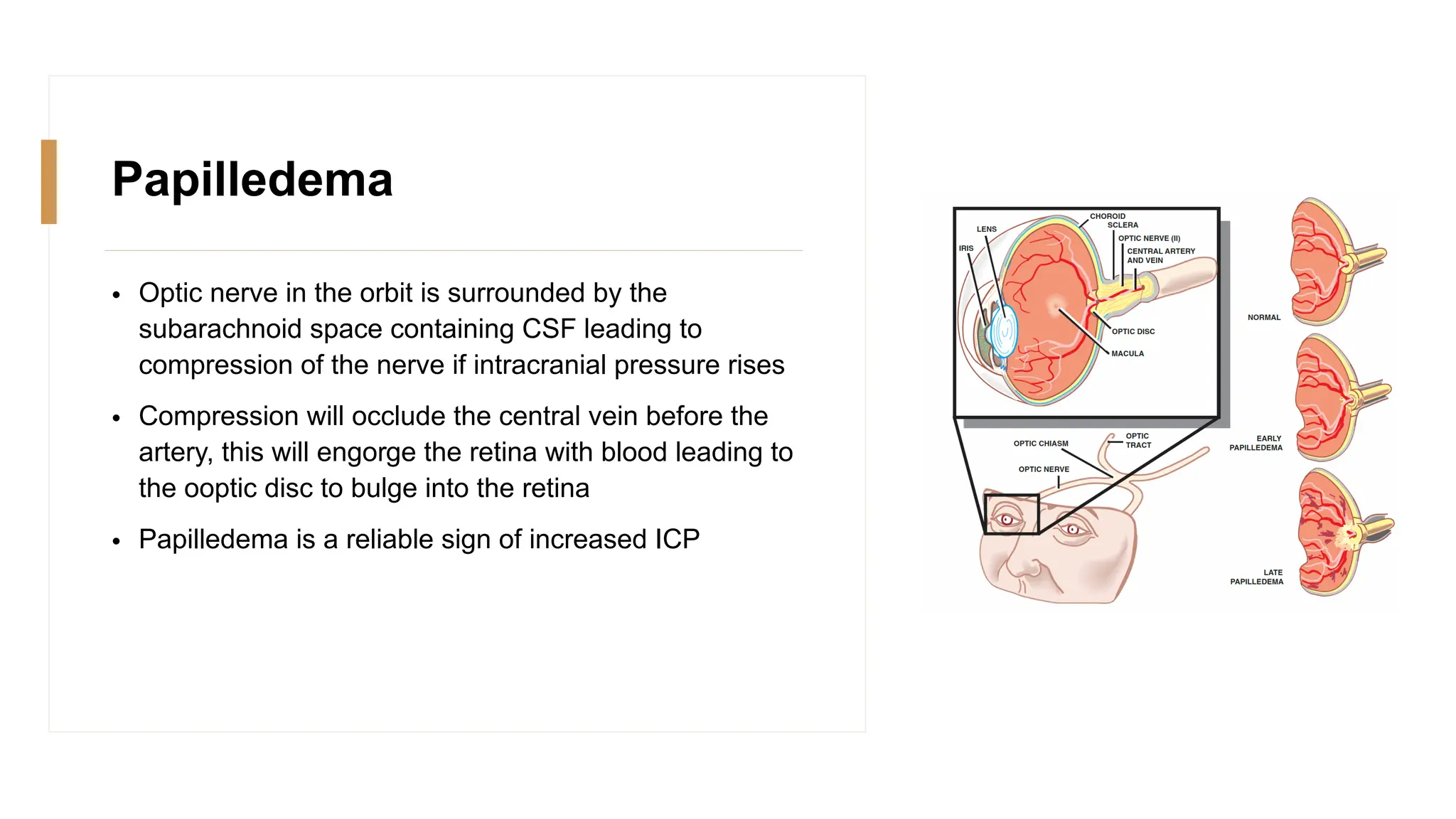
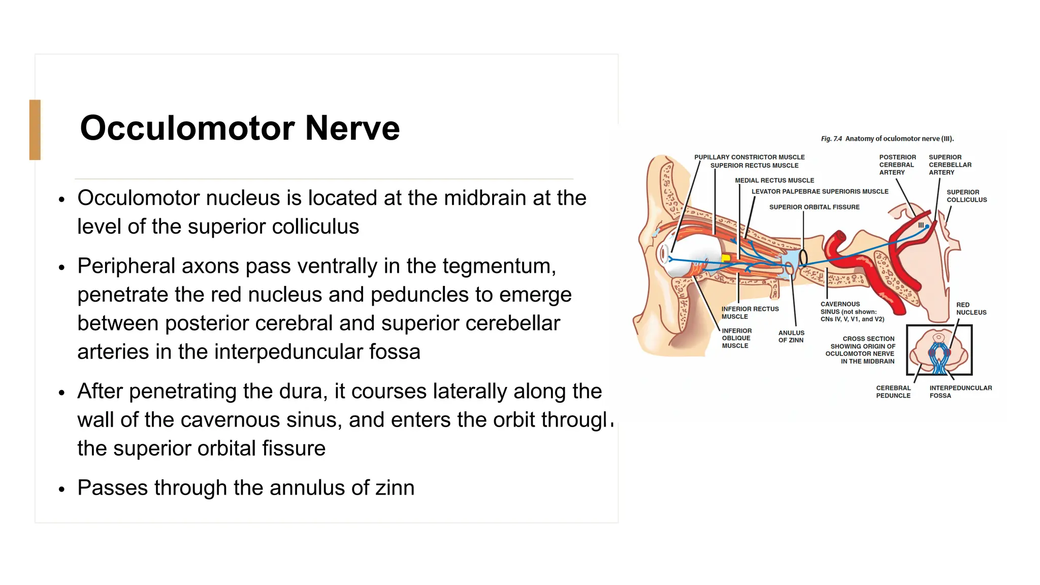
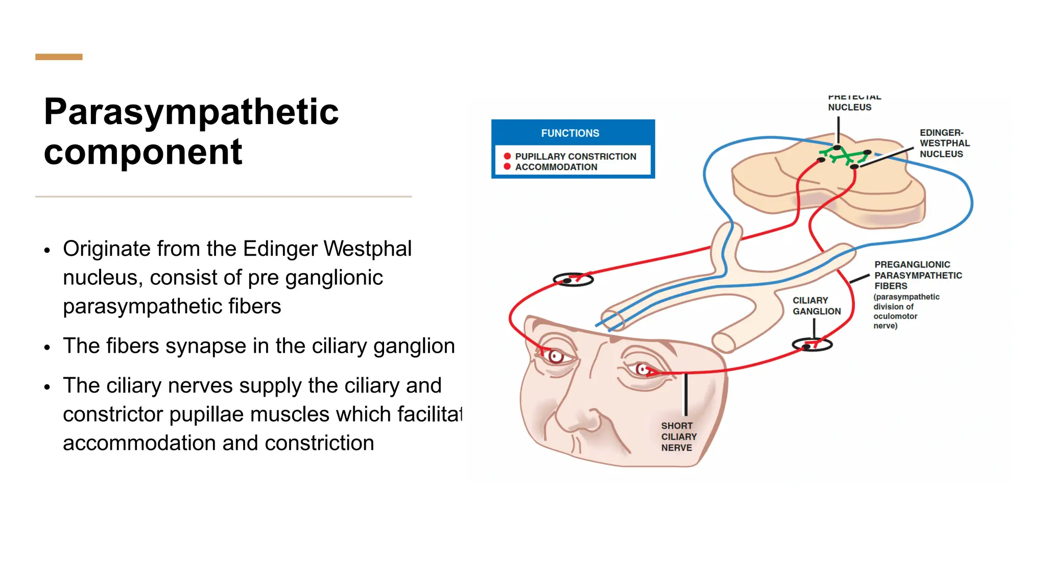
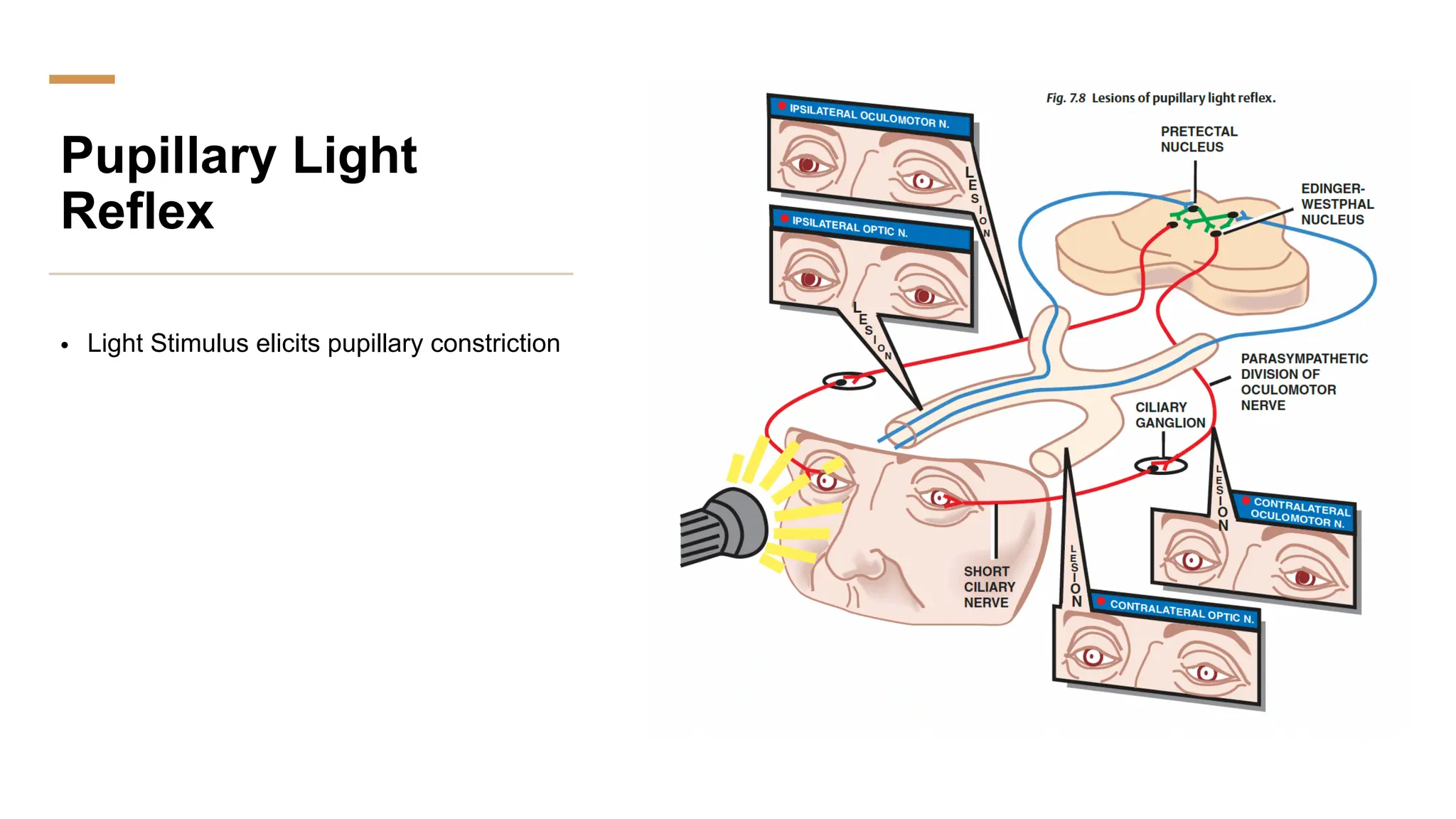
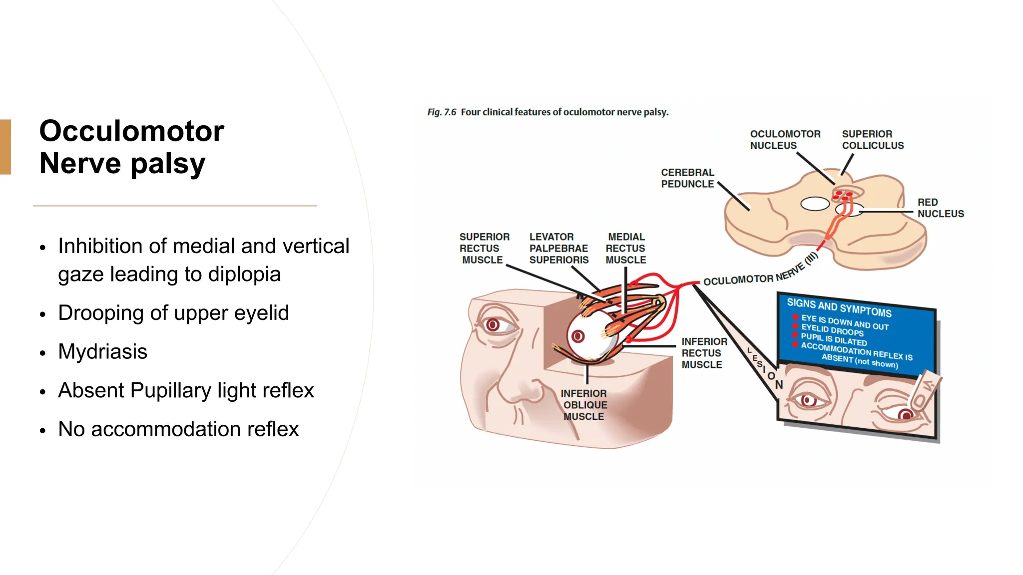
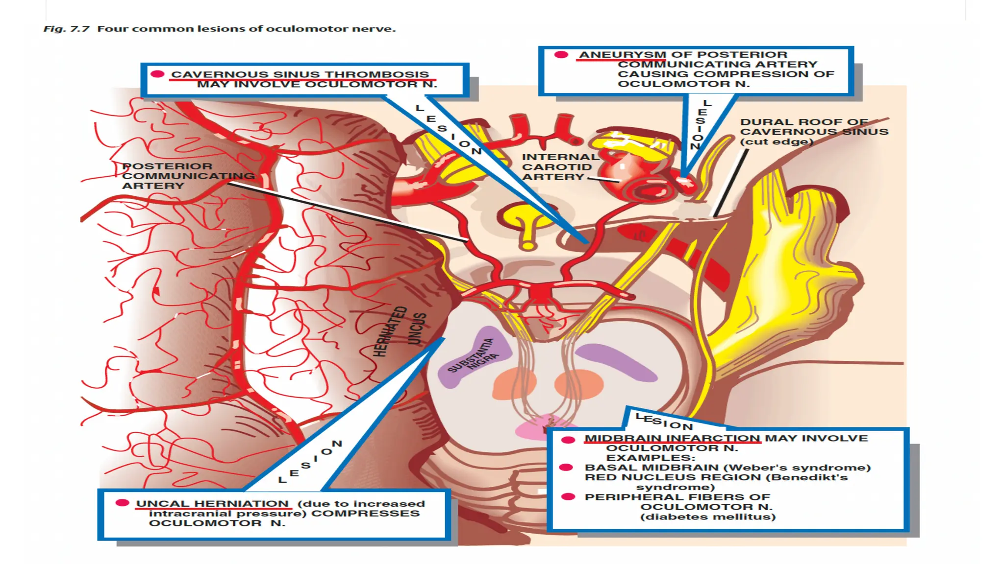
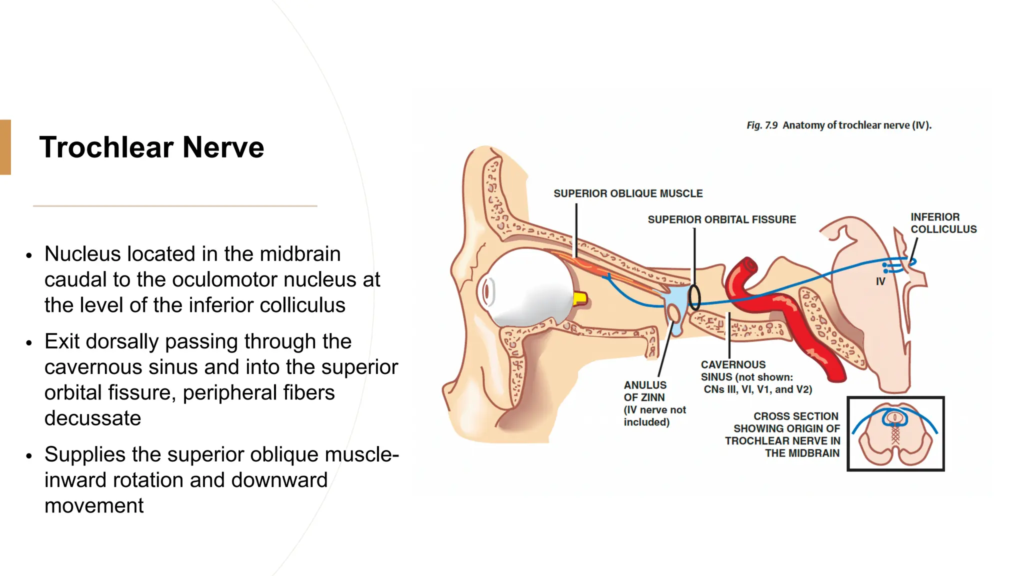
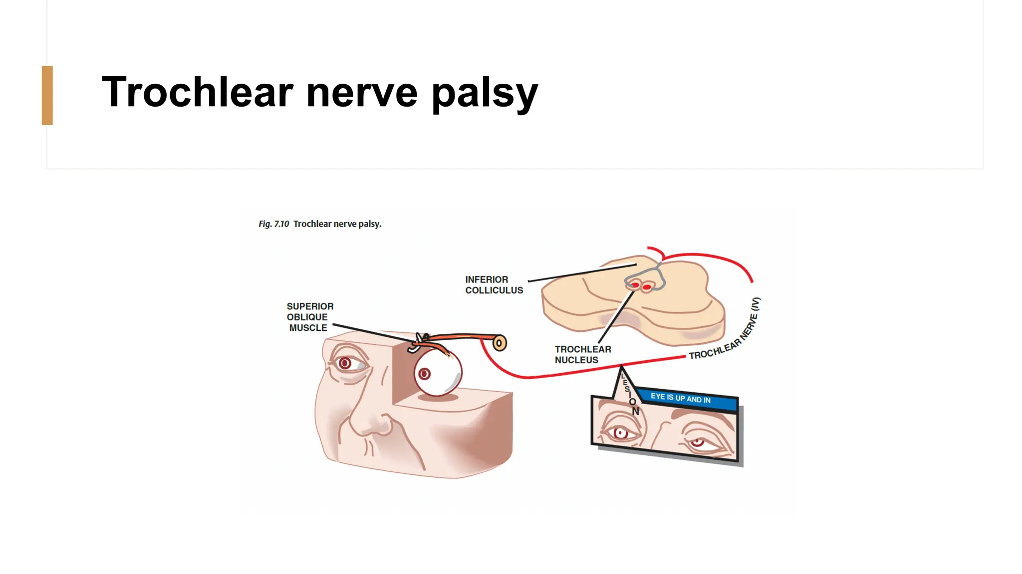
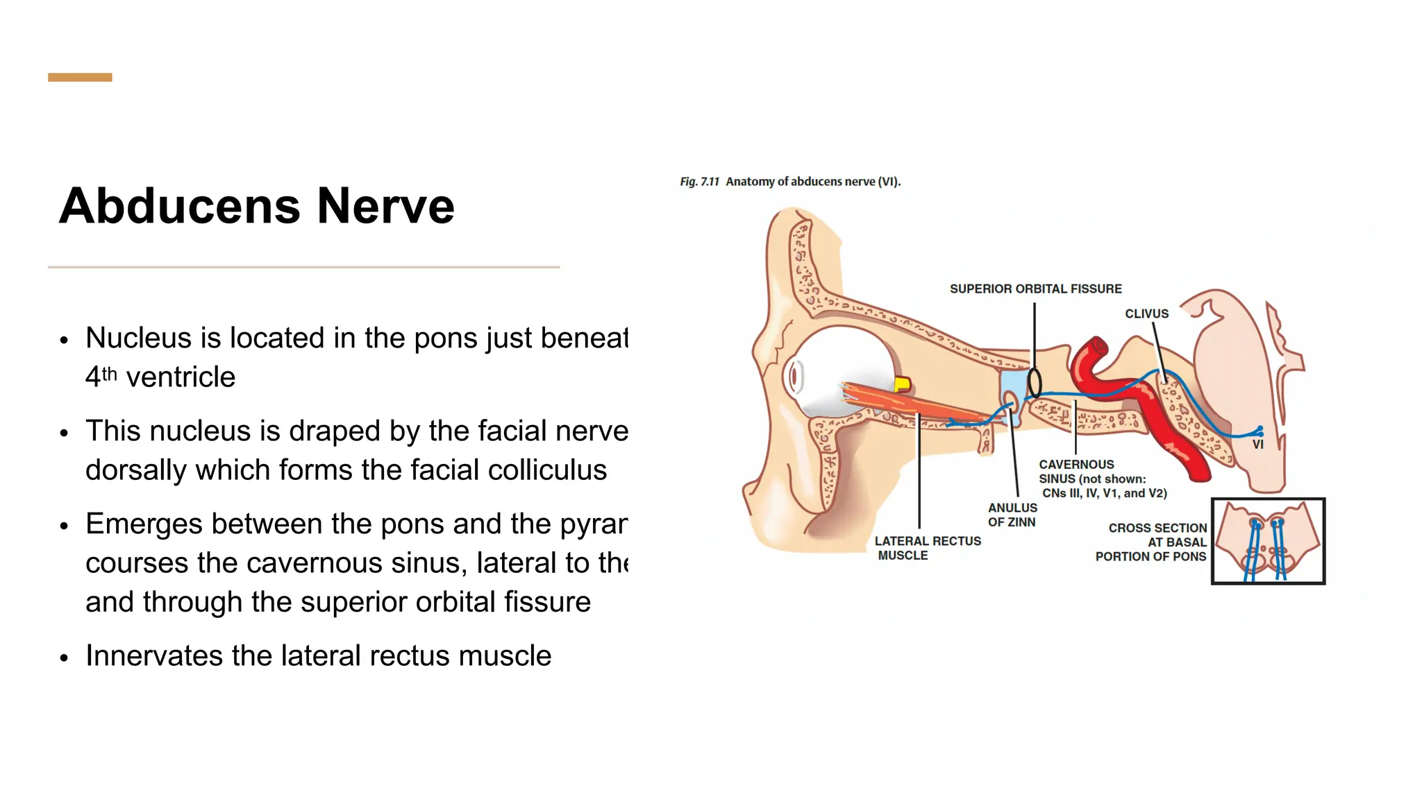
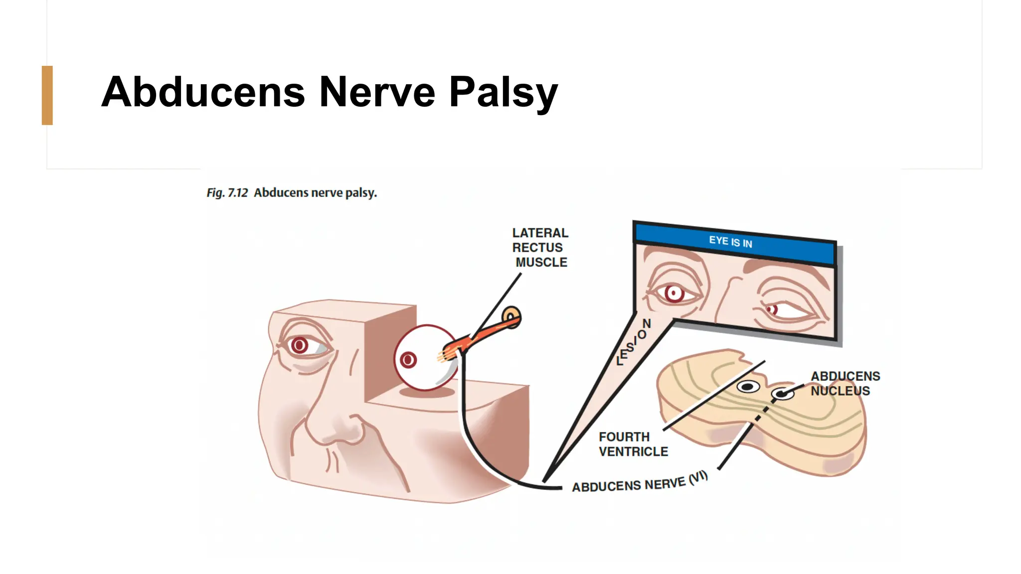
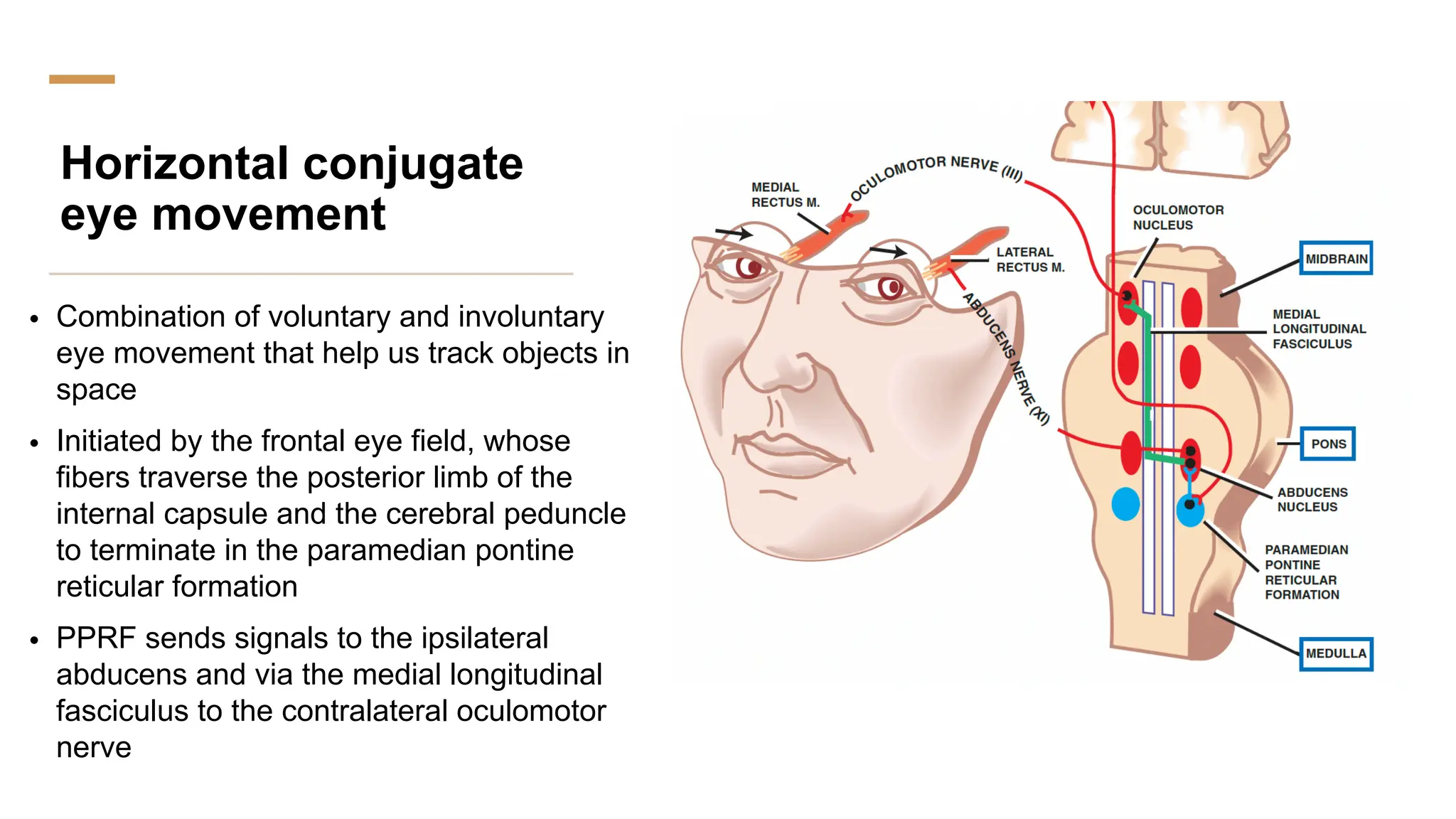
Cranial Nerve
2,3,4,6
Sight
• Is not only dependent on the cerebal cortex but also on cranial nerves
• Perception is a function of the retina, optic nerve, tract, radiation and
cortex
• The optic pathway transmits impulses from the retina to the brain
CN 2: Optic
• A CNS Tract
• Primary sensory neurons are bipolar and confined to the retina
• Axons of the secondary sensory neurons form the optic nerve, chiasm
and tract
• Pathway from the eyeball to the optic chiasm
• Purely sensory
CN 2:Optic
• Retina
• Two Layers: Neural and pigment
• Neural layer: rods and cones
• Terminal portioin of the neural layer is in contact with the
pigment layer
• Rods and cones synapse with the bipolar cells
• Bipolar cells synapse with the ganglion cells
• Axons of the ganglion cells form the optic nerve
Optic Nerve
• Passes posteriorly from the eyeball
• Surrounded by meninges, subarachnoid space and CSF
• Passes through the common tendinous ring and leaves the orbit through the
optic canal
• Nerve is surrounded by the tube like extension of the 3 meningeal layers and
CSF
• The Paired optics communicate at the chiasm
• Nasal Portion of the nerve decussate at the chiasm
• From the chiasm the optic tract extend to the lateral geniculate body
Optic Nerve
• From the LGB some axons bifurcate sending signals to the midbrain
from the pupillary light reflex
• Inferior optic radiations pass through the temporal lobe (Meyers loop)
• Superior optic radiations pass by the parietal lobe
• Majority of axons from the LGB make its way to the occipital lobe as the
optic radiation
Optic Nerve
• Connection to the midbrain
• To the pretectal nuclei of the midbrain to facilitate pupillary
light reflex
• Connected to the superior colliculus and medial
longitudinal fasciculus to facilitate eye accommodation and
conjugate eye movement
Visual
pathway
Papilledema
• Optic nerve in the orbit is surrounded by the
subarachnoid space containing CSF leading to
compression of the nerve if intracranial pressure rises
• Compression will occlude the central vein before the
artery, this will engorge the retina with blood leading to
the ooptic disc to bulge into the retina
• Papilledema is a reliable sign of increased ICP
Cranial Nerves: 3 4 6
• Innervate the extraocular muscles which move the eyeball
• Function in conjugate eye movement
Occulomotor Nerve
• Innervate the levator palpebra superioris
• Innervate the superior, inferior, medial and inferior oblique
• Through its parasympathetic component it constricts the pupil
Occulomotor Nerve
• Occulomotor nucleus is located at the midbrain at the
level of the superior colliculus
• Peripheral axons pass ventrally in the tegmentum,
penetrate the red nucleus and peduncles to emerge
between posterior cerebral and superior cerebellar
arteries in the interpeduncular fossa
• After penetrating the dura, it courses laterally along the
wall of the cavernous sinus, and enters the orbit through
the superior orbital fissure
• Passes through the annulus of zinn
Parasympathetic
component
• Originate from the Edinger Westphal
nucleus, consist of pre ganglionic
parasympathetic fibers
• The fibers synapse in the ciliary ganglion
• The ciliary nerves supply the ciliary and
constrictor pupillae muscles which facilitate
accommodation and constriction
Pupillary Light
Reflex
• Light Stimulus elicits pupillary constriction
Accommodation Reflex
• Part of the adaptation of the eyes to near vision
• Afferent limb terminates in the visual cortex
• Efferent limb terminates in the pupillary constrictors, medial rectus and
ciliary muscles via the pretectal region
• Constriction, convergence and change in lens size
Occulomotor
Nerve palsy
• Inhibition of medial and vertical
gaze leading to diplopia
• Drooping of upper eyelid
• Mydriasis
• Absent Pupillary light reflex
• No accommodation reflex
Trochlear Nerve
• Nucleus located in the midbrain
caudal to the oculomotor nucleus at
the level of the inferior colliculus
• Exit dorsally passing through the
cavernous sinus and into the superior
orbital fissure, peripheral fibers
decussate
• Supplies the superior oblique muscle-
inward rotation and downward
movement
Trochlear nerve palsy
Abducens Nerve
• Nucleus is located in the pons just beneath the
4th ventricle
• This nucleus is draped by the facial nerve
dorsally which forms the facial colliculus
• Emerges between the pons and the pyramids,
courses the cavernous sinus, lateral to the ICA
and through the superior orbital fissure
• Innervates the lateral rectus muscle
Abducens Nerve Palsy
Horizontal conjugate
eye movement
• Combination of voluntary and involuntary
eye movement that help us track objects in
space
• Initiated by the frontal eye field, whose
fibers traverse the posterior limb of the
internal capsule and the cerebral peduncle
to terminate in the paramedian pontine
reticular formation
• PPRF sends signals to the ipsilateral
abducens and via the medial longitudinal
fasciculus to the contralateral oculomotor
nerve

Cranial Nerve 2,3,4,6 function, loci.pdf

  • 1.
  • 2.
    Sight • Is notonly dependent on the cerebal cortex but also on cranial nerves • Perception is a function of the retina, optic nerve, tract, radiation and cortex • The optic pathway transmits impulses from the retina to the brain
  • 3.
    CN 2: Optic •A CNS Tract • Primary sensory neurons are bipolar and confined to the retina • Axons of the secondary sensory neurons form the optic nerve, chiasm and tract • Pathway from the eyeball to the optic chiasm • Purely sensory
  • 4.
    CN 2:Optic • Retina •Two Layers: Neural and pigment • Neural layer: rods and cones • Terminal portioin of the neural layer is in contact with the pigment layer • Rods and cones synapse with the bipolar cells • Bipolar cells synapse with the ganglion cells • Axons of the ganglion cells form the optic nerve
  • 5.
    Optic Nerve • Passesposteriorly from the eyeball • Surrounded by meninges, subarachnoid space and CSF • Passes through the common tendinous ring and leaves the orbit through the optic canal • Nerve is surrounded by the tube like extension of the 3 meningeal layers and CSF • The Paired optics communicate at the chiasm • Nasal Portion of the nerve decussate at the chiasm • From the chiasm the optic tract extend to the lateral geniculate body
  • 6.
    Optic Nerve • Fromthe LGB some axons bifurcate sending signals to the midbrain from the pupillary light reflex • Inferior optic radiations pass through the temporal lobe (Meyers loop) • Superior optic radiations pass by the parietal lobe • Majority of axons from the LGB make its way to the occipital lobe as the optic radiation
  • 7.
    Optic Nerve • Connectionto the midbrain • To the pretectal nuclei of the midbrain to facilitate pupillary light reflex • Connected to the superior colliculus and medial longitudinal fasciculus to facilitate eye accommodation and conjugate eye movement
  • 8.
  • 9.
    Papilledema • Optic nervein the orbit is surrounded by the subarachnoid space containing CSF leading to compression of the nerve if intracranial pressure rises • Compression will occlude the central vein before the artery, this will engorge the retina with blood leading to the ooptic disc to bulge into the retina • Papilledema is a reliable sign of increased ICP
  • 10.
    Cranial Nerves: 34 6 • Innervate the extraocular muscles which move the eyeball • Function in conjugate eye movement
  • 11.
    Occulomotor Nerve • Innervatethe levator palpebra superioris • Innervate the superior, inferior, medial and inferior oblique • Through its parasympathetic component it constricts the pupil
  • 12.
    Occulomotor Nerve • Occulomotornucleus is located at the midbrain at the level of the superior colliculus • Peripheral axons pass ventrally in the tegmentum, penetrate the red nucleus and peduncles to emerge between posterior cerebral and superior cerebellar arteries in the interpeduncular fossa • After penetrating the dura, it courses laterally along the wall of the cavernous sinus, and enters the orbit through the superior orbital fissure • Passes through the annulus of zinn
  • 13.
    Parasympathetic component • Originate fromthe Edinger Westphal nucleus, consist of pre ganglionic parasympathetic fibers • The fibers synapse in the ciliary ganglion • The ciliary nerves supply the ciliary and constrictor pupillae muscles which facilitate accommodation and constriction
  • 14.
    Pupillary Light Reflex • LightStimulus elicits pupillary constriction
  • 15.
    Accommodation Reflex • Partof the adaptation of the eyes to near vision • Afferent limb terminates in the visual cortex • Efferent limb terminates in the pupillary constrictors, medial rectus and ciliary muscles via the pretectal region • Constriction, convergence and change in lens size
  • 16.
    Occulomotor Nerve palsy • Inhibitionof medial and vertical gaze leading to diplopia • Drooping of upper eyelid • Mydriasis • Absent Pupillary light reflex • No accommodation reflex
  • 18.
    Trochlear Nerve • Nucleuslocated in the midbrain caudal to the oculomotor nucleus at the level of the inferior colliculus • Exit dorsally passing through the cavernous sinus and into the superior orbital fissure, peripheral fibers decussate • Supplies the superior oblique muscle- inward rotation and downward movement
  • 19.
  • 20.
    Abducens Nerve • Nucleusis located in the pons just beneath the 4th ventricle • This nucleus is draped by the facial nerve dorsally which forms the facial colliculus • Emerges between the pons and the pyramids, courses the cavernous sinus, lateral to the ICA and through the superior orbital fissure • Innervates the lateral rectus muscle
  • 21.
  • 22.
    Horizontal conjugate eye movement •Combination of voluntary and involuntary eye movement that help us track objects in space • Initiated by the frontal eye field, whose fibers traverse the posterior limb of the internal capsule and the cerebral peduncle to terminate in the paramedian pontine reticular formation • PPRF sends signals to the ipsilateral abducens and via the medial longitudinal fasciculus to the contralateral oculomotor nerve