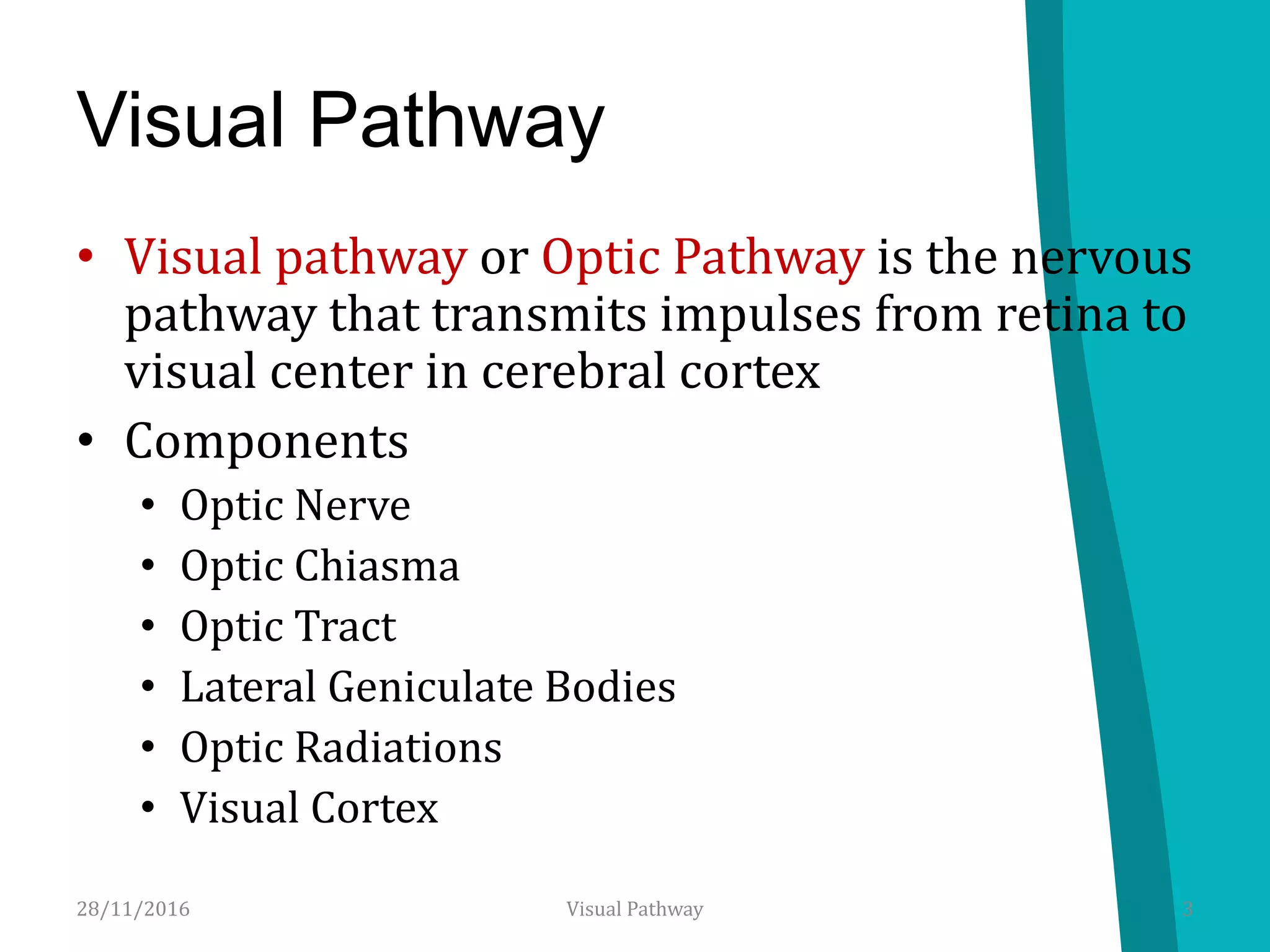
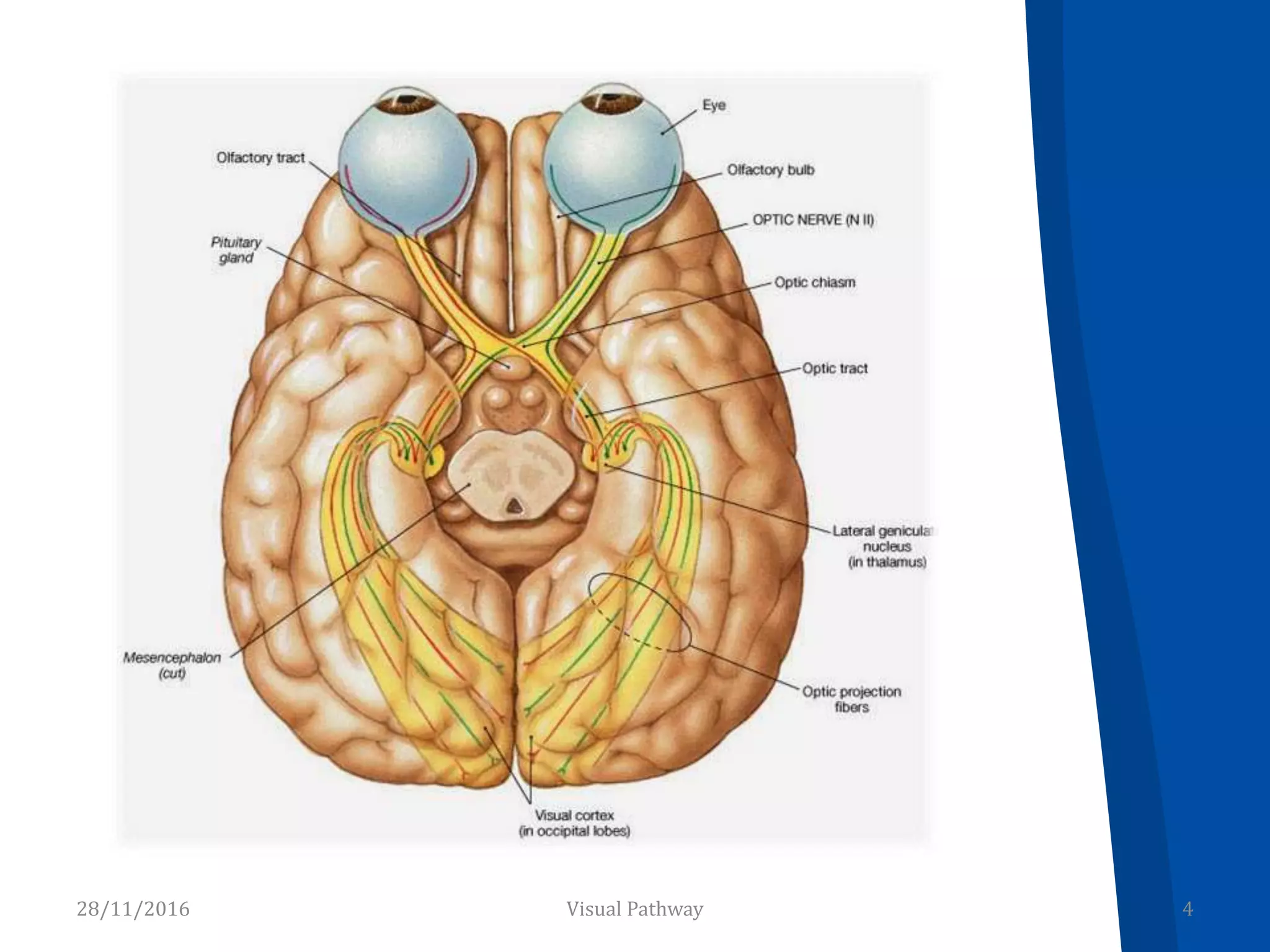
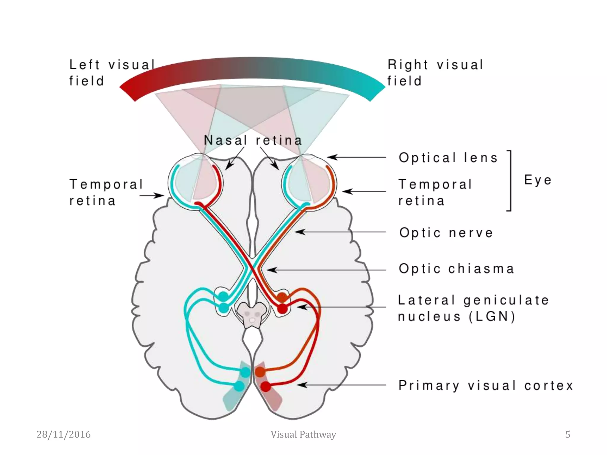
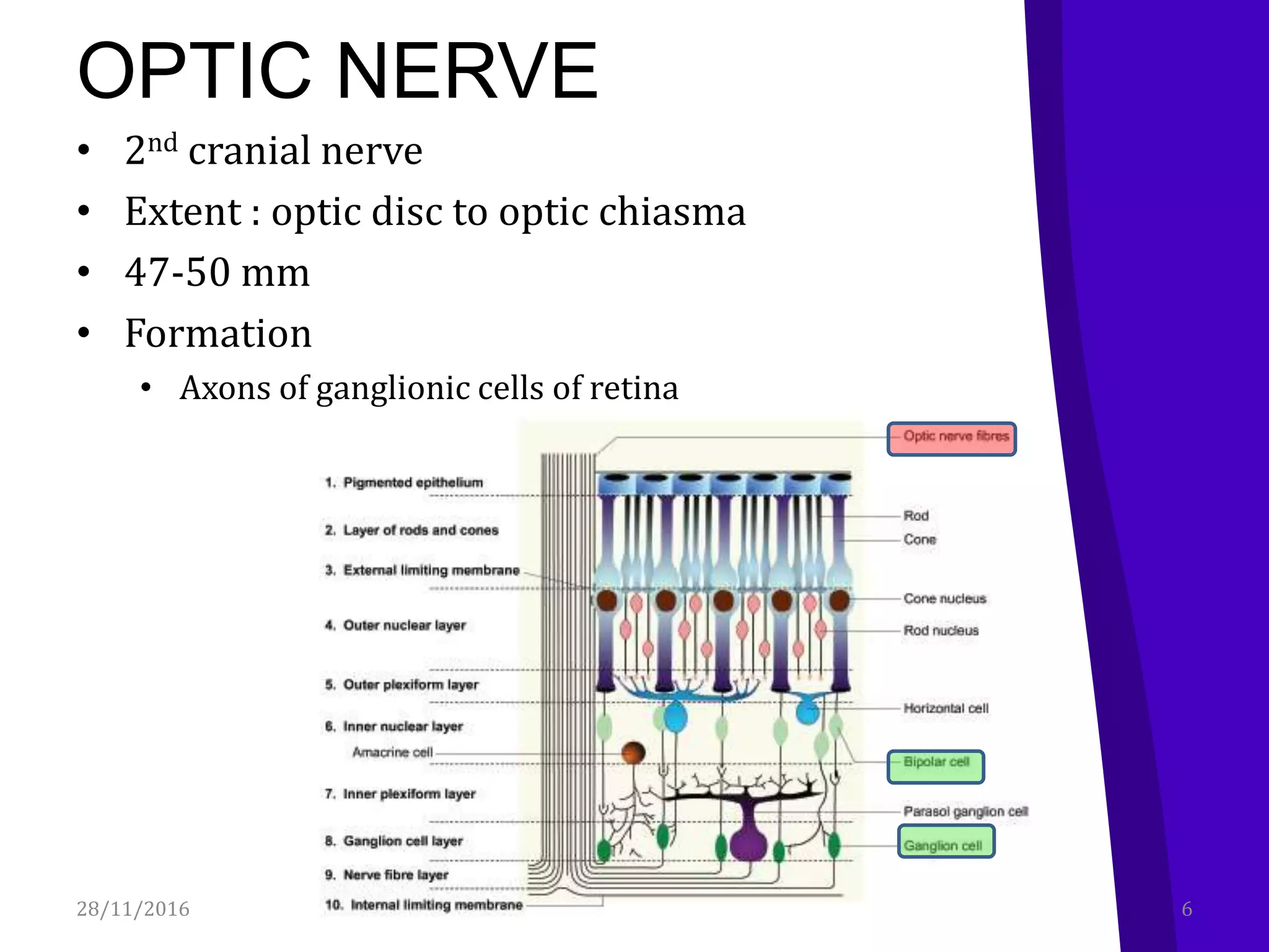
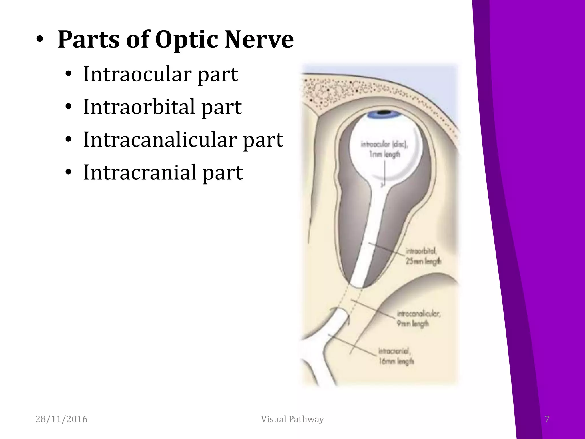
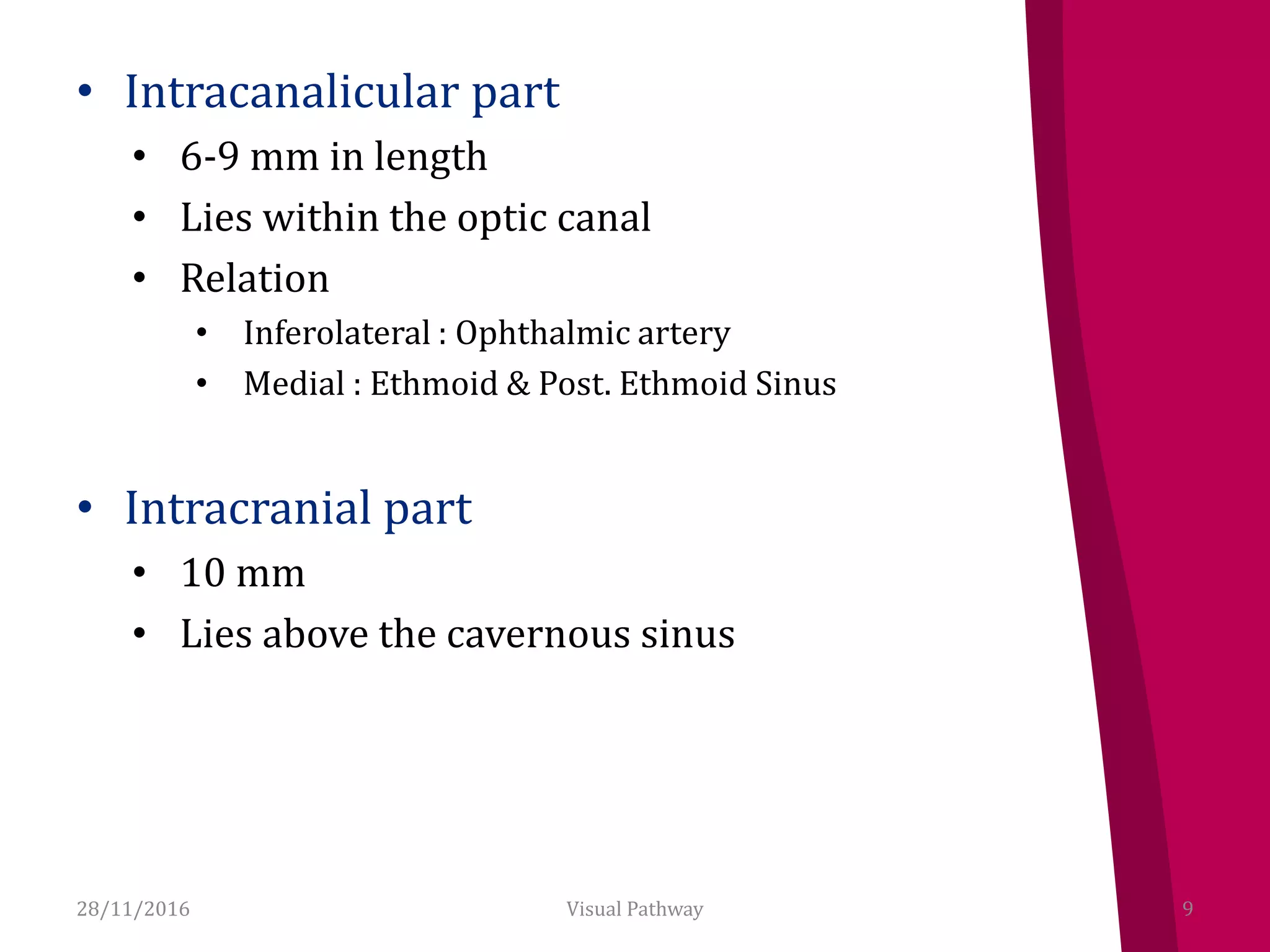
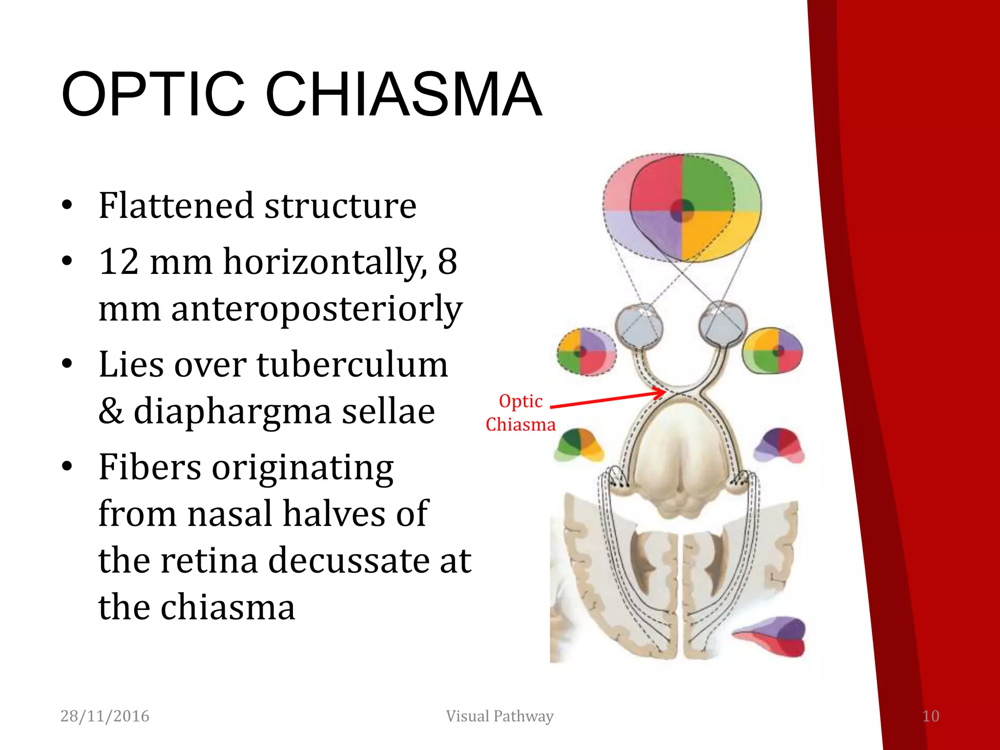
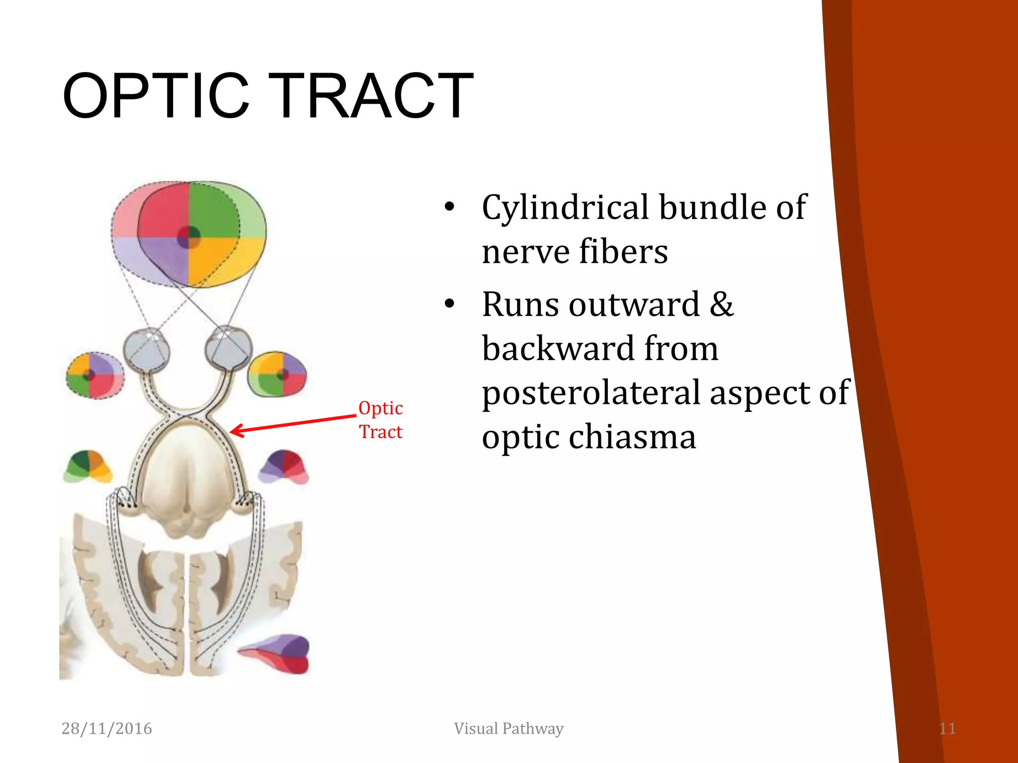
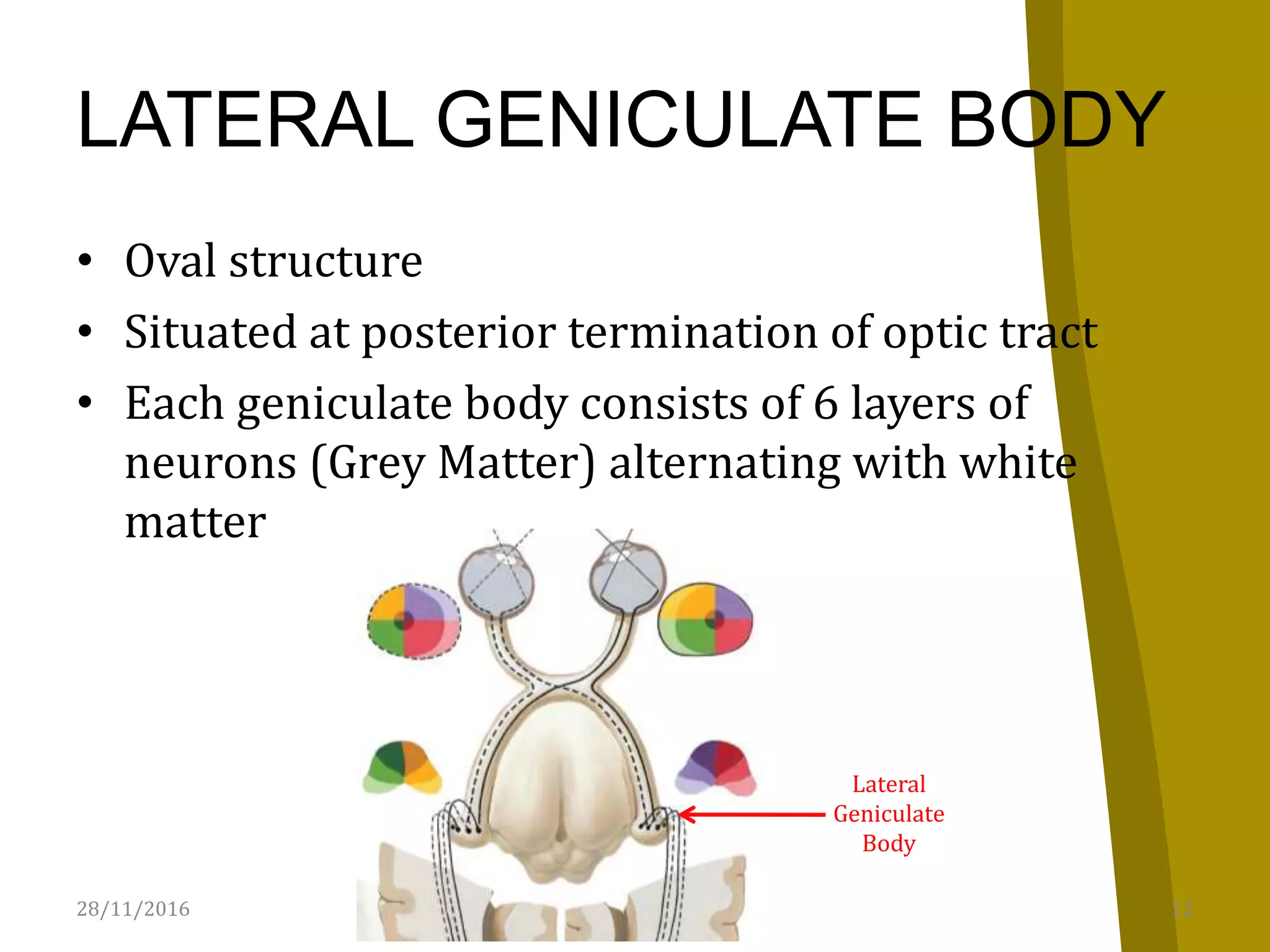
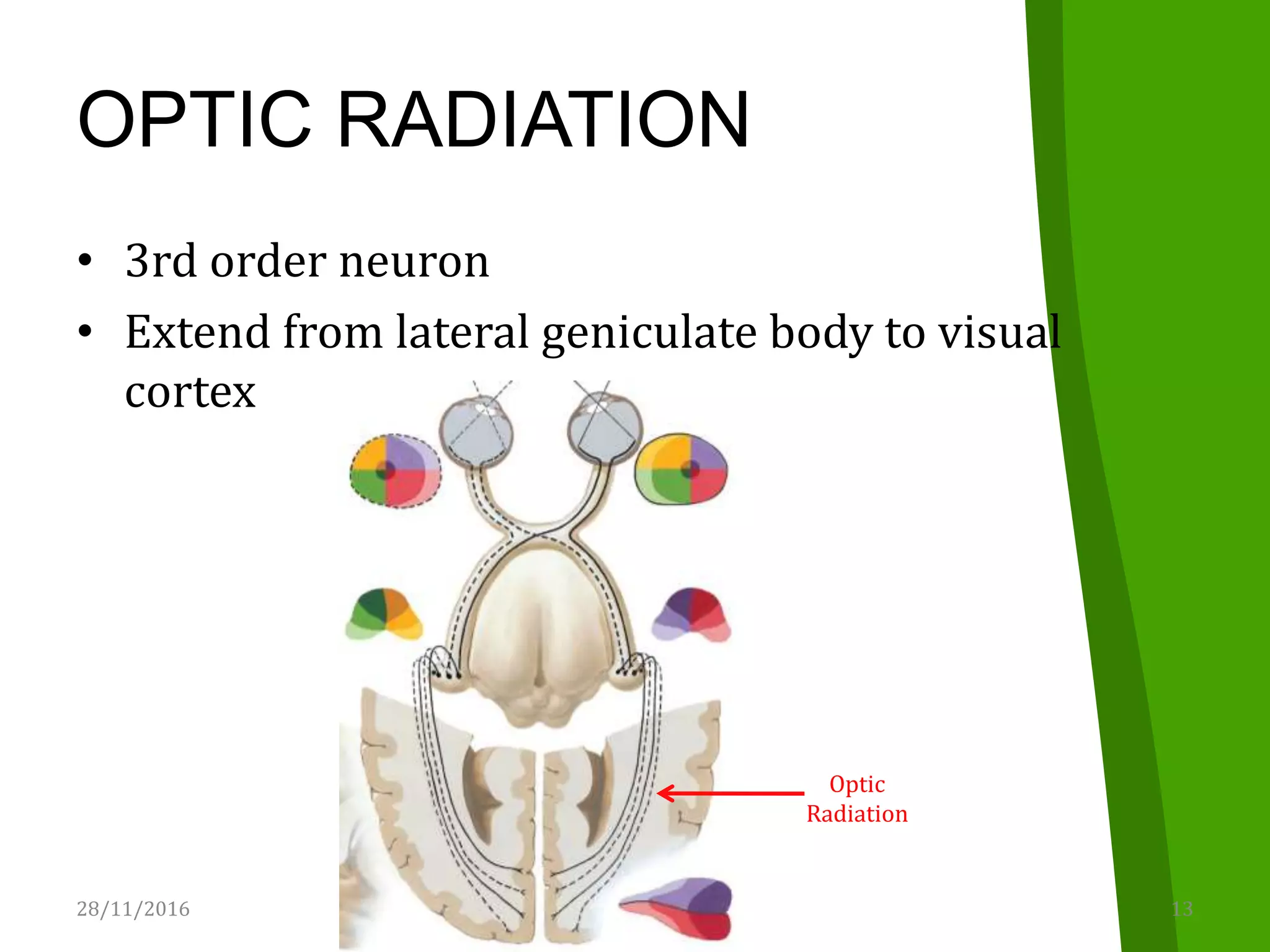
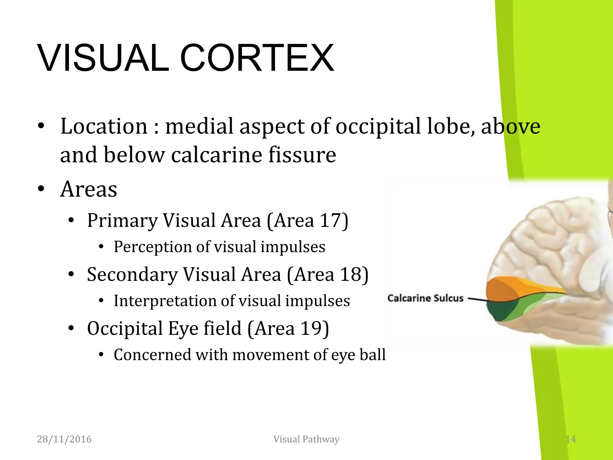
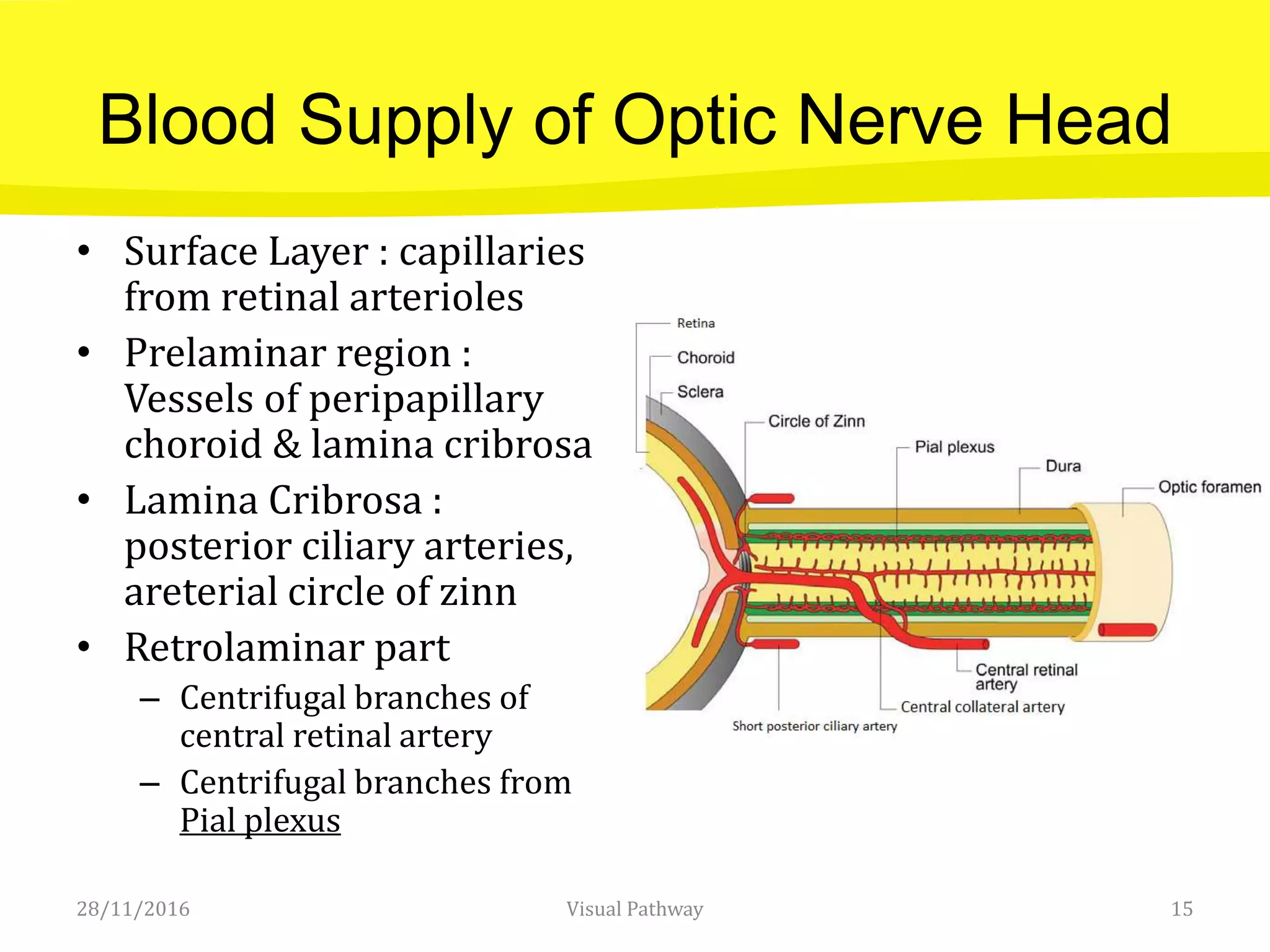
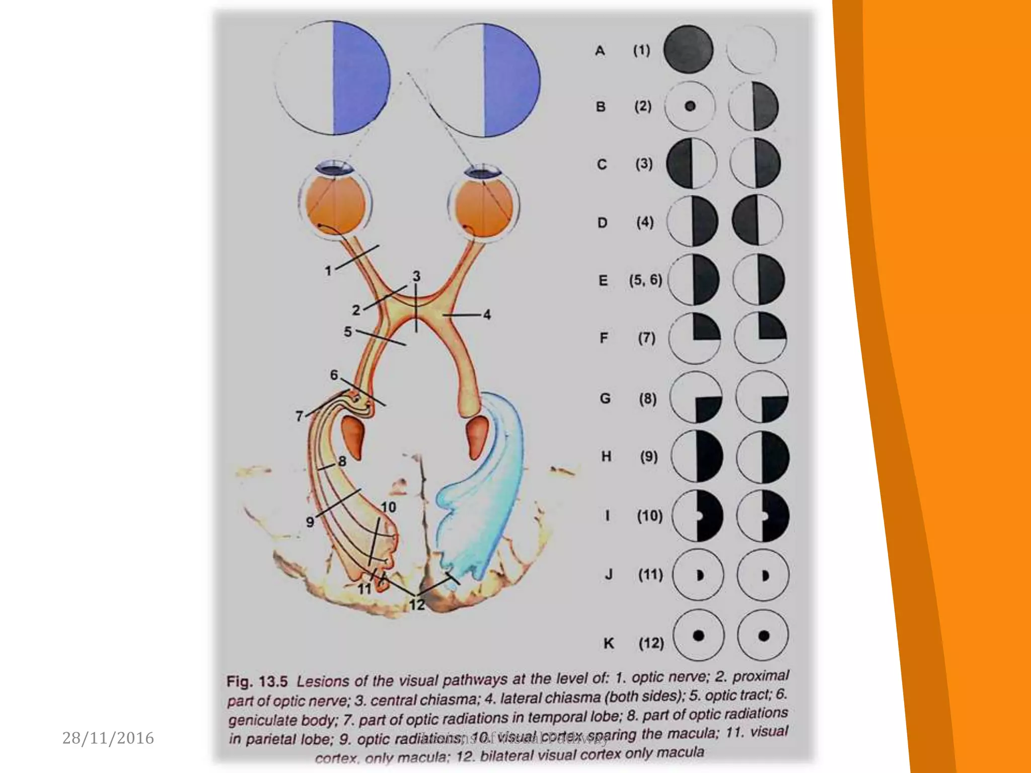
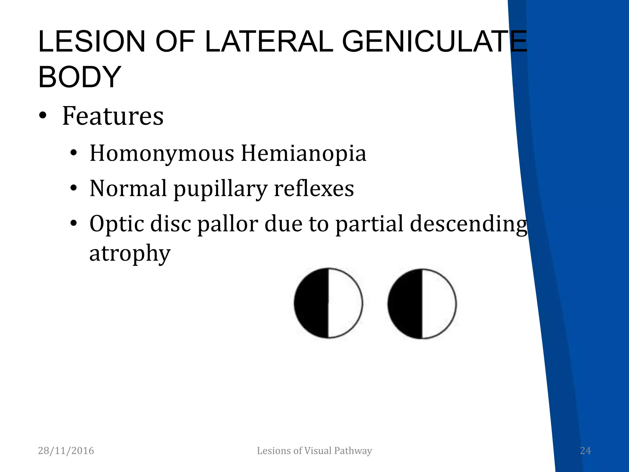
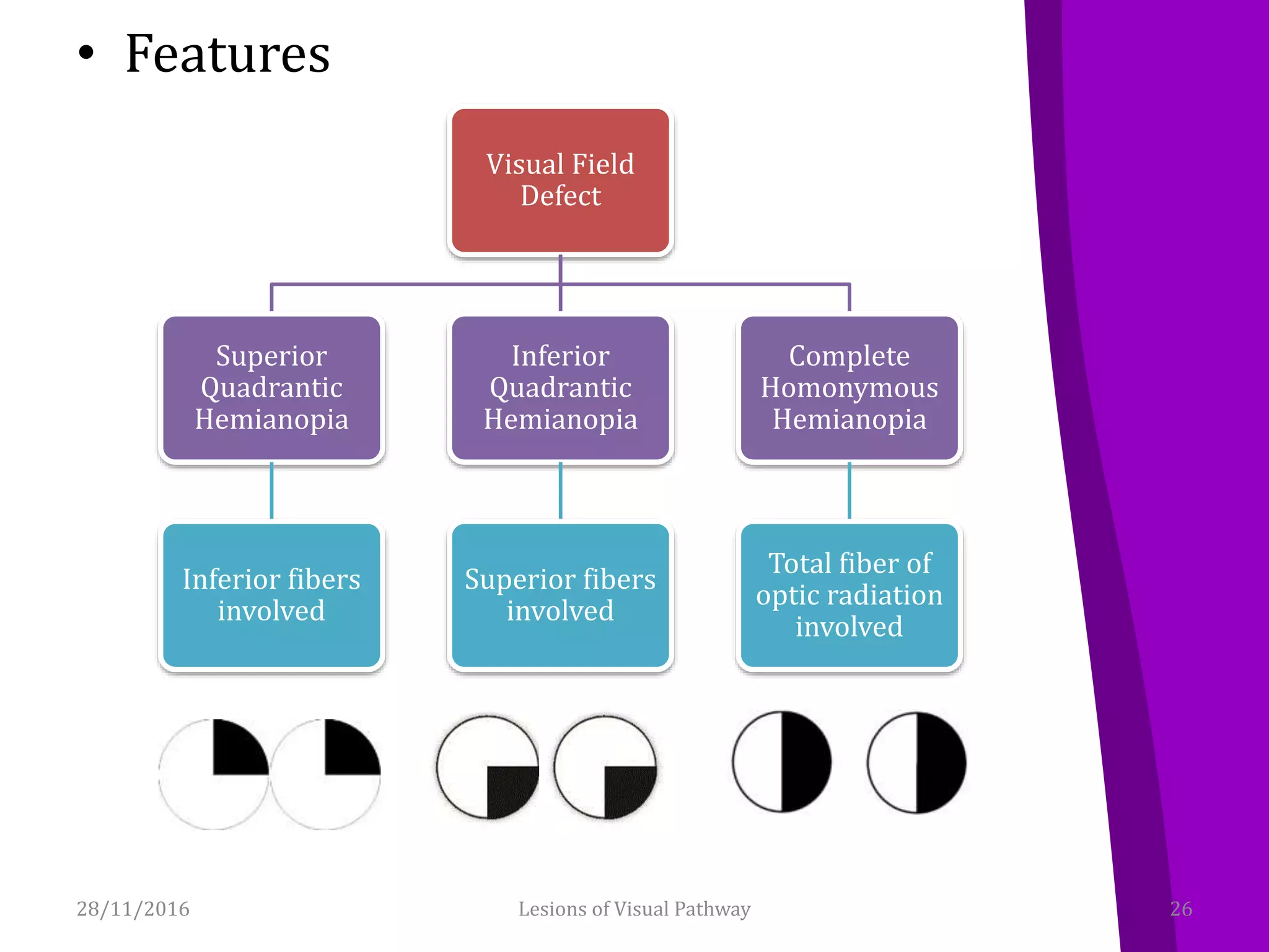
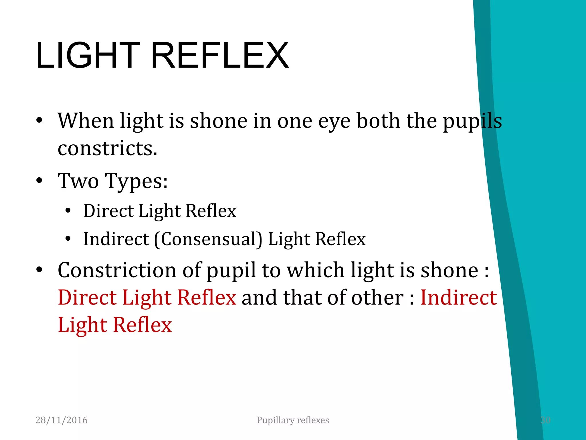
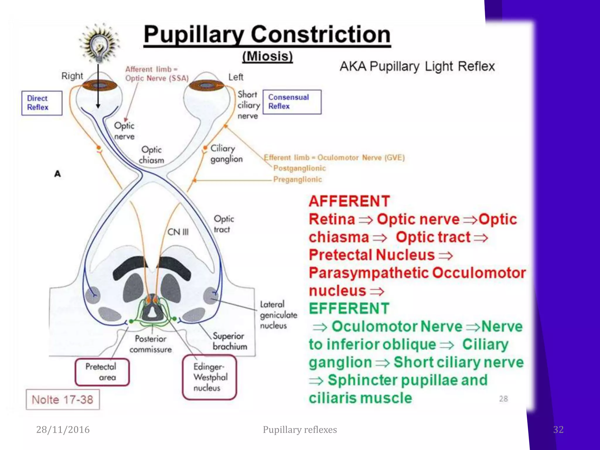
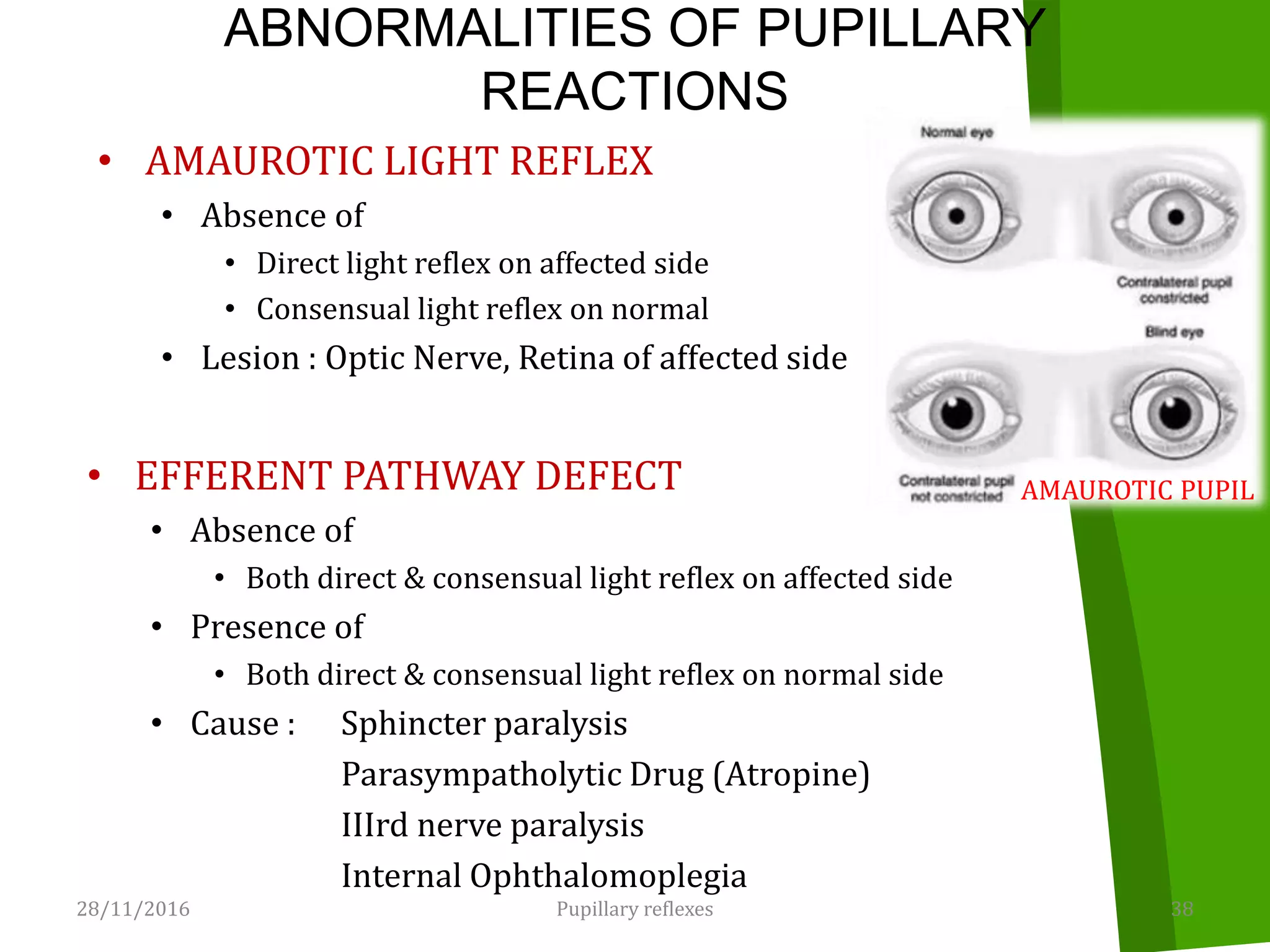
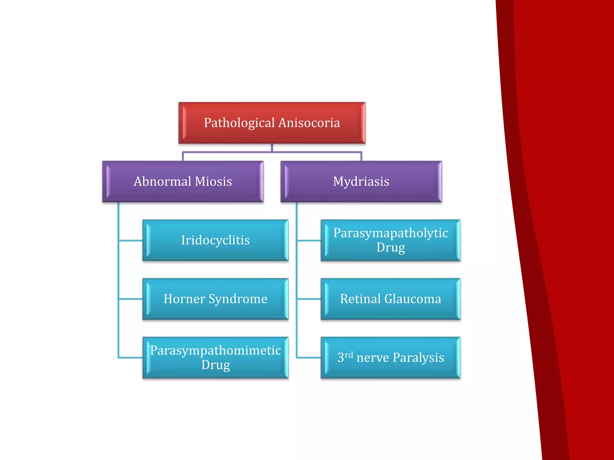
Presented By:
Khemchand Sahu
MBBS Final Part I
Guided By:
Dr. Divya Verma
Asst. Professor
Dept. of Ophthalmology
Lt. Shri Lakhiram Agrawal Memorial
Medical College, Raigarh
Contents
• Anatomy & Physiology of Visual Pathway
• Lesions of the Visual Pathway
• Pupillary Reflexes
• Abnormalities of Pupillary Reflexes
28/11/2016 2Visual Pathway
Visual Pathway
• Visual pathway or Optic Pathway is the nervous
pathway that transmits impulses from retina to
visual center in cerebral cortex
• Components
• Optic Nerve
• Optic Chiasma
• Optic Tract
• Lateral Geniculate Bodies
• Optic Radiations
• Visual Cortex
28/11/2016 3Visual Pathway
28/11/2016 4Visual Pathway
28/11/2016 5Visual Pathway
OPTIC NERVE
• 2nd cranial nerve
• Extent : optic disc to optic chiasma
• 47-50 mm
• Formation
• Axons of ganglionic cells of retina
28/11/2016 6Visual Pathway
• Parts of Optic Nerve
• Intraocular part
• Intraorbital part
• Intracanalicular part
• Intracranial part
28/11/2016 7Visual Pathway
• Intraocular part
• 1 mm
• Starts from optic disc, pierces the choroid & sclera,
converting it into sieve like structure Lamina
Cribrosa
• Intraorbital part
• 30 mm
• From back of the eyeball to optic foramina
• Relation
• Ant : Seperated from the ocular muscle by the orbital fat
• Post : Closely surrounded by Annulus of Zinn
• Retro bulbar Neuritis : Painful ocular movements
• Intracanalicular part
• 6-9 mm in length
• Lies within the optic canal
• Relation
• Inferolateral : Ophthalmic artery
• Medial : Ethmoid & Post. Ethmoid Sinus
• Intracranial part
• 10 mm
• Lies above the cavernous sinus
28/11/2016 9Visual Pathway
OPTIC CHIASMA
• Flattened structure
• 12 mm horizontally, 8
mm anteroposteriorly
• Lies over tuberculum
& diaphargma sellae
• Fibers originating
from nasal halves of
the retina decussate at
the chiasma
28/11/2016 Visual Pathway 10
Optic
Chiasma
OPTIC TRACT
• Cylindrical bundle of
nerve fibers
• Runs outward &
backward from
posterolateral aspect of
optic chiasma
28/11/2016 Visual Pathway 11
Optic
Tract
LATERAL GENICULATE BODY
• Oval structure
• Situated at posterior termination of optic tract
• Each geniculate body consists of 6 layers of
neurons (Grey Matter) alternating with white
matter
28/11/2016 12Visual Pathway
Lateral
Geniculate
Body
OPTIC RADIATION
• 3rd order neuron
• Extend from lateral geniculate body to visual
cortex
28/11/2016 13Visual Pathway
Optic
Radiation
VISUAL CORTEX
• Location : medial aspect of occipital lobe, above
and below calcarine fissure
• Areas
• Primary Visual Area (Area 17)
• Perception of visual impulses
• Secondary Visual Area (Area 18)
• Interpretation of visual impulses
• Occipital Eye field (Area 19)
• Concerned with movement of eye ball
28/11/2016 14Visual Pathway
Blood Supply of Optic Nerve Head
• Surface Layer : capillaries
from retinal arterioles
• Prelaminar region :
Vessels of peripapillary
choroid & lamina cribrosa
• Lamina Cribrosa :
posterior ciliary arteries,
areterial circle of zinn
• Retrolaminar part
– Centrifugal branches of
central retinal artery
– Centrifugal branches from
Pial plexus
28/11/2016 Visual Pathway 15
LESIONS OF VISUAL
PATHWAY
28/11/2016 17Lesions of Visual Pathway
OPTIC NERVE LESION
• Causes
• Optic atrophy
• Traumatic avulsion of optic nerve
• Indirect optic neuropathy
• Ischemic optic neuropathy
• Acute optic neuritis
• Clinical features
28/11/2016 18
Proximal Optic Nerve Lesion Distal Optic Nerve Lesion
•Ipsilateral Blindness
•Contralateral hemianopia
•Abolition of direct light reflex on
affected side & consensual on
contralateral side
•Near reflex is intact
•Ipsilateral blindness
•Abolition of direct light reflex on
affected side & consensual on
contralateral side
•Near reflex is intact
Lesions of Visual Pathway
CHIASMAL LESION
• Causes
– Intrinsic
• Gliomas
• Multiple sclerosis
– Extrinsic : compressive lesion
• Pituitary adenoma
• Craniopharyngiomas
• Meningiomas
– Other
• Metabolic
• Toxic
• Inflammatory
28/11/2016 19Lesions of Visual Pathway
Clinical feature of chiasmal lesion
• CHIASMAL SYNDROME
28/11/2016 20
Anterior Chiasmal Syndrome
Affected part : Ipsilateral
Optic Nerve fiber &
Contralateral inferonasal fiber
Junctional Scotoma
Middle Chiasmal
Syndrome
Affected part :
Decussating fibers in
the body of chiasma
Bitemporal
hemianopia
Posterior Chiasmal Syndrome
Affected part : Caudal fiber
Paracenbitemporal field defect
Homonymous hemianopia on
contralateral side
Lesions of Visual Pathway
• LATERAL CHIASMAL LESION
• Causes
• Distension of 3rd ventricle
• Atheroma of the carotid or posterior
communicating arteries
• Features
• Binasal Hemianopia
28/11/2016 21Lesions of Visual Pathway
OPTIC TRACT LESION
• Causes
• Intrinsic
• Demyelinating diseases
• Infarction
• Extrinsic
• Pituitary adenoma
• Craniopharyngioma
• Others
• Syphilitic meningitis
• Tubercular meningitis
28/11/2016 22Lesions of Visual Pathway
• Clinical Feature
• Incongruous Homonymous Hemianopia
• Wernicke’s reaction
• Optic disc changes
• Descending partial optic atropy : temporal pallor on
side of lesion & bow-tie atrophy on contralateral side
28/11/2016 23Lesions of Visual Pathway
LESION OF LATERAL GENICULATE
BODY
• Features
• Homonymous Hemianopia
• Normal pupillary reflexes
• Optic disc pallor due to partial descending
atrophy
28/11/2016 24Lesions of Visual Pathway
LESION OF OPTIC RADIATION
• Causes
• Vascular occlusion
• Tumor
• Trauma
• Temporal lobectomy
28/11/2016 25Lesions of Visual Pathway
• Features
Visual Field
Defect
Superior
Quadrantic
Hemianopia
Inferior fibers
involved
Inferior
Quadrantic
Hemianopia
Superior fibers
involved
Complete
Homonymous
Hemianopia
Total fiber of
optic radiation
involved
28/11/2016 26Lesions of Visual Pathway
LESION OF VISUAL CORTEX
• Causes
• Occlusion of posterior cerebral artery
• Head injury
28/11/2016 27Lesions of Visual Pathway
• Visual Field Defect
• Congruous Homonymous
hemianopia (sparing the
macula)
• Congruous Homonymous
Macular defect
• Bilateral Homonymous
macular defect
• Bilateral Homonymous
Hemianopia with macula
sparing
• Other Manifestation
• Cortical Blindness
• Dyschromatopsia
• Visual Hallucination
• Palinopsia
• Polyopsia
Features
28/11/2016 28Lesions of Visual Pathway
PUPILLARY REFLEXES
LIGHT REFLEX
• When light is shone in one eye both the pupils
constricts.
• Two Types:
• Direct Light Reflex
• Indirect (Consensual) Light Reflex
• Constriction of pupil to which light is shone :
Direct Light Reflex and that of other : Indirect
Light Reflex
28/11/2016 30Pupillary reflexes
Afferent Fiber
From retina to
pretectal nucleus
Internuncial
Fiber
Connects each
pretectal nucleus
with Edinger-
Westphal Nuclei of
both side
Efferent
Pathway
From Edinger-
Westphal nucleus
to sphincter
pupillae
28/11/2016 31Pupillary reflexes
Pathway of light Reflex
28/11/2016 32Pupillary reflexes
NEAR REFLEX
• Occurs on looking at near object
• Two components
• Convergence Reflex : Contraction of pupil on
convergence
• Accommodation Reflex : Contraction of pupil associated
with accommodation
28/11/2016 33Pupillary reflexes
Pathway of Near Reflex
28/11/2016 34Pupillary reflexes
PSYCHOSENSORY REFLEX
• Dilatation of pupil in response to sensory &
psychic stimuli
28/11/2016 35Pupillary reflexes
EXAMINATION OF PUPILLARY REFLEXES
• Direct Light Reflex
• Normal pupil reacts briskly &
its constriction to light is well
maintained
• Consensual Light Reflex
• Normally contralateral pupil
should also constrict when
light is thrown on to one pupil
28/11/2016 Pupillary reflexes 36
• Swinging Flash Light Test
• Performed when relative
afferent pathway defect is
suspected in one eye
• Normally, both pupil
constrict equally & pupil to
which light is transferred is
tightly constricted
• MARCUS-GUNN Pupil
28/11/2016 37Pupillary reflexes
• Near Reflex
• Pupil constrict while looking at near object
ABNORMALITIES OF PUPILLARY
REACTIONS
• AMAUROTIC LIGHT REFLEX
• Absence of
• Direct light reflex on affected side
• Consensual light reflex on normal
• Lesion : Optic Nerve, Retina of affected side
28/11/2016 38Pupillary reflexes
• EFFERENT PATHWAY DEFECT
• Absence of
• Both direct & consensual light reflex on affected side
• Presence of
• Both direct & consensual light reflex on normal side
• Cause : Sphincter paralysis
Parasympatholytic Drug (Atropine)
IIIrd nerve paralysis
Internal Ophthalomoplegia
AMAUROTIC PUPIL
• WERNICKE’S HEMIANOPIC PUPIL
• Light reflex is ABSENT
• When light is thrown on temporal half retina of affected side & nasal
half of opposite side
• Light reflex is PRESENT
• When light is thrown on nasal half of affected side & temporal half of
opposite side
• Lesion of optic tract
• MARCUS-GUNN PUPIL
• Seen in Relative Afferent Pupillary Defect (RAPD)
• Affected pupil will dilate when light is moved from normal to
abnormal eye
• Earliest indication of optic nerve disease
• Cause : Incomplete optic nerve lesion, Severe retinal disease
• Test : Swinging Flash Light Test
28/11/2016 39Pupillary reflexes
• ARGYLL ROBERTSON PUPIL (ARP)
• For both pupil
• Reaction to near reflex : Present
• But light reflex : Absent
• Both pupil slightly small in size
• Cause – Neurosyphilis in region of tectum
28/11/2016 40Pupillary reflexes
• ADIE’S TONIC PUPIL
• Reaction to
• Light reflex : Absent
• Near reflex : Very slow & tonic
• Affected pupil is larger (Anisocoria) initially
• Cause : Postganglionic parasympathetic pupillomotor damage
• Constricts with weak Pilocarpine (0.125%), while normal does not
28/11/2016 41Pupillary reflexes
ANISOCORIA
• Normal size pupil : 3-4 mm
• Difference between size of two pupil is called
Anisocoria
• Causes
• Physiological
• Pathological
• Difference of 2 mm or more
28/11/2016 Pupillary reflexes 42
Pathological Anisocoria
Abnormal Miosis
Iridocyclitis
Horner Syndrome
Parasympathomimetic
Drug
Mydriasis
Parasymapatholytic
Drug
Retinal Glaucoma
3rd nerve Paralysis
• Evaluation of Anisocoria
• Pupil Size
• Anisocoria in dark & bright illumination :
Physiological
• Anisocoria greater in dim illumination : Horner
Syndrome
• Pupillary Light Reflex
• Normal light reaction followed by dilatation lag :
Horner Syndrome
• Poor light reaction : Parasympathetic System Defect
(3rd nerve palsy, tonic pupil, anti-cholinergic drug)
28/11/2016 44Pupillary reflexes
– Pharmacological Test :
28/11/2016 45
For suspected
parasympathetic palsy
: PILOCARPINE TEST
Pupil Constrict to low conc. of
pilocarpine (0.125%)
• Adie’s Tonic Pupil
Pupil Constrict to usual conc. of
pilocarpine (1-2%)
• 3rd nerve palsy
Pupil does not constrict to
pilocarpine
• Pharmacological mydriasis
For suspected
sympathetic palsy
Cocaine test (2-4%)
• Small pupil fails to dilate
in Horner Syndrome
Hydroxyamphetamine test
(1%)
• Small pupil Dilate in
Central & preganglionic
and does not dilate in post
ganglionic Horner
syndrome
Pupillary reflexes
28/11/2016 46

Visual pathway kHem

  • 1.
    Presented By: Khemchand Sahu MBBSFinal Part I Guided By: Dr. Divya Verma Asst. Professor Dept. of Ophthalmology Lt. Shri Lakhiram Agrawal Memorial Medical College, Raigarh
  • 2.
    Contents • Anatomy &Physiology of Visual Pathway • Lesions of the Visual Pathway • Pupillary Reflexes • Abnormalities of Pupillary Reflexes 28/11/2016 2Visual Pathway
  • 3.
    Visual Pathway • Visualpathway or Optic Pathway is the nervous pathway that transmits impulses from retina to visual center in cerebral cortex • Components • Optic Nerve • Optic Chiasma • Optic Tract • Lateral Geniculate Bodies • Optic Radiations • Visual Cortex 28/11/2016 3Visual Pathway
  • 4.
  • 5.
  • 6.
    OPTIC NERVE • 2ndcranial nerve • Extent : optic disc to optic chiasma • 47-50 mm • Formation • Axons of ganglionic cells of retina 28/11/2016 6Visual Pathway
  • 7.
    • Parts ofOptic Nerve • Intraocular part • Intraorbital part • Intracanalicular part • Intracranial part 28/11/2016 7Visual Pathway
  • 8.
    • Intraocular part •1 mm • Starts from optic disc, pierces the choroid & sclera, converting it into sieve like structure Lamina Cribrosa • Intraorbital part • 30 mm • From back of the eyeball to optic foramina • Relation • Ant : Seperated from the ocular muscle by the orbital fat • Post : Closely surrounded by Annulus of Zinn • Retro bulbar Neuritis : Painful ocular movements
  • 9.
    • Intracanalicular part •6-9 mm in length • Lies within the optic canal • Relation • Inferolateral : Ophthalmic artery • Medial : Ethmoid & Post. Ethmoid Sinus • Intracranial part • 10 mm • Lies above the cavernous sinus 28/11/2016 9Visual Pathway
  • 10.
    OPTIC CHIASMA • Flattenedstructure • 12 mm horizontally, 8 mm anteroposteriorly • Lies over tuberculum & diaphargma sellae • Fibers originating from nasal halves of the retina decussate at the chiasma 28/11/2016 Visual Pathway 10 Optic Chiasma
  • 11.
    OPTIC TRACT • Cylindricalbundle of nerve fibers • Runs outward & backward from posterolateral aspect of optic chiasma 28/11/2016 Visual Pathway 11 Optic Tract
  • 12.
    LATERAL GENICULATE BODY •Oval structure • Situated at posterior termination of optic tract • Each geniculate body consists of 6 layers of neurons (Grey Matter) alternating with white matter 28/11/2016 12Visual Pathway Lateral Geniculate Body
  • 13.
    OPTIC RADIATION • 3rdorder neuron • Extend from lateral geniculate body to visual cortex 28/11/2016 13Visual Pathway Optic Radiation
  • 14.
    VISUAL CORTEX • Location: medial aspect of occipital lobe, above and below calcarine fissure • Areas • Primary Visual Area (Area 17) • Perception of visual impulses • Secondary Visual Area (Area 18) • Interpretation of visual impulses • Occipital Eye field (Area 19) • Concerned with movement of eye ball 28/11/2016 14Visual Pathway
  • 15.
    Blood Supply ofOptic Nerve Head • Surface Layer : capillaries from retinal arterioles • Prelaminar region : Vessels of peripapillary choroid & lamina cribrosa • Lamina Cribrosa : posterior ciliary arteries, areterial circle of zinn • Retrolaminar part – Centrifugal branches of central retinal artery – Centrifugal branches from Pial plexus 28/11/2016 Visual Pathway 15
  • 16.
  • 17.
  • 18.
    OPTIC NERVE LESION •Causes • Optic atrophy • Traumatic avulsion of optic nerve • Indirect optic neuropathy • Ischemic optic neuropathy • Acute optic neuritis • Clinical features 28/11/2016 18 Proximal Optic Nerve Lesion Distal Optic Nerve Lesion •Ipsilateral Blindness •Contralateral hemianopia •Abolition of direct light reflex on affected side & consensual on contralateral side •Near reflex is intact •Ipsilateral blindness •Abolition of direct light reflex on affected side & consensual on contralateral side •Near reflex is intact Lesions of Visual Pathway
  • 19.
    CHIASMAL LESION • Causes –Intrinsic • Gliomas • Multiple sclerosis – Extrinsic : compressive lesion • Pituitary adenoma • Craniopharyngiomas • Meningiomas – Other • Metabolic • Toxic • Inflammatory 28/11/2016 19Lesions of Visual Pathway
  • 20.
    Clinical feature ofchiasmal lesion • CHIASMAL SYNDROME 28/11/2016 20 Anterior Chiasmal Syndrome Affected part : Ipsilateral Optic Nerve fiber & Contralateral inferonasal fiber Junctional Scotoma Middle Chiasmal Syndrome Affected part : Decussating fibers in the body of chiasma Bitemporal hemianopia Posterior Chiasmal Syndrome Affected part : Caudal fiber Paracenbitemporal field defect Homonymous hemianopia on contralateral side Lesions of Visual Pathway
  • 21.
    • LATERAL CHIASMALLESION • Causes • Distension of 3rd ventricle • Atheroma of the carotid or posterior communicating arteries • Features • Binasal Hemianopia 28/11/2016 21Lesions of Visual Pathway
  • 22.
    OPTIC TRACT LESION •Causes • Intrinsic • Demyelinating diseases • Infarction • Extrinsic • Pituitary adenoma • Craniopharyngioma • Others • Syphilitic meningitis • Tubercular meningitis 28/11/2016 22Lesions of Visual Pathway
  • 23.
    • Clinical Feature •Incongruous Homonymous Hemianopia • Wernicke’s reaction • Optic disc changes • Descending partial optic atropy : temporal pallor on side of lesion & bow-tie atrophy on contralateral side 28/11/2016 23Lesions of Visual Pathway
  • 24.
    LESION OF LATERALGENICULATE BODY • Features • Homonymous Hemianopia • Normal pupillary reflexes • Optic disc pallor due to partial descending atrophy 28/11/2016 24Lesions of Visual Pathway
  • 25.
    LESION OF OPTICRADIATION • Causes • Vascular occlusion • Tumor • Trauma • Temporal lobectomy 28/11/2016 25Lesions of Visual Pathway
  • 26.
    • Features Visual Field Defect Superior Quadrantic Hemianopia Inferiorfibers involved Inferior Quadrantic Hemianopia Superior fibers involved Complete Homonymous Hemianopia Total fiber of optic radiation involved 28/11/2016 26Lesions of Visual Pathway
  • 27.
    LESION OF VISUALCORTEX • Causes • Occlusion of posterior cerebral artery • Head injury 28/11/2016 27Lesions of Visual Pathway
  • 28.
    • Visual FieldDefect • Congruous Homonymous hemianopia (sparing the macula) • Congruous Homonymous Macular defect • Bilateral Homonymous macular defect • Bilateral Homonymous Hemianopia with macula sparing • Other Manifestation • Cortical Blindness • Dyschromatopsia • Visual Hallucination • Palinopsia • Polyopsia Features 28/11/2016 28Lesions of Visual Pathway
  • 29.
  • 30.
    LIGHT REFLEX • Whenlight is shone in one eye both the pupils constricts. • Two Types: • Direct Light Reflex • Indirect (Consensual) Light Reflex • Constriction of pupil to which light is shone : Direct Light Reflex and that of other : Indirect Light Reflex 28/11/2016 30Pupillary reflexes
  • 31.
    Afferent Fiber From retinato pretectal nucleus Internuncial Fiber Connects each pretectal nucleus with Edinger- Westphal Nuclei of both side Efferent Pathway From Edinger- Westphal nucleus to sphincter pupillae 28/11/2016 31Pupillary reflexes Pathway of light Reflex
  • 32.
  • 33.
    NEAR REFLEX • Occurson looking at near object • Two components • Convergence Reflex : Contraction of pupil on convergence • Accommodation Reflex : Contraction of pupil associated with accommodation 28/11/2016 33Pupillary reflexes
  • 34.
    Pathway of NearReflex 28/11/2016 34Pupillary reflexes
  • 35.
    PSYCHOSENSORY REFLEX • Dilatationof pupil in response to sensory & psychic stimuli 28/11/2016 35Pupillary reflexes
  • 36.
    EXAMINATION OF PUPILLARYREFLEXES • Direct Light Reflex • Normal pupil reacts briskly & its constriction to light is well maintained • Consensual Light Reflex • Normally contralateral pupil should also constrict when light is thrown on to one pupil 28/11/2016 Pupillary reflexes 36
  • 37.
    • Swinging FlashLight Test • Performed when relative afferent pathway defect is suspected in one eye • Normally, both pupil constrict equally & pupil to which light is transferred is tightly constricted • MARCUS-GUNN Pupil 28/11/2016 37Pupillary reflexes • Near Reflex • Pupil constrict while looking at near object
  • 38.
    ABNORMALITIES OF PUPILLARY REACTIONS •AMAUROTIC LIGHT REFLEX • Absence of • Direct light reflex on affected side • Consensual light reflex on normal • Lesion : Optic Nerve, Retina of affected side 28/11/2016 38Pupillary reflexes • EFFERENT PATHWAY DEFECT • Absence of • Both direct & consensual light reflex on affected side • Presence of • Both direct & consensual light reflex on normal side • Cause : Sphincter paralysis Parasympatholytic Drug (Atropine) IIIrd nerve paralysis Internal Ophthalomoplegia AMAUROTIC PUPIL
  • 39.
    • WERNICKE’S HEMIANOPICPUPIL • Light reflex is ABSENT • When light is thrown on temporal half retina of affected side & nasal half of opposite side • Light reflex is PRESENT • When light is thrown on nasal half of affected side & temporal half of opposite side • Lesion of optic tract • MARCUS-GUNN PUPIL • Seen in Relative Afferent Pupillary Defect (RAPD) • Affected pupil will dilate when light is moved from normal to abnormal eye • Earliest indication of optic nerve disease • Cause : Incomplete optic nerve lesion, Severe retinal disease • Test : Swinging Flash Light Test 28/11/2016 39Pupillary reflexes
  • 40.
    • ARGYLL ROBERTSONPUPIL (ARP) • For both pupil • Reaction to near reflex : Present • But light reflex : Absent • Both pupil slightly small in size • Cause – Neurosyphilis in region of tectum 28/11/2016 40Pupillary reflexes
  • 41.
    • ADIE’S TONICPUPIL • Reaction to • Light reflex : Absent • Near reflex : Very slow & tonic • Affected pupil is larger (Anisocoria) initially • Cause : Postganglionic parasympathetic pupillomotor damage • Constricts with weak Pilocarpine (0.125%), while normal does not 28/11/2016 41Pupillary reflexes
  • 42.
    ANISOCORIA • Normal sizepupil : 3-4 mm • Difference between size of two pupil is called Anisocoria • Causes • Physiological • Pathological • Difference of 2 mm or more 28/11/2016 Pupillary reflexes 42
  • 43.
    Pathological Anisocoria Abnormal Miosis Iridocyclitis HornerSyndrome Parasympathomimetic Drug Mydriasis Parasymapatholytic Drug Retinal Glaucoma 3rd nerve Paralysis
  • 44.
    • Evaluation ofAnisocoria • Pupil Size • Anisocoria in dark & bright illumination : Physiological • Anisocoria greater in dim illumination : Horner Syndrome • Pupillary Light Reflex • Normal light reaction followed by dilatation lag : Horner Syndrome • Poor light reaction : Parasympathetic System Defect (3rd nerve palsy, tonic pupil, anti-cholinergic drug) 28/11/2016 44Pupillary reflexes
  • 45.
    – Pharmacological Test: 28/11/2016 45 For suspected parasympathetic palsy : PILOCARPINE TEST Pupil Constrict to low conc. of pilocarpine (0.125%) • Adie’s Tonic Pupil Pupil Constrict to usual conc. of pilocarpine (1-2%) • 3rd nerve palsy Pupil does not constrict to pilocarpine • Pharmacological mydriasis For suspected sympathetic palsy Cocaine test (2-4%) • Small pupil fails to dilate in Horner Syndrome Hydroxyamphetamine test (1%) • Small pupil Dilate in Central & preganglionic and does not dilate in post ganglionic Horner syndrome Pupillary reflexes
  • 46.