1. The eye receives its blood supply from branches of the ophthalmic artery, which branches directly from the internal carotid artery.
2. The central retinal artery supplies the retina, while the ciliary arteries supply the choroid, iris, and ciliary body.
3. Veins drain blood from the eye and empty into the superior and inferior ophthalmic veins, which drain into the cavernous sinus. The cavernous sinus contains several cranial nerves.
INTRODUCTIONThe clear fluid filling the space in front of the eyeball between lens and cornea.The aqueous humour supplies nutrition and removes waste from the clear structure in the anterior eye(cornea and lens)The balance between aqueous production and outflow determines the intraocular pressure.
INTRODUCTION
The clear fluid filling the space in front of the eyeball between lens and cornea.
The aqueous humour supplies nutrition and removes waste from the clear structure in the anterior eye(cornea and lens)
The balance between aqueous production and outflow determines the intraocular pressure.
The aqueous humour is a transparent, watery fluid similar to plasma, but containing low protein concentrations. It is secreted from the ciliary epithelium, a structure supporting the lens
The retina is the sensory membrane that lines the inner surface of the back of the eyeball. It's composed of several layers, including one that contains specialized cells called photoreceptors.
Photoreceptor cells take light focused by the cornea and lens and convert it into chemical and nervous signals which are transported to visual centers in the brain by way of the optic nerve.
In the visual cortex of the brain (which, ironically, is located in the back of the brain), these signals are converted into images and visual perceptions.
The lacrimal apparatus is the physiological system containing the orbital structures for tear production and drainage. It consists of: The lacrimal gland, which secretes the tears, and its excretory ducts, which convey the fluid to the surface of the human eye;it is a serous gland located in lacrimal fossa.
Each eyelid contains a fibrous plate, called a tarsus, that gives it structure and shape; muscles, which move the eyelids; and meibomian (or tarsal) glands, which secrete lubricating fluids. The lids are covered with skin, lined with mucous membrane, and bordered with a fringe of hairs, the eyelashes.
Direct Download Link ❤❤https://healthkura.com/eye-ppt/28/❤❤
Dear viewers Check Out my other piece of works at ❤❤❤ https://healthkura.com/eye-ppt/❤❤❤
anatomy of optic nerve and its blood supply and clinical corelation
Presentation Layout: optic nerve anatomy
Embryology of optic nerve
Introduction
Parts of optic nerve
Blood supply
Clinical significance
For Further Reading
Wolff’s Anatomy of the eye and orbit by Bron, Tripathi and Tripathi
Anatomy and Physiology of eye by A.K. Khurana 2nd edition
Comprehensive Ophthalmology by A.K. Khurana 5th edition
AAO- Fundamentals & Principles of Ophthalmology : sec 2
Walsh and Hoyt’s Clinical Ophthalmology
Internet
• All structures are supplied by branches of
Internal Carotid Artery
• Except eyelids and conjunctiva which receives
blood supply from the branches of both
internal and external carotid artery
Central retinal artery
• First branch from the ophthalmic artery
• End arteries
• Divides into equal superior & inferior branches,
then another division (nasal & temporal)
The brain receives blood from two sources: the internal carotid arteries, which arise at the point in the neck where the common carotid arteries bifurcate, and the vertebral arteries . The internal carotid arteries branch to form two major cerebral arteries, the anterior and middle cerebral arteries. The right and left vertebral arteries come together at the level of the pons on the ventral surface of the brainstem to form the midline basilar artery. The basilar artery joins the blood supply from the internal carotids in an arterial ring at the base of the brain (in the vicinity of the hypothalamus and cerebral peduncles) called the circle of Willis. The posterior cerebral arteries arise at this confluence, as do two small bridging arteries, the anterior and posterior communicating arteries. Conjoining the two major sources of cerebral vascular supply via the circle of Willis presumably improves the chances of any region of the brain continuing to receive blood if one of the major arteries becomes occluded
The carotid arteries are the primary vessels supplying blood to the brain and face. The right common carotid artery (RCCA) originates in the neck from the brachiocephalic artery while the left common carotid artery (LCCA) arises in the thorax from the arch of the aorta.
Instructions for Submissions thorugh G- Classroom.pptxJheel Barad
This presentation provides a briefing on how to upload submissions and documents in Google Classroom. It was prepared as part of an orientation for new Sainik School in-service teacher trainees. As a training officer, my goal is to ensure that you are comfortable and proficient with this essential tool for managing assignments and fostering student engagement.
The French Revolution, which began in 1789, was a period of radical social and political upheaval in France. It marked the decline of absolute monarchies, the rise of secular and democratic republics, and the eventual rise of Napoleon Bonaparte. This revolutionary period is crucial in understanding the transition from feudalism to modernity in Europe.
For more information, visit-www.vavaclasses.com
2024.06.01 Introducing a competency framework for languag learning materials ...Sandy Millin
http://sandymillin.wordpress.com/iateflwebinar2024
Published classroom materials form the basis of syllabuses, drive teacher professional development, and have a potentially huge influence on learners, teachers and education systems. All teachers also create their own materials, whether a few sentences on a blackboard, a highly-structured fully-realised online course, or anything in between. Despite this, the knowledge and skills needed to create effective language learning materials are rarely part of teacher training, and are mostly learnt by trial and error.
Knowledge and skills frameworks, generally called competency frameworks, for ELT teachers, trainers and managers have existed for a few years now. However, until I created one for my MA dissertation, there wasn’t one drawing together what we need to know and do to be able to effectively produce language learning materials.
This webinar will introduce you to my framework, highlighting the key competencies I identified from my research. It will also show how anybody involved in language teaching (any language, not just English!), teacher training, managing schools or developing language learning materials can benefit from using the framework.
The Art Pastor's Guide to Sabbath | Steve ThomasonSteve Thomason
What is the purpose of the Sabbath Law in the Torah. It is interesting to compare how the context of the law shifts from Exodus to Deuteronomy. Who gets to rest, and why?
How to Make a Field invisible in Odoo 17Celine George
It is possible to hide or invisible some fields in odoo. Commonly using “invisible” attribute in the field definition to invisible the fields. This slide will show how to make a field invisible in odoo 17.
Ethnobotany and Ethnopharmacology:
Ethnobotany in herbal drug evaluation,
Impact of Ethnobotany in traditional medicine,
New development in herbals,
Bio-prospecting tools for drug discovery,
Role of Ethnopharmacology in drug evaluation,
Reverse Pharmacology.
Students, digital devices and success - Andreas Schleicher - 27 May 2024..pptxEduSkills OECD
Andreas Schleicher presents at the OECD webinar ‘Digital devices in schools: detrimental distraction or secret to success?’ on 27 May 2024. The presentation was based on findings from PISA 2022 results and the webinar helped launch the PISA in Focus ‘Managing screen time: How to protect and equip students against distraction’ https://www.oecd-ilibrary.org/education/managing-screen-time_7c225af4-en and the OECD Education Policy Perspective ‘Students, digital devices and success’ can be found here - https://oe.cd/il/5yV
This is a presentation by Dada Robert in a Your Skill Boost masterclass organised by the Excellence Foundation for South Sudan (EFSS) on Saturday, the 25th and Sunday, the 26th of May 2024.
He discussed the concept of quality improvement, emphasizing its applicability to various aspects of life, including personal, project, and program improvements. He defined quality as doing the right thing at the right time in the right way to achieve the best possible results and discussed the concept of the "gap" between what we know and what we do, and how this gap represents the areas we need to improve. He explained the scientific approach to quality improvement, which involves systematic performance analysis, testing and learning, and implementing change ideas. He also highlighted the importance of client focus and a team approach to quality improvement.
Welcome to TechSoup New Member Orientation and Q&A (May 2024).pdfTechSoup
In this webinar you will learn how your organization can access TechSoup's wide variety of product discount and donation programs. From hardware to software, we'll give you a tour of the tools available to help your nonprofit with productivity, collaboration, financial management, donor tracking, security, and more.
Read| The latest issue of The Challenger is here! We are thrilled to announce that our school paper has qualified for the NATIONAL SCHOOLS PRESS CONFERENCE (NSPC) 2024. Thank you for your unwavering support and trust. Dive into the stories that made us stand out!
2. Arterial blood supply:
• Internal carotid artery:
• External carotid artery:
Note:
(eyelids and conjunctiva from both internal and external carotid
arteries)
3. Internal carotid artery:
• First intracranial branch of the internal carotid just
as the artery exits from the cavernous sinus
• optic foramen below and lateral to the optic nerve
• Pass over the optic nerve to its medial side
• Between the MR and SO
• Terminates by dividing into dorsonasal and
supratrochlear
Ophthalmic artery:
4. Branches of ophthalmic artery:
• Central retinal a.
• Supra-orbital artery
• Posterior ciliary artery
– Long posterior ciliary a. (2 arteries)
– Short post. Ciliary a. (10-20 arteries)
• Muscular arteries
– Anterior ciliary a. (7 arteries)
• Lacrimal artery (terminate into zygomatic branches)
• Ant. And post. Ethmoidal arteries
• Superior and inferior palpebral arteries
• Dorsonasal artery
• Supratrochlear artery
5.
6. Central retinal artery
• pierces the dural sheath of the optic
n. 12 mm behind the globe
• It gives off small meningeal branches
to supply the pial sheath of the optic
nerve.
• It’s functionally as end artery.
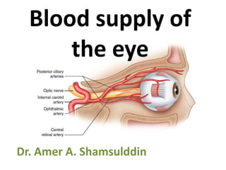
7. 1. ophthalmic artery.
2. central retinal artery
3. ciliary arteries (on each side of the
optic nerve). These vessels divide
into 2 long posterior ciliary
arteries(#4 in Figure) and ~20 short
posterior ciliary arteries (#5 in
Figure) that enter the eye
immediately adjacent and around the
optic nerve . The short posterior
ciliary arteries directly supply the
choroid and the long posterior ciliary
arteries travel in the suprachoroidal
space anteriorly (#6 in Figure) then
supply the choroid anteriorly via
recurrent branches.
posterior (#7 in Figure) and anterior (#8
in Figure) ethmoidal vessels.
The superior oblique muscle is shown for
orientation ( #9 in Figure).
8. Ciliary arteries
• Long post, short post. And ant. ciliary aa. are the major blood supply to the
globe
• Long post. Ciliary aa:
– Paired arteries that pierce the sclera outside the circle of Zinn snd
– travel forward in the suprachoroidal space to the ciliary body,
– Gives recurrent branches that supply the choroid anterior to the
equator and anastomose with short post ciliary aa.
• Short post. Ciliary aa:
– 10- 20 branches pierces the sclera around the optic n
– This anastomotic circle of Zinn supplies the optic n. head (optic disc)
– Supplies the choroid to the equator
• Ant. Ciliary aa:
– 7 arteries 2 for each rectus m. exept LR m. only 1
– Supply the sclera and conjunctiva and the iris
12. Blood supply of the
eyelids
• The main arterial blood
supply:
– Lat. Palpebral a. (from
lacrimal artery)
– med. Palpebral a. ( from
ophthalmic artery)
• These palpebral aa. (from
internal carotid a.)
anastamose with facial artery
(external carotid) and its
branch (zygomatico-temporal a.
and angular a.) at the lat.
aspect of the lid and form the
Marginal and peripheral
palpebral arcades
14. Lymph drainage
• Lat. 2/3 of upper
and lower lids
superficail parotid
nodes (pre-
auricular nn.)
• Medial
submandibular
nodes
15. Nerve supply:
• Lower lid:
– infra-orbital (from V2)
– Med. Aspect infra-
trochlear n. (V1)
• Upper lid:
– Supra-orbital n.
– Supra-trochlear n.
– Lacrimal n. (v1)
16. Blood supply to the conjunctiva
• Palpebral conjunctiva marginal and peripheral
arcades (from med. And lat. Palpebral aa.)
• Limbal conjunctiva ant. ciliary art.
• Venous drainage:
The conj. veins sup. and inf. ophthalmic vv.
• Nerve supply:
Bulbar conj. long ciliary nn. (nasocialiary of ophth. n.)
17. Blood supply of the AC
Long post. ciliary aa. anastomose with ant. ciliary aa.
– Major arterial arcade (ciliary stroma)
– Minor arterial arcade (At the collarete of the iris)
• They are the major blood supply to the iris and ciliary body
• Iridial vessels are non-fenestrated (endothilial tight junctions)
• Vessels in the ciliary body are fenestrated and no tight junctions
• Blood-aqueous barrier tight junctions in the ciliary epithelium
• Venous drainage: follow the aa. minor venous circle directly
into the vortex veins (not into the corresponding major circle)
18.
19. retinal blood supply:
• The outer plexiform layer is
the watershed region
• Outer to this layer
(neuroreceptors and RPE)
choroidal circulation by
diffusion
• Inner 2/3 central retinal
a. directly
• Retinal aa. run in the nerve
fiber layer with 4 branches
for each quadrant
20. • Retinal capillaries are concentrated in the macula
but are absent from the fovea centralis (FAZ)
• In 20% of people there’s cilioretinal a. (from post.
ciliary a.) supplies the macula
21. Blood retinal barrier
• Inner BRB:
– Tight junctions between the endothelial cells or
retinal blood vessels
• Outer BRB:
– Tight junctions between
RPE cells
22. Blood supply to the visual pathway
• Intra-ocular optic n. short post. ciliary aa.
That form the circle of Zinn.
• The central retinal a. pierces the dural sheaths
of the orbital optic n. but doest supply it, it’s
supplied by pial plexus of vessels
• Intracraneal optic n. is supplied by the
superior hypophyseal and ophthalmic aa.
• A rise in the ICP may compromise blood flow to these vessels
papilloedema
23.
24. • Optic tracts pial arteries (from ant.
Choroidal and post. Communicating aa)
• Lat. Geniculate body and post. aspect of
optic radiations the middle cerebral a. with
the post. Cerebral a.
• Visual cortex:
– Most of its blood supply post. Cerebral a.
– Occipital pole (macular area)Middle cerebral a.
(causing macular sparing in some of occipital lobe
strokes)
26. • There 4 vortex veins
(2 on each side)
exit the eyeball
post. to the equator.
• Sup. Vortex vv. and central retinal v. Superior
ophthalmic v. (which is from supra-orbital and facial vv.)
• Inferior vortex vv. Inf. Ophthalmic v. which:
– may drain to the sup. Ophthalmic v or directly to
cavernous sinus
– communicates with the pterygoid venous plexus (and
because of the reason the inf. From this plexus may reach
cavernous sinus cavernous sinus thrombosis)
27.
28. Cavernous sinus
• Extends from the sup. orbital fissure ant.ly to
the apex of petrous bone (temporal bone)
• Floor dural coverings of greater wing of
sphenoid
• Med body of sphenoid and pitiutary fossa
29. • Internal carotid a.
grooving the medial wall
of the sinus then pierces
its roof.
• The abducent nerve (VI)
travels within it
(first nerve to damage by cavernous
sinus thrombosis)
• Its lat. Wall encloses III,
IV, V1 & V2 nerves.