
More Related Content
What's hot
What's hot (20)
Similar to ANATOMY OF VISUAL PATHWAY - DR.RUTHRA.pptx
Similar to ANATOMY OF VISUAL PATHWAY - DR.RUTHRA.pptx (20)
Recently uploaded
Recently uploaded (20)
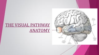
ANATOMY OF VISUAL PATHWAY - DR.RUTHRA.pptx
- 2. • Visual Pathway refers to Neuronal networks that extend from the Retina to Visual cortex • It comprises of • Optic Nerve • Optic Chiasma • Optic Tract • Lateral Geniculate Body • Optic Radiations • Visual cortex
- 3. OPTIC NERVE • 2nd cranialnerve • Optic disc to Optic chiasma • It contains 1.2 million axons • Afferent fibres for light reflex • About 47 – 50 mm length, having four parts 1)Intra ocular (1mm) 2)Intra orbital (30mm) 3)Intra canalicular (6-9mm) 4)Intra cranial (10mm)
- 4. OPTIC NERVE HEAD – INTRAOCULAR PART • About 1mm in size, passes through sclera,choroid & finally appears in eye as optic disc. • Divided in 4 portions (ant – post) 1)Surface nerve fibre layer 2)Prelaminar region 3)Lamina cribrosa 4)Retrolaminar region
- 5. 1)Surface nerve fibre layer • Composed of axonal bundles, covered by thin layer of Astrocytes INT. LIM. MEM. ELSCHING- thickened portion MENISCUS OF KUHNT • Retinal layers separated from OPT. N/V by INTERMEDIARY TISSUE OF KUHNT 2)Prelaminar region • > Astroglial tissue, separated from choroid by TISSUE OF JCOBY 3)Lamina cribrosa • Sieve structure by scleral connective tissue • Rim of connective tissue fibre btw S,C,Opt. N/V – BORDER TISSUE OF ELSHING 4)Retrolaminar region • < in astrocytes, Myelination by oligodents (1.5mm ->3mm)
- 6. INTRA ORBITAL PART • About 25-30 mm in length , from globe to orbital apex • Takes slightly curved paths for the eye movements. • Here optic nerve is surrounded by all 3 layers of meninges & subarachnoid space. • Some fibres of medial and superior rectus are adherent to its sheath (retrobulbar neuritis) • The Central retinal artery along with vein enters the subarachnoid space to enter the nerve on its inferomedial aspect • Optic n/v and Lateral rectus – Ciliary Ganglion • L -> M cross over superiorly – Ophth.artery, vein and Nasociliary n/v
- 7. INTRACANALICULAR PART • 6- 9mm long • This part is closely related to Ophthalmic artery.(crosses the nerve from lateral to medial side in Dural sheath) • Sphenoid & posterior ethmoidal sinuses lie medial to it & separated by thin bony lamina, this relation accounts for retrobulbar neuritis following infection of sinuses. • Frequently affected in Traumatic neuropathies
- 8. INTRACRANIAL PART • About 10mm • Lies above cavernous sinus & converges with its fellow to form chiasma. • Ensheathed in pia mater. • Internal carotid artery runs below then lateral to it. (Aneurysm of ICA may compress)
- 9. AXONS IN OPTIC NERVE IN THE OPTIC NERVE – BEHIND THE EYEBALL IN THE OPTIC NERVE – NEAR THE CHIASMA
- 10. OPTIC CHIASMA • Flattened structure,12 mm horizontally and 8mm anteroposteriorly • Cross over of two Optic nerves covered by PIA and CSF • Nerve fibres arising from nasal half of two retina decussate at the chiasma. • Lies over diaphragma sellae so visual field defects seen in patient with pituitary tumour having suprasellar extension • Posteriorly chiasma continuous with the optic tracts & form the anterior wall of 3rd ventricle
- 11. RELATIONS OF OPTIC CHIASM • Anterior - Anterior cerebral arteries & its communicating arteries • Posterior- Tuber cinereum, pituitary body, posterior perforated substance. • Superior- Third ventricle. • Inferior- Hypophysis • Lateral- Extra cavernous part of internal carotid artery
- 12. ANATOMICAL VARITAIONS IN POSITION a)Central(80%): lies directly over Sella, expanding pituitary tumour involves chiasma first. b)Pre-fixed(10%) : lies more anteriorly over tuberculum sellae, pituitary tumour involves optic tract first. c)Post-fixed(10%): lies more posterior over dorsum sellae, pituitary tumour damage optic nerve ORGANISATION OF OPTIC CHIASMA Fibres from the Inferior Nasal Retina cross and loop anteriorly in the C/L Optic nerve before heading to the optic tract – VON WILLEBRANDS KNEE
- 13. OPTIC TRACTS • 5mm long, Cylindrical bundle of nerve fibres, that run from chiasm to LGB • Fibres from temporal half of retina of same eye & nasal half of opposite eye. • Run outwards & backwards from posterolateral aspect of optic chiasma , between tuber cinereum & anterior perforated substance to unite with LGB • Pupillary Reflex fibres end at Superior Colliculi through the Superior Brachium • FIBRES IN OPTIC TRACT Macular fibres (C&UC) grey – Dorso lateral of O.Tract Superior fibres (C&UC) pink – Medial part of O.Tract Inferior fibres (C&UC) orange– Lateral part of O.Tract
- 14. LATERAL GENICULATE BODY • Oval structures situated at termination of the optic tracts (lateral mid brain) • Fibres of 2nd order neuron coming via optic tract relay here • Each consist of 6 layers (Laminas) of neurons(grey matter) alternating with white matter (optic fibres) • Crossed fibres end in Lamina 1,4,6 & Uncrossed end in 2,3,5.
- 15. • Layers 1 & 2 Magnocellular – Large cells, Input from Y-Ganglion cells. Transmit signals for movement • Layers 3 - 6 Parvocellular – Small cells, X- Ganglion cells. Transmit signals for Color vision, Depth ARRANGEMENT OF FIBRES • Macular – Posterior 2/3 of LGB • Upper retinal – Medial ½ of Anterior 1/3 • Lower retinal – Lateral ½ of Anterior 1/3
- 16. OPTIC RADIATIONS (GENICULO CALCARINE TRACT) • From LGB to the occipital cortex • Pass forwards then laterally through the area of Wernicke as OPTIC PEDUNCLE • Inferior fibres subserve upper visual fields , sweep anteroinferiorly in MEYER'S LOOP & then to Temporal lobe to visual cortex • Superior fibres subserve inferior visual field directly proceed posteriorly through Parietal lobe to visual cortex
- 17. ARRANGEMENT OF FIBRES Fibres then spread out fanwise to form medullary optic lamina -Temporal rotation of fibres
- 18. VISUAL CORTEX (cortical retina) Located on the medial aspect of occipital lobe, near calcarine fissure ARRANGEMENT OF FIBRES MODIFIED NOMENCLATURE Visual cortex Peristriate area 18 Parastriate area 19 Visuopsychic area Visuosensory area Striate area 17
- 19. BLOOD SUPPLY OF VISUAL PATHWAY ARTERIAL CIRCLE OF WILLIS CAROTID ARTERIAL SYSTEM VERTEBRAL ARTERIAL SYSTEM
- 20. BLOOD SUPPLY OF OPTIC NERVE INTRAOCULAR PART • PERIPAPILLARY CHOROIDAL VESSELS PRELAMINAR • POSTERIOR CHOROIDAL VESSELS – CIRCLE OF ZINN LAMINA CRIBROSA REGION • CENTRIFUGAL BRANCHES FROM CENTRAL RETINAL ARTERY • CENTRIPETAL BRANCHES FROM PIAL VESSELS RETROLAMINAR
- 21. • 6 B/O INTERNAL CAROTID ARTERY OPHTHALMIC, LONG & SHORT POSTERIOR CILIARY ARTERY. LACRIMAL ARTERY. CENTRAL ARTERY OF RETINA • INTRANEURAL B/O CENTRAL RETINAL ARTERY. • COLLATERAL B/O CENTRAL RETINAL ARTERY. • CENTRAL ARTERY OF OPTIC NERVE PERIAXIAL SYSTEM OF VESSELS AXIAL SYSTEM OF VESSELS INTRAORBITAL PART INTRACANALICULAR PART - BY PERIAXIAL SYSTEM OF VESSELS
- 22. B/O INTERNAL CAROTID ARTERY B/O ANTERIOR CEREBRALARTERY B/O OPHTHALMIC ARTERY TWIGS FROM ANTERIOR COMMUNICATING ARTERY INTRACRANIAL PART – BY PIAL PLEXUS VENOUS DRAINAGE OF OPTIC NERVE OPTIC NERVE HEAD • Central Retinal vein ORBITAL PART • Peripheral Pial plexus • Central Retinal vein INTRACRANIAL PART • Pial plexus which ends in Anterior Cerebral & Basal vein
- 23. BLOOD SUPPLY OF OPTIC CHIASM •B/O ANTERIOR CEREBRAL & ANTERIOR COMMUNICATING ARTERY SUPERIOR ASPECT • B/O INTERNAL CAROTID ARTERY ,POSTERIOR COMMUNICATING ARTERY,ANTERIOR HYPOPHYSEAL ARTERY INFERIOR ASPECT • SUPERIOR CHIASMAL VEIN DRAINS INTO ANTERIOR CEREBRAL VEIN SUPERIOR ASPECT • PRE-INFUNDIBULAR VEIN DRAINING INTO BASILAR VEIN INFERIOR ASPECT VENOUS ARTERIAL
- 24. BLOOD SUPPLY OF LGB • ARTERIAL : Pial plexus receiving contribution from Posterior Communicating Artery, Anterior Choroidal Artery & Middle Cerebral Artery. • VENOUS DRAINAGE: Anterior cerebral vein & Basal vein. BLOOD SUPPLY OF OPTIC TRACT • SUPERIOR FIBRES OF RETINA – by Post. Cerebral Artery • INFERIOR FIBRES OF RETINA – by Ant. Choroidal Artery • MACULAR FIBRES - by Anast. Btw PCA & Ant. Choroidal
- 25. • Anterior part – ANTERIOR CHOROIDAL ARTERY • Middle part – DEEP OPTIC ARTERY (B/o MCA) • Posterior part – CALCARINE BRANCH (B/o PCA) BLOOD SUPPLY OF OPTIC RADIATIONS BLOOD SUPPLY OF VISUAL CORTEX ARTERIAL • CALCARINE ARTERY (OTHER B/o Posterior cerebral artery) (HENCE SPARING MACULA) • B/O MIDDLE CEREBRAL ARTERY & Anast. Btwn MCA & PCA VENOUS • Medial aspect – INTERNAL CEREBRAL VEIN -> GREAT CEREBRAL VEIN OF GALEN & STRAIGHT SINUS • Superolateral aspect – INFERIOR CEREBRAL VEIN -> CAVERNAL SINUS
- 26. LESIONS IN VISUAL PATHWAY
- 27. LESIONS OF THE OPTIC NERVE Causes • Optic atrophy • Acute optic neuritis • Traumatic avulsion of optic nerve Characterised by • Complete blindness in affected eye with loss of both direct on ipsilateral & consensual light reflex on contralateral side. • Near reflex is preserved. LEFT OPTIC NERVE INVLOVED
- 28. LESIONS AT OPTIC CHIASMA CENTRAL LESION CAUSES • Suprasellar aneurysm • Tumors of pituitary gland • Craniopharyngioma • Third ventricle dilation • Obstructive Characterised by • Bitemporal hemianopia LATERAL LESION CAUSES • Distension of 3rd ventricle(side) • Atheroma of carotids & posterior communicating artery Characterised by • Binasal hemianopia
- 29. CAUSES • Syphilitic meningitis/ gumma. • Tuberculosis • Tumours of optic thalamus • Aneurysm of superior cerebellar or posterior cerebral arteries Characterised by • Incongruous homonymous hemianopia with C/L hemianopia pupillary reaction( Wernicke's reaction) LESIONS AT OPTIC TRACT LESIONS AT LGB • Homonymous hemianopia • Sparing of pupillary reflexes
- 30. CAUSES • Vascular occlusion • Primary & secondary tumors • Trauma Characterised by • Total optic radiation involvement leading HOMONYMOUS HEMIANOPIA (may spare macula) • Lesion involving inferior fibres (TEMPORAL LOBE) - SUPERIOR QUADRANTIC HEMIANOPIA(PIE ON THE ROOF) • Lesion involving superior fibres (PARIETAL LOBE) - INFERIOR QUADRANTIC HEMIANOPIA(PIE ON THE FLOOR) NORMAL PUPILARRY REACTIONS LESIONS AT OPTIC RADIATIONS
- 31. • Congruous homonymous hemianopia(sparing macula) - Occlusion of posterior cerebral artery supplying anterior part of occipital cortex • Congruous homonymous macular defect - Head injury/gun shot injury leading to lesions of tip of occipital cortex • Pupillary light reflex is normal LESIONS AT VISUAL CORTEX
- 32. THANKYOU