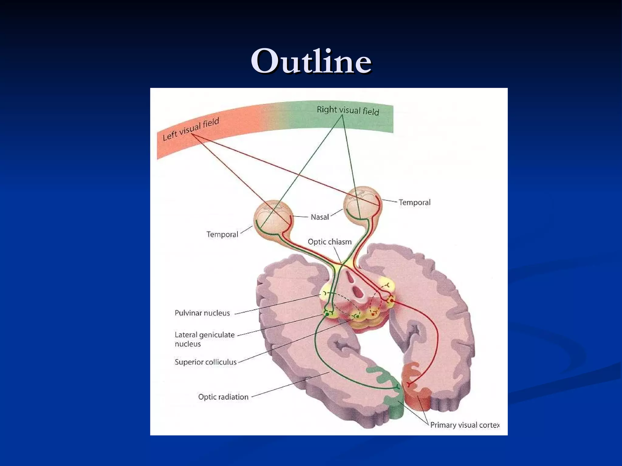
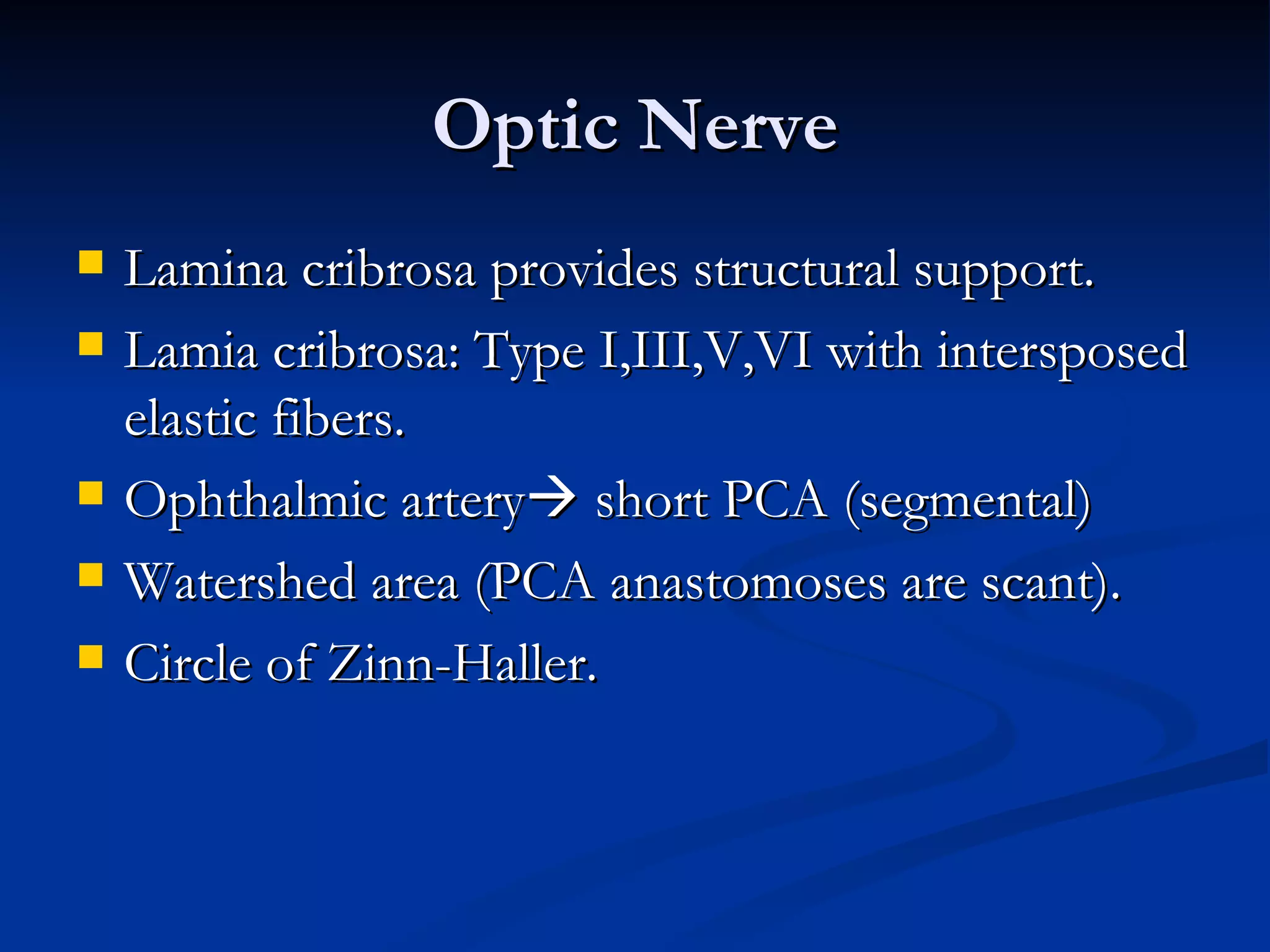
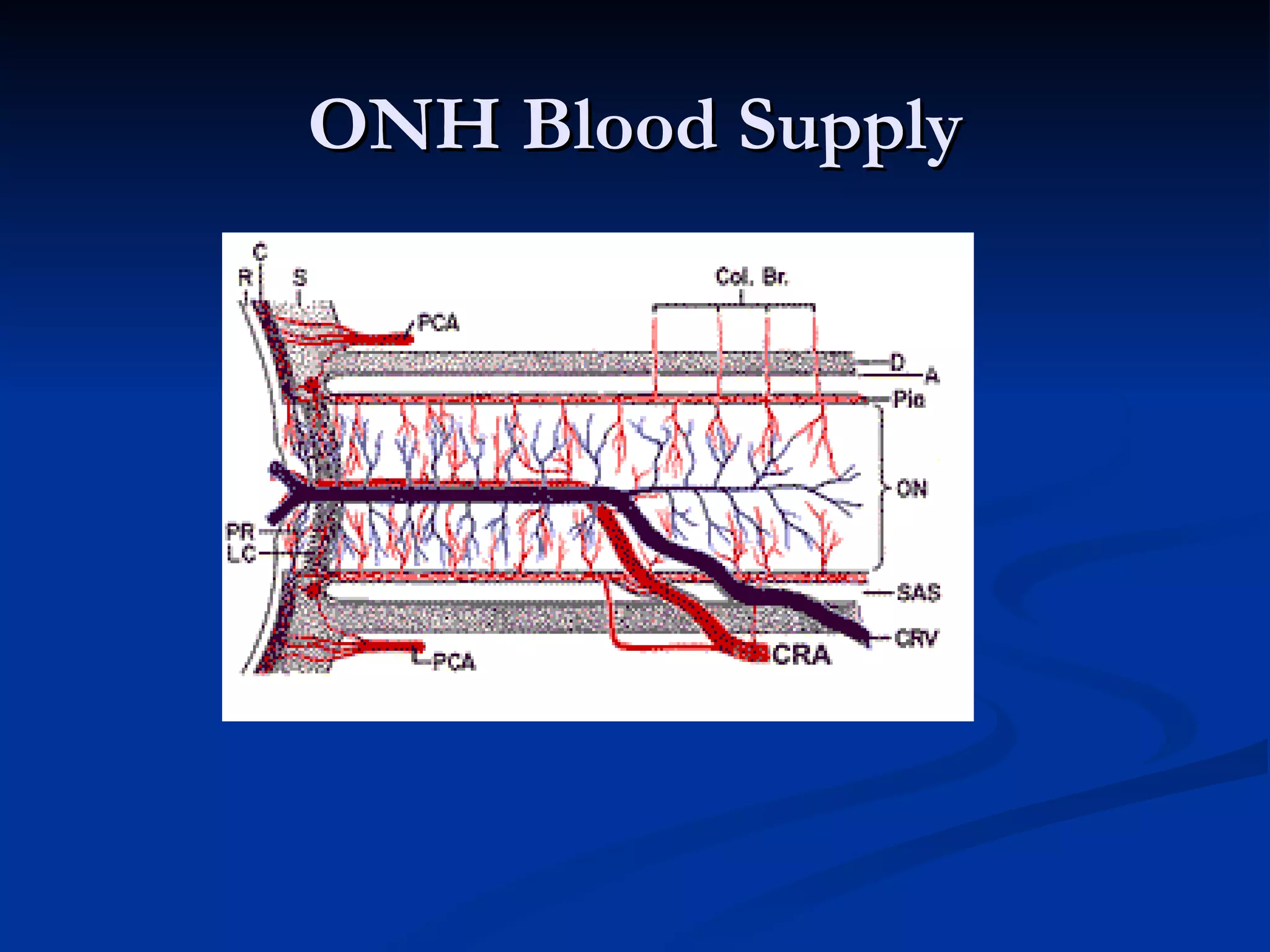
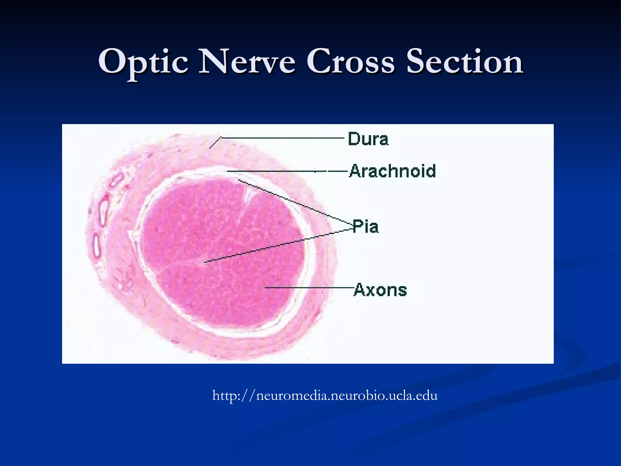
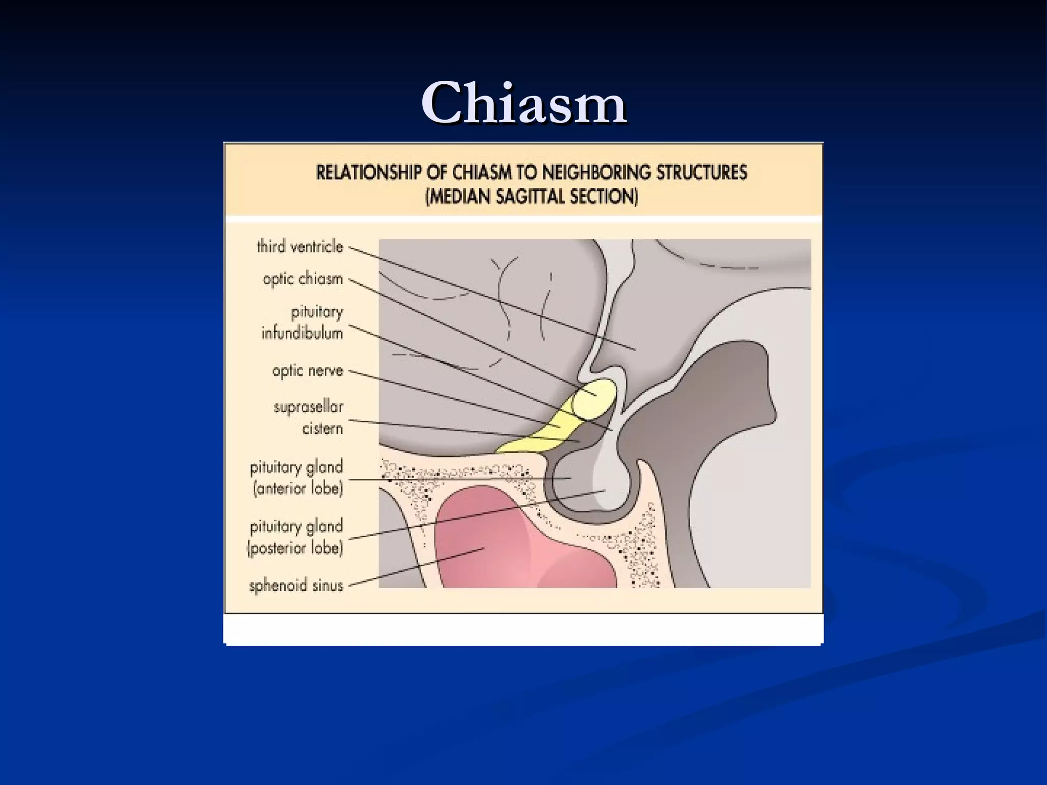
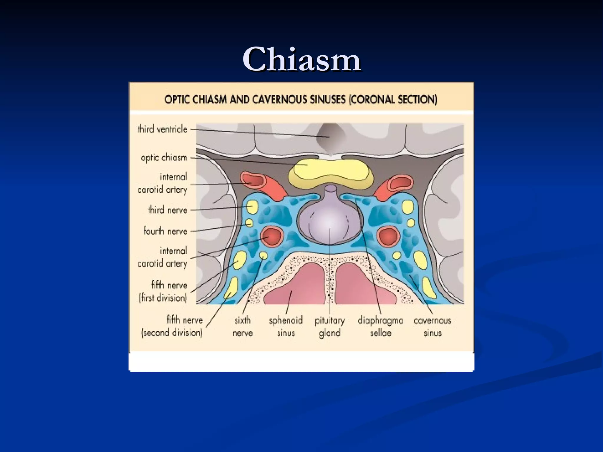
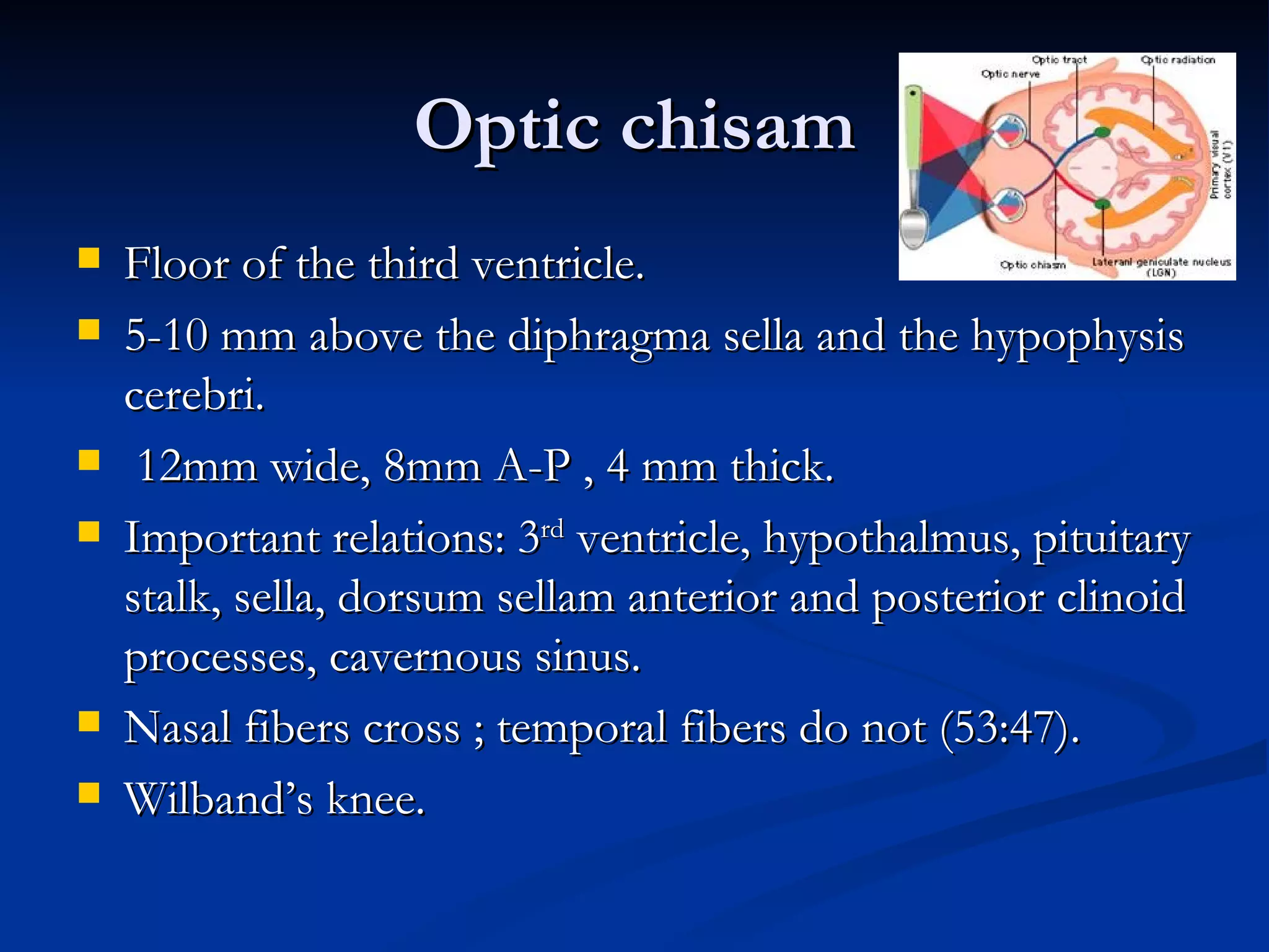
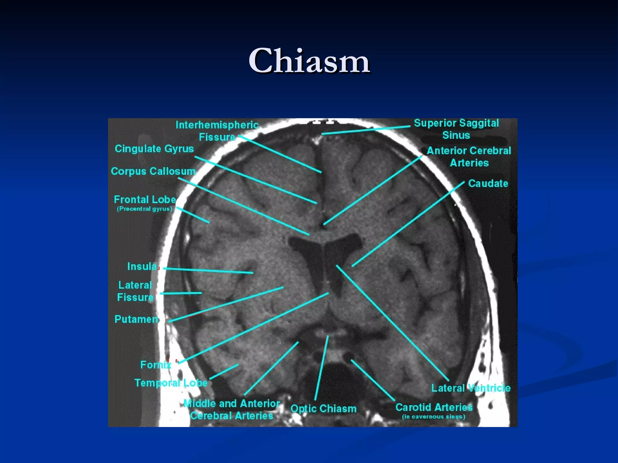
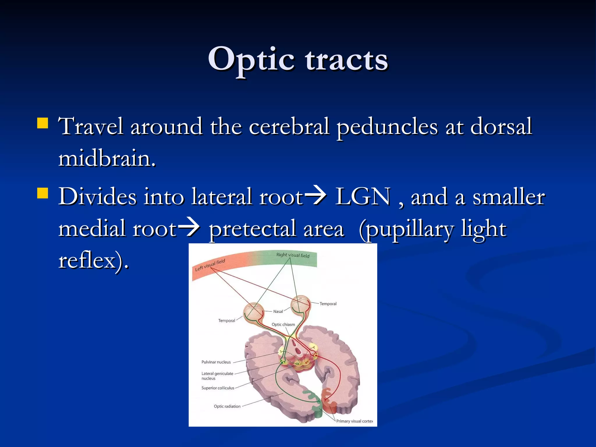
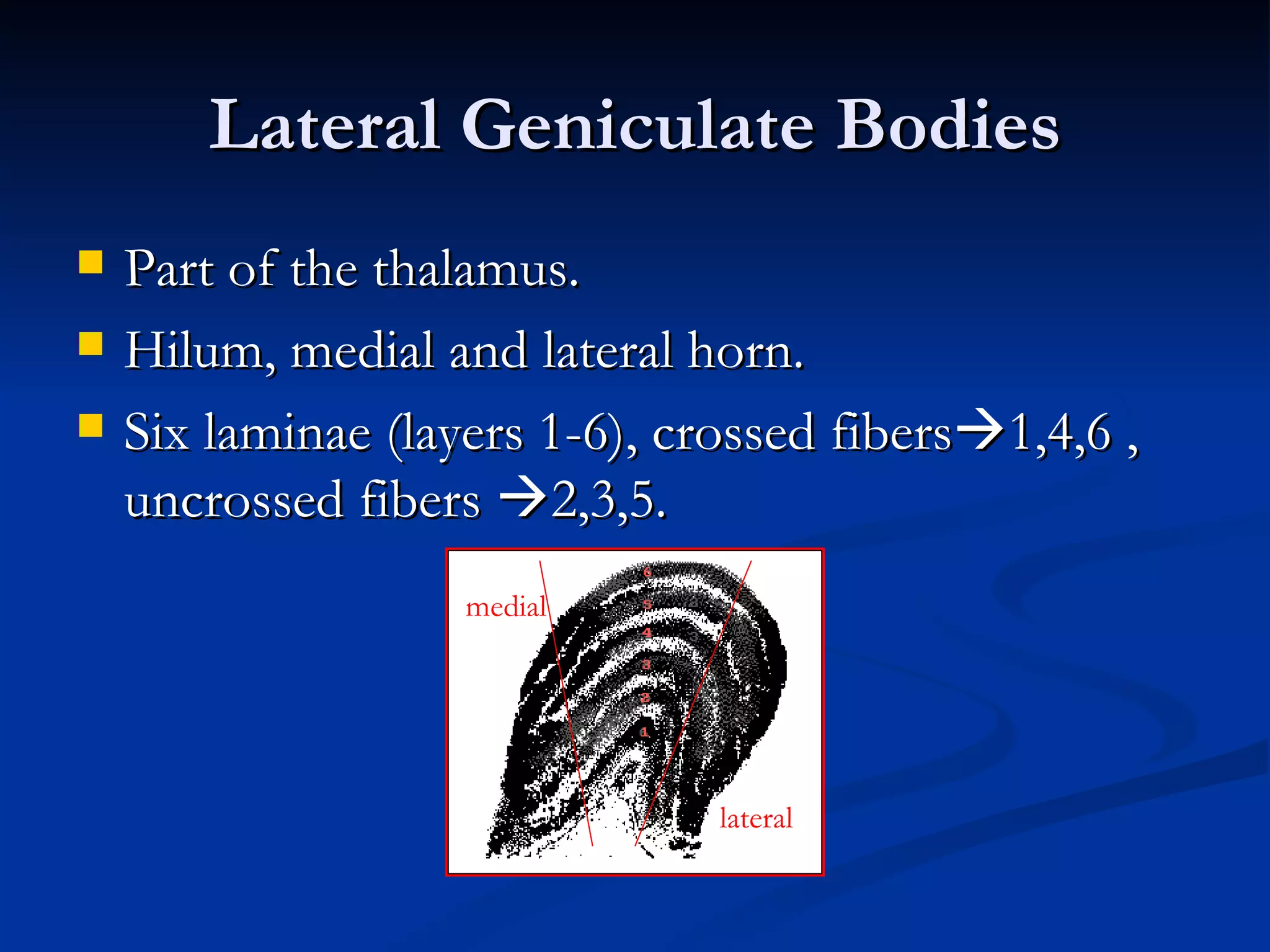
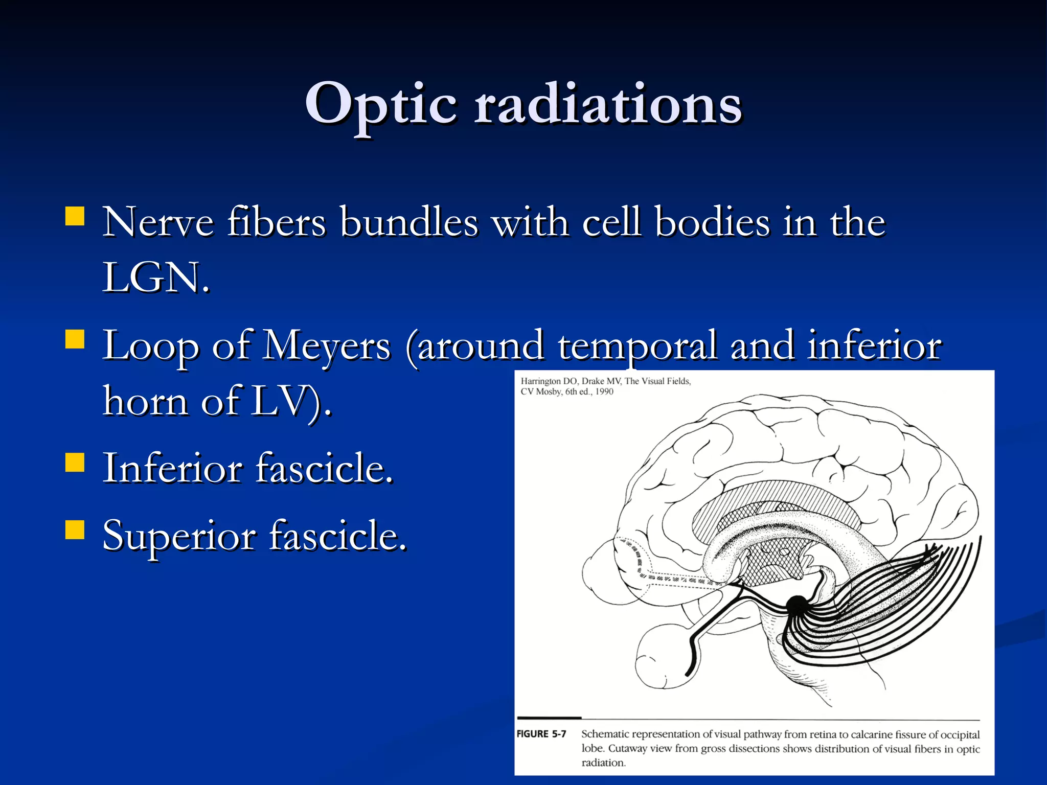
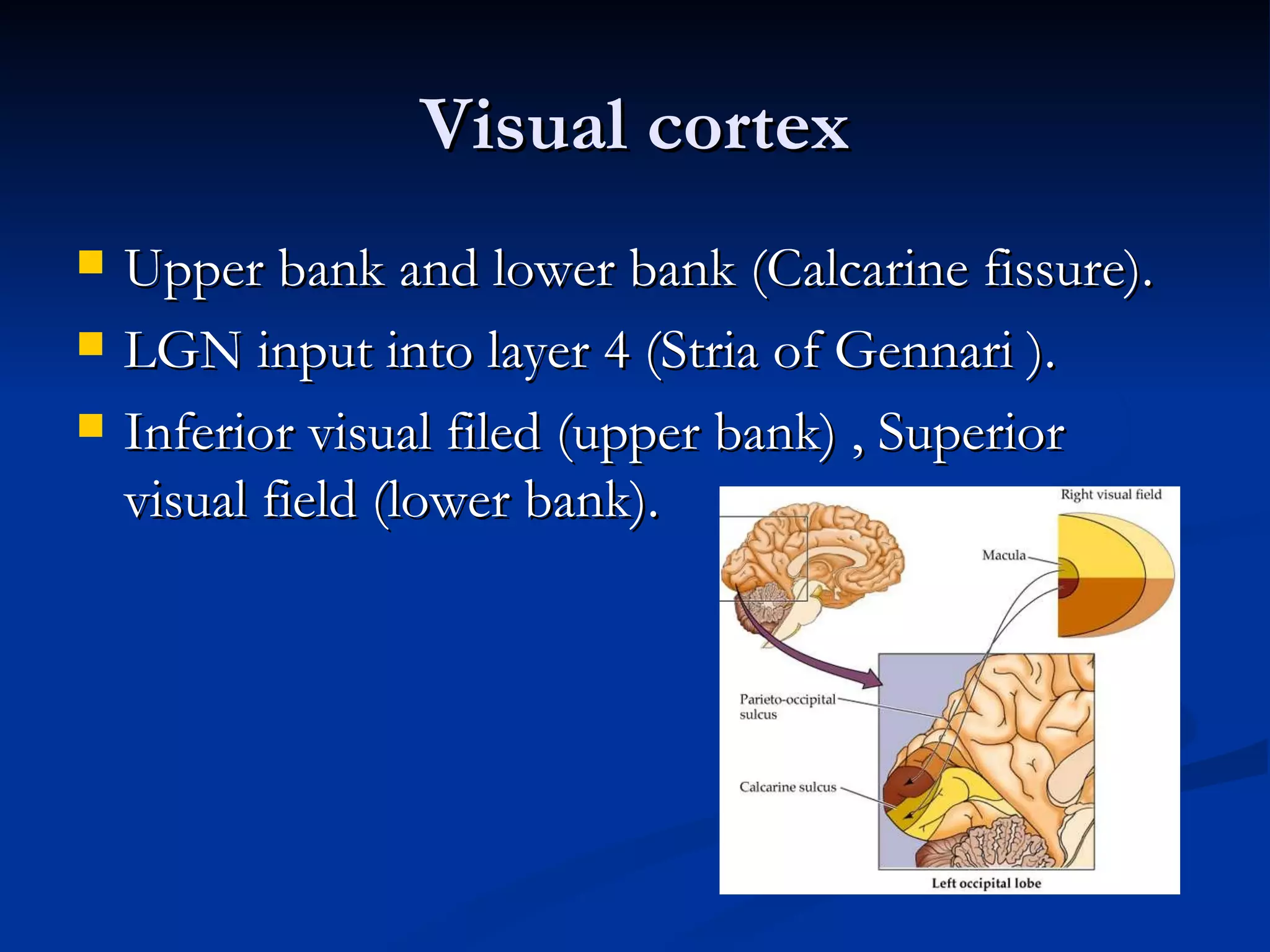
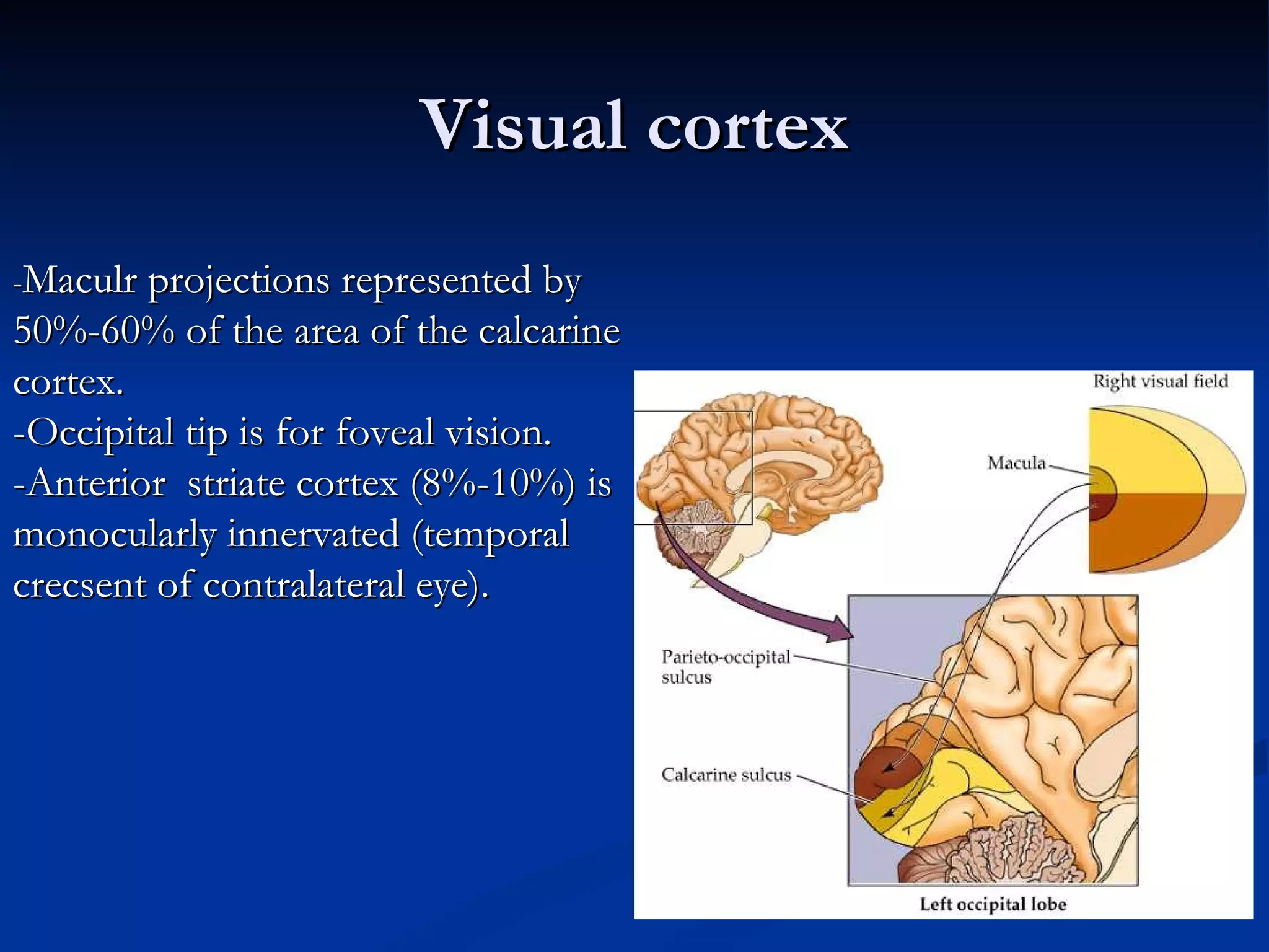
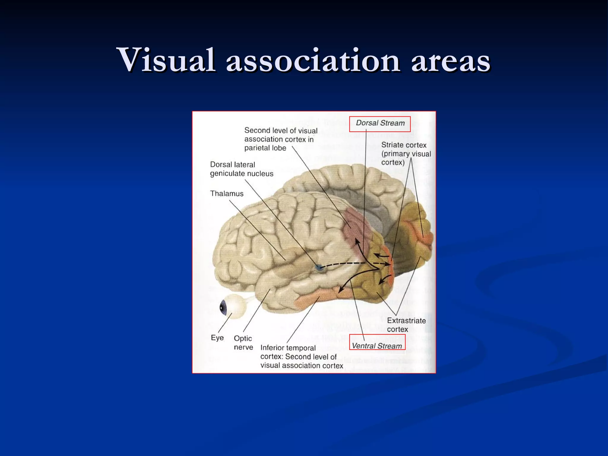
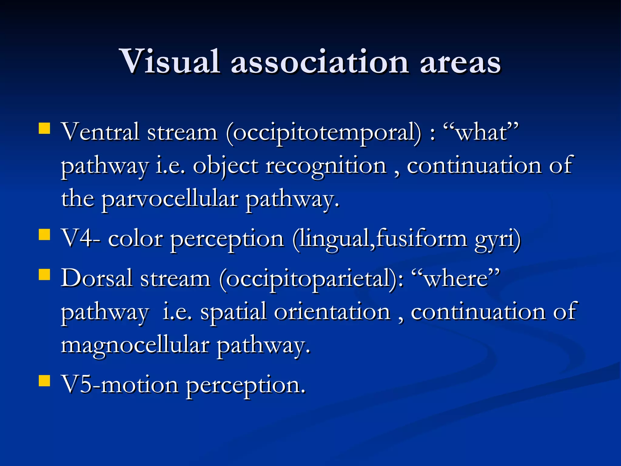
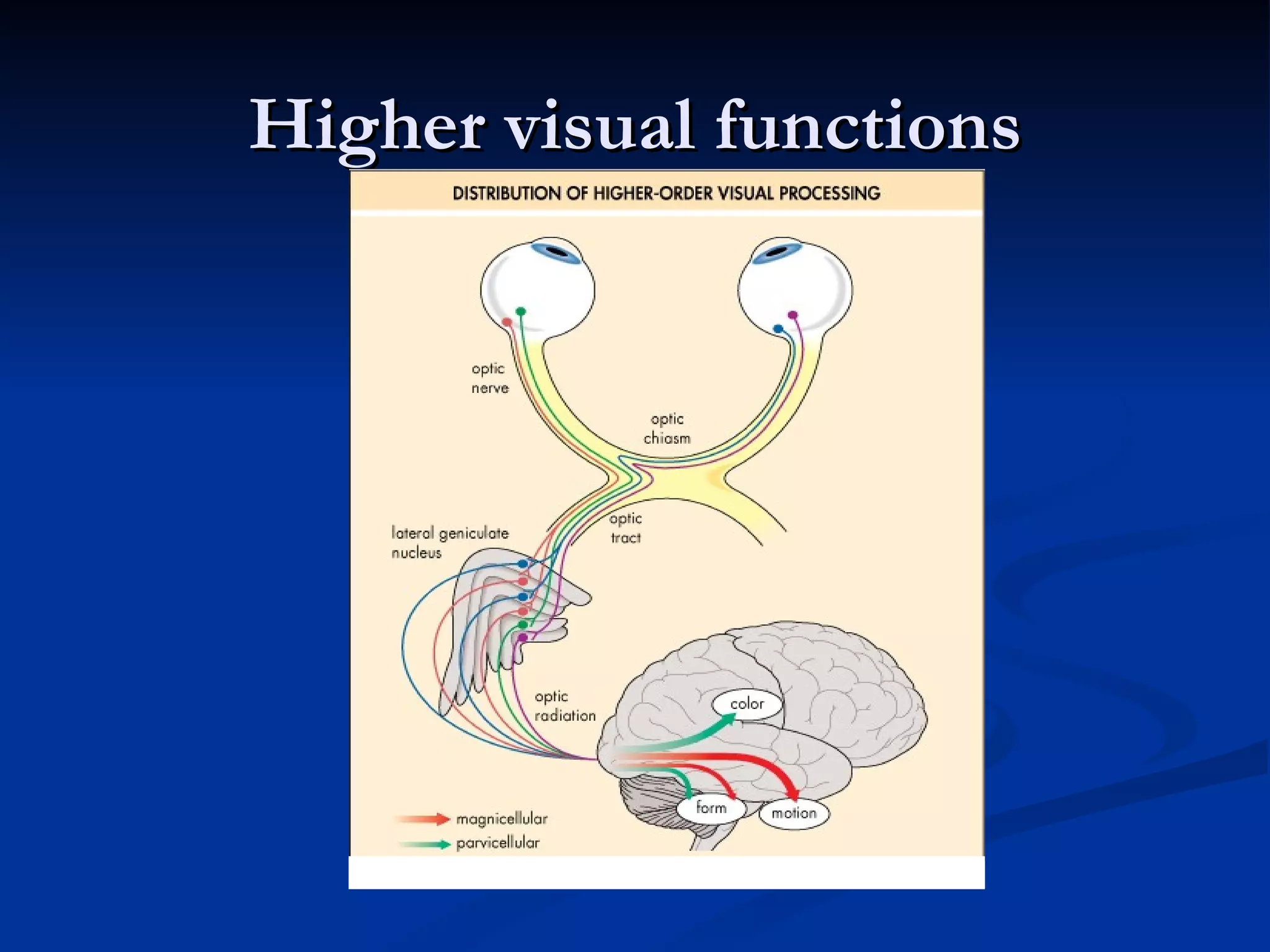
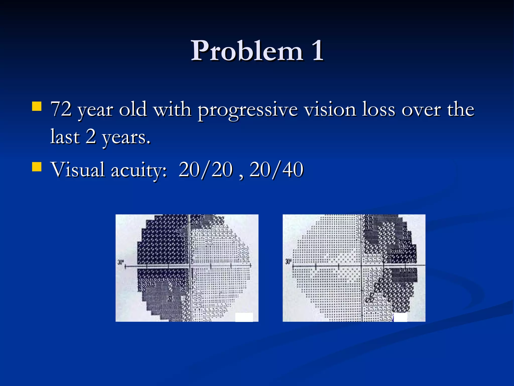
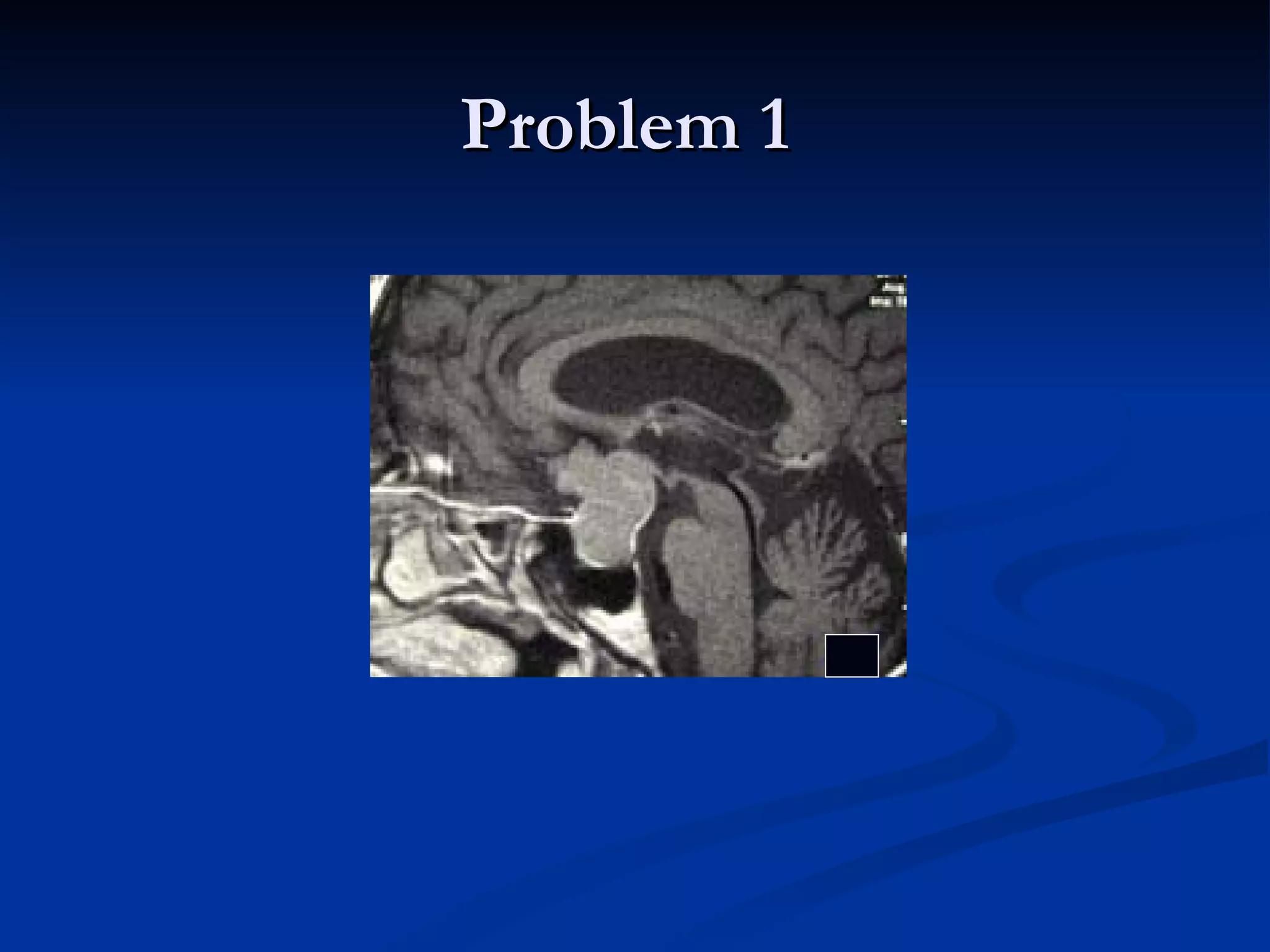
Anatomy and Physiology
 of the Visual pathways
    Raed Behbehani , MD, ABO
Outline
The optic nerve
   1.2 Milllion RGC axons.
   Different types of RGC (parvocellular,
    magnocellular).
   Parvocellular : high spatial resolution, color
    vision, fine stereopsis.
   Magnocellular : low spatial resolution, motion,
    gorss stereopsis.
   Parvocellulae layer 4c-alpha
   Magnocellular layer 4c beta.
RGC axons


http://www.city.ac.uk
Optic Nerve
   Lamina cribrosa provides structural support.
   Lamia cribrosa: Type I,III,V,VI with intersposed
    elastic fibers.
   Ophthalmic artery short PCA (segmental)
   Watershed area (PCA anastomoses are scant).
   Circle of Zinn-Haller.
ONH Blood Supply
Optic Nerve Cross Section


      http://neuromedia.neurobio.ucla.edu
Intracanalicular Optic Nerve
   Within the two bases of the LWS.
   Medial wall of canal forms lateral wall of
    sphenoid sinus (can be absent !).
   Within canal : meninges, ophthalmic artery and
    sympathetic plexus.
   10 mm length.
   Tight space !
   Internal carotid artery.
Intra-orbital Optic Nerve
   Myelination (oligodendrocytes).
   20-30 mm Long.
   Axons: mylein and glial cell (metabolic support
    at the nodes of Ranvier).
Optic nerve (Intracranial)
   Leaves the cranial end of the optic canal (medially,
    backwards, upwards).
   4-15 m (depending on the position of chiasm).
   Upward 45 degree-angle.
   Anterior cerebral and anterior comunicating artery lie
    superior.
Physiology of the Optic Nerve
   Axoplasmic transport : clearance of expired
    organelles, structural maintainance, and energy
    requirements.
   Interruption of axoplasmic transport : ischemia,
    compression, inflammation.
   Orthograde axonal transport : away from the cell
    body LGN.
   Retrograde axonal transport : toward cell body.
Physiology of the Optic Nerve
   Orthograde  fast and slow speeds.
   Proteins, transmitters transported in smooth
    surface vesicles at 400 mm/day (5 hours to
    LGB).
   Elements of cytoskeleton (microtubules,
    neurofilaments) dependant on actin, kinesin and
    dynein.
   Mitochondria moving in both orthograde and
    retrograde fashion.
Chiasm
Chiasm
Optic chisam
   Floor of the third ventricle.
   5-10 mm above the diphragma sella and the hypophysis
    cerebri.
    12mm wide, 8mm A-P , 4 mm thick.
   Important relations: 3rd ventricle, hypothalmus, pituitary
    stalk, sella, dorsum sellam anterior and posterior clinoid
    processes, cavernous sinus.
   Nasal fibers cross ; temporal fibers do not (53:47).
   Wilband’s knee.
Chiasm
Chiasm
Chiasm
Chiasm
   90% of chiasmal fibers have macular origin
    (superior and posterior portions of chiasm).
   Supraclinoid portions of carotid artery lie lateral
    to chiasm.
Wilbrand’s knee
Optic tracts
   Travel around the cerebral peduncles at dorsal
    midbrain.
   Divides into lateral root LGN , and a smaller
    medial root pretectal area (pupillary light
    reflex).
Lateral Geniculate Bodies
   Part of the thalamus.
   Hilum, medial and lateral horn.
   Six laminae (layers 1-6), crossed fibers1,4,6 ,
    uncrossed fibers 2,3,5.
                    medial


                                lateral
LGN
   Upper quadrant medial aspect of LGN,
    Lower quadrant lateral aspect of LGN.
   Macular fibers central wedge of LGN.
   Layers 1,2: magnocellular. (motion)
   Layers 3-6: Parvocellular. (color)
Optic radiations
   Nerve fibers bundles with cell bodies in the
    LGN.
   Loop of Meyers (around temporal and inferior
    horn of LV).
   Inferior fascicle.
   Superior fascicle.
Optic radiations
   Inferior fascicle anterior pole of temporal
    lobe lower calcarine cortex.
   Superior fascicle parietal lobe upper
    calacrine cortex.
Visual cortex
   Upper bank and lower bank (Calcarine fissure).
   LGN input into layer 4 (Stria of Gennari ).
   Inferior visual filed (upper bank) , Superior
    visual field (lower bank).
Visual cortex
-Maculr projections represented by
50%-60% of the area of the calcarine
cortex.
-Occipital tip is for foveal vision.
-Anterior striate cortex (8%-10%) is
monocularly innervated (temporal
crecsent of contralateral eye).
Visual association areas
   Ventral stream (occipitotemporal) : “what”
    pathway i.e. object recognition , continuation of
    the parvocellular pathway.
   V4- color perception (lingual,fusiform gyri)
   Dorsal stream (occipitoparietal): “where”
    pathway i.e. spatial orientation , continuation of
    magnocellular pathway.
   V5-motion perception.
Higher visual functions
Problem 1
   72 year old with progressive vision loss over the
    last 2 years.
   Visual acuity: 20/20 , 20/40
Problem 1
Problem 2
   A 55 year old patient with sudden, pain less
    vision loss in her left eye.
   Visual acuity : OD-20/200 OS-20/25
   Color vision : OD- Nil OS-8/8
Problem 2
Problem 2
Problem 2
Problem 3

Anatomy of the visual pathways and visual cortex

  • 1.
    Anatomy and Physiology of the Visual pathways Raed Behbehani , MD, ABO
  • 2.
  • 3.
    The optic nerve  1.2 Milllion RGC axons.  Different types of RGC (parvocellular, magnocellular).  Parvocellular : high spatial resolution, color vision, fine stereopsis.  Magnocellular : low spatial resolution, motion, gorss stereopsis.  Parvocellulae layer 4c-alpha  Magnocellular layer 4c beta.
  • 4.
  • 5.
    Optic Nerve  Lamina cribrosa provides structural support.  Lamia cribrosa: Type I,III,V,VI with intersposed elastic fibers.  Ophthalmic artery short PCA (segmental)  Watershed area (PCA anastomoses are scant).  Circle of Zinn-Haller.
  • 6.
  • 7.
    Optic Nerve CrossSection http://neuromedia.neurobio.ucla.edu
  • 8.
    Intracanalicular Optic Nerve  Within the two bases of the LWS.  Medial wall of canal forms lateral wall of sphenoid sinus (can be absent !).  Within canal : meninges, ophthalmic artery and sympathetic plexus.  10 mm length.  Tight space !  Internal carotid artery.
  • 10.
    Intra-orbital Optic Nerve  Myelination (oligodendrocytes).  20-30 mm Long.  Axons: mylein and glial cell (metabolic support at the nodes of Ranvier).
  • 11.
    Optic nerve (Intracranial)  Leaves the cranial end of the optic canal (medially, backwards, upwards).  4-15 m (depending on the position of chiasm).  Upward 45 degree-angle.  Anterior cerebral and anterior comunicating artery lie superior.
  • 12.
    Physiology of theOptic Nerve  Axoplasmic transport : clearance of expired organelles, structural maintainance, and energy requirements.  Interruption of axoplasmic transport : ischemia, compression, inflammation.  Orthograde axonal transport : away from the cell body LGN.  Retrograde axonal transport : toward cell body.
  • 13.
    Physiology of theOptic Nerve  Orthograde  fast and slow speeds.  Proteins, transmitters transported in smooth surface vesicles at 400 mm/day (5 hours to LGB).  Elements of cytoskeleton (microtubules, neurofilaments) dependant on actin, kinesin and dynein.  Mitochondria moving in both orthograde and retrograde fashion.
  • 14.
  • 15.
  • 16.
    Optic chisam  Floor of the third ventricle.  5-10 mm above the diphragma sella and the hypophysis cerebri.  12mm wide, 8mm A-P , 4 mm thick.  Important relations: 3rd ventricle, hypothalmus, pituitary stalk, sella, dorsum sellam anterior and posterior clinoid processes, cavernous sinus.  Nasal fibers cross ; temporal fibers do not (53:47).  Wilband’s knee.
  • 17.
  • 18.
  • 19.
  • 20.
    Chiasm  90% of chiasmal fibers have macular origin (superior and posterior portions of chiasm).  Supraclinoid portions of carotid artery lie lateral to chiasm.
  • 21.
  • 22.
    Optic tracts  Travel around the cerebral peduncles at dorsal midbrain.  Divides into lateral root LGN , and a smaller medial root pretectal area (pupillary light reflex).
  • 23.
    Lateral Geniculate Bodies  Part of the thalamus.  Hilum, medial and lateral horn.  Six laminae (layers 1-6), crossed fibers1,4,6 , uncrossed fibers 2,3,5. medial lateral
  • 24.
    LGN  Upper quadrant medial aspect of LGN, Lower quadrant lateral aspect of LGN.  Macular fibers central wedge of LGN.  Layers 1,2: magnocellular. (motion)  Layers 3-6: Parvocellular. (color)
  • 25.
    Optic radiations  Nerve fibers bundles with cell bodies in the LGN.  Loop of Meyers (around temporal and inferior horn of LV).  Inferior fascicle.  Superior fascicle.
  • 26.
    Optic radiations  Inferior fascicle anterior pole of temporal lobe lower calcarine cortex.  Superior fascicle parietal lobe upper calacrine cortex.
  • 27.
    Visual cortex  Upper bank and lower bank (Calcarine fissure).  LGN input into layer 4 (Stria of Gennari ).  Inferior visual filed (upper bank) , Superior visual field (lower bank).
  • 28.
    Visual cortex -Maculr projectionsrepresented by 50%-60% of the area of the calcarine cortex. -Occipital tip is for foveal vision. -Anterior striate cortex (8%-10%) is monocularly innervated (temporal crecsent of contralateral eye).
  • 29.
  • 30.
    Visual association areas  Ventral stream (occipitotemporal) : “what” pathway i.e. object recognition , continuation of the parvocellular pathway.  V4- color perception (lingual,fusiform gyri)  Dorsal stream (occipitoparietal): “where” pathway i.e. spatial orientation , continuation of magnocellular pathway.  V5-motion perception.
  • 31.
  • 32.
    Problem 1  72 year old with progressive vision loss over the last 2 years.  Visual acuity: 20/20 , 20/40
  • 33.
  • 34.
    Problem 2  A 55 year old patient with sudden, pain less vision loss in her left eye.  Visual acuity : OD-20/200 OS-20/25  Color vision : OD- Nil OS-8/8
  • 35.
  • 36.
  • 37.
  • 38.