1. Fungi are eukaryotic organisms that can exist as molds or yeasts and reproduce both sexually and asexually. They have cell walls made of chitin and can cause superficial infections of the skin and nails as well as some systemic infections.
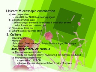
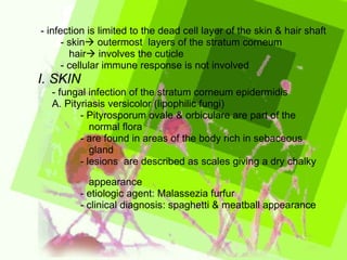
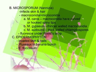
2. Common superficial fungal infections include ringworm, athlete's foot, and nail fungus. These are caused by dermatophyte fungi and generally only infect the outer layers of skin, hair, and nails. Diagnosis involves microscopic examination of skin and nail samples.
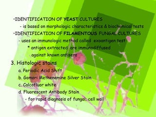
3. Systemic fungal infections are diagnosed through culture, histology, antigen detection and PCR. Treatment involves topical or oral antifungal medications such as azo