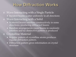
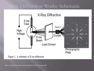
X-ray diffraction is used to determine the atomic structure of crystalline solids by analyzing the diffraction patterns produced when X-rays interact with a crystal. The regular arrangement of atoms in a crystal causes the X-rays to diffract into specific patterns determined by the spacing of crystal planes and the X-ray wavelength. This technique is useful for solving structures in fields like solid-state physics, biophysics, and biochemistry.
X ray crystallography and X ray DiffractionFaisal Hussain
This is the short description about x ray crystallography.
simplest and easy to understand.
Procedure of X ray Diffraction.
Pros and Cons of X ray Crystallography
X ray crystallography and X ray DiffractionFaisal Hussain
This is the short description about x ray crystallography.
simplest and easy to understand.
Procedure of X ray Diffraction.
Advantages and Disadvantages of X ray Crystallography
X ray crystallography and X ray DiffractionFaisal Hussain
This is the short description about x ray crystallography.
simplest and easy to understand.
Procedure of X ray Diffraction.
Pros and Cons of X ray Crystallography
X ray crystallography and X ray DiffractionFaisal Hussain
This is the short description about x ray crystallography.
simplest and easy to understand.
Procedure of X ray Diffraction.
Advantages and Disadvantages of X ray Crystallography
X ray
Md. Waliullah Wali
Dept. of pharmacy
Southeast University
Outline
XRD
X-ray diffraction (XRD) is an analytical technique looking at X-ray scattering from crystalline materials. Each material produces a unique X-ray "fingerprint" of X-ray intensity versus scattering angle that is characteristic of it's crystalline atomic structure.
X-ray diffraction procedures
apply only to crystalline
Materials.
Principles of XRD
X-ray diffraction is based on constructive interference of monochromatic X-rays and a crystalline sample.
The interaction of the incident rays with the sample produces constructive interference (and a diffracted ray) when conditions satisfy Bragg's Law (nλ=2d sin θ).
XRD Techniques
XRD Techniques
Applications of XRD
Limitations of XRD
XRF
X-Ray Fluorescence is defined as “The emission of characteristic "secondary" (or fluorescent) X-rays from a material that has been excited by bombarding with high-energy X-rays. The phenomenon is widely used for elemental analysis.”
X-ray fluorescence procedures
applied to the material
in any physical state,
solid, liquid and gas.
Principles of XRF
The XRF method depends on fundamental principles that are common to several other instrumental methods involving interactions between electron beams and X-rays with samples, including, X-ray spectroscopy (e.g. SEM – EDS), X-ray diffraction (XRD) and wavelength dispersive spectroscopy (microprobe WDS).
XRF Techniques
Applications of XRF
Advantages of XRF
Limitation of XRF
0
References
1. Elements of physical chemistry by S Glasstone
2. Atkins physical chemistry
3. Pharmaceutical chemistry by LG Chattem
4. Brady, John B., and Boardman, Shelby J., 1995, Introducing Mineralogy Students to X-ray Diffraction Through Optical Diffraction Experiments Using Lasers. Jour. Geol. Education, v. 43 #5, 471-476.
5. Brady, John B., Newton, Robert M., and Boardman, Shelby J., 1995, New Uses for Powder X-ray Diffraction Experiments in the Undergraduate Curriculum. Jour. Geol. Education, v. 43 #5, 466-470.
6. Buhrke, V. E., Jenkins, R., Smith, D. K., A Practical Guide for the Preparation of Specimens for XRF and XRD Analysis, Wiley, 1998.
X-Ray Crystallography is a technique used to determine the atomic and molecular structure of a crystal, in which the crystalline atoms cause a beam of incident X-rays to diffract into many specific directions.
X-ray Crystallography is a scientific method used to determine the arrangement of atoms of a crystalline solid in three dimension. It is based on x ray diffraction. Reveals structure of a crystal at atomic level.
Machine Learning and Deep Learning from Foundations to Applications Excel, R,...Narendra Ashar
Preparing stakeholders across the organization in Advanced Machine learning, Deep Learning, Algorithms, Machine Learning for Image Processing, Machine Learning for Text Processing, Deep Learning Applications.
Courses can be tailored for
Freshers in a corporate
Senior Executives
Marketing, Business Development and other staff. who want to get a simpler view on these newer and apparently complex topics.
X ray, invisible, highly penetrating electromagnetic radiation of much shorter wavelength (higher frequency) than visible light. The wavelength range for X rays is from about 10-8 m to about 10-11 m, the corresponding frequency range is from about 3 × 10¹⁶ Hz to about 3 × 10¹⁹ Hz.
X-ray crystallography is the experimental science determining the atomic and molecular structure of a crystal, in which the crystalline structure causes a beam of incident X-rays to diffract into many specific directions. By measuring the angles and intensities of these diffracted beams, a crystallographer can produce a three-dimensional picture of the density of electrons within the crystal. From this electron density, the mean positions of the atoms in the crystal can be determined, as well as their chemical bonds, their crystallographic disorder, and various other information.
X- Rays were discovered by Wilhelm Roentgen, so x-rays are also called Roentgen rays.
X-ray diffraction in crystals was discovered by Max von Laue. The wavelength range is 10-7 to about 10-15 m.
The penetrating power of x-rays depends on energy-
Hard x-rays: High frequency & More energy
Soft x-rays: Less penetrating & Low energy
X-rays are short-wavelength electromagnetic radiations produced by the deceleration of high energy electrons or by electronic transitions of electrons in the inner orbital of atoms.
X-ray region- 0.1-100 A˚
Analytical purpose- 0.7-2 A˚
Properties: Highly penetrating invisible rays
Liberate minute amounts of heat on passing through matter
Not deflected by electric and magnetic fields
Poly energetic, having widespread energies and wavelengths
Cause ionization (adding or removing electrons in atoms and molecules)
Transmitted by (pass-through) healthy body tissue
Principle: X-ray diffraction is based on constructive interference of monochromatic x-rays and a crystalline sample.
The interaction of incident rays with the sample produces constructive interference when conditions satisfy Bragg’s law.
Production of x rays: X- Rays are generated when the high velocity of electrons impinge on a metal target.
1% of total energy of the electron beam is converted into X –radiation.
This presentation explores a brief idea about the structural and functional attributes of nucleotides, the structure and function of genetic materials along with the impact of UV rays and pH upon them.
What is greenhouse gasses and how many gasses are there to affect the Earth.moosaasad1975
What are greenhouse gasses how they affect the earth and its environment what is the future of the environment and earth how the weather and the climate effects.
Comparing Evolved Extractive Text Summary Scores of Bidirectional Encoder Rep...University of Maribor
Slides from:
11th International Conference on Electrical, Electronics and Computer Engineering (IcETRAN), Niš, 3-6 June 2024
Track: Artificial Intelligence
https://www.etran.rs/2024/en/home-english/
Slide 1: Title Slide
Extrachromosomal Inheritance
Slide 2: Introduction to Extrachromosomal Inheritance
Definition: Extrachromosomal inheritance refers to the transmission of genetic material that is not found within the nucleus.
Key Components: Involves genes located in mitochondria, chloroplasts, and plasmids.
Slide 3: Mitochondrial Inheritance
Mitochondria: Organelles responsible for energy production.
Mitochondrial DNA (mtDNA): Circular DNA molecule found in mitochondria.
Inheritance Pattern: Maternally inherited, meaning it is passed from mothers to all their offspring.
Diseases: Examples include Leber’s hereditary optic neuropathy (LHON) and mitochondrial myopathy.
Slide 4: Chloroplast Inheritance
Chloroplasts: Organelles responsible for photosynthesis in plants.
Chloroplast DNA (cpDNA): Circular DNA molecule found in chloroplasts.
Inheritance Pattern: Often maternally inherited in most plants, but can vary in some species.
Examples: Variegation in plants, where leaf color patterns are determined by chloroplast DNA.
Slide 5: Plasmid Inheritance
Plasmids: Small, circular DNA molecules found in bacteria and some eukaryotes.
Features: Can carry antibiotic resistance genes and can be transferred between cells through processes like conjugation.
Significance: Important in biotechnology for gene cloning and genetic engineering.
Slide 6: Mechanisms of Extrachromosomal Inheritance
Non-Mendelian Patterns: Do not follow Mendel’s laws of inheritance.
Cytoplasmic Segregation: During cell division, organelles like mitochondria and chloroplasts are randomly distributed to daughter cells.
Heteroplasmy: Presence of more than one type of organellar genome within a cell, leading to variation in expression.
Slide 7: Examples of Extrachromosomal Inheritance
Four O’clock Plant (Mirabilis jalapa): Shows variegated leaves due to different cpDNA in leaf cells.
Petite Mutants in Yeast: Result from mutations in mitochondrial DNA affecting respiration.
Slide 8: Importance of Extrachromosomal Inheritance
Evolution: Provides insight into the evolution of eukaryotic cells.
Medicine: Understanding mitochondrial inheritance helps in diagnosing and treating mitochondrial diseases.
Agriculture: Chloroplast inheritance can be used in plant breeding and genetic modification.
Slide 9: Recent Research and Advances
Gene Editing: Techniques like CRISPR-Cas9 are being used to edit mitochondrial and chloroplast DNA.
Therapies: Development of mitochondrial replacement therapy (MRT) for preventing mitochondrial diseases.
Slide 10: Conclusion
Summary: Extrachromosomal inheritance involves the transmission of genetic material outside the nucleus and plays a crucial role in genetics, medicine, and biotechnology.
Future Directions: Continued research and technological advancements hold promise for new treatments and applications.
Slide 11: Questions and Discussion
Invite Audience: Open the floor for any questions or further discussion on the topic.
Introduction:
RNA interference (RNAi) or Post-Transcriptional Gene Silencing (PTGS) is an important biological process for modulating eukaryotic gene expression.
It is highly conserved process of posttranscriptional gene silencing by which double stranded RNA (dsRNA) causes sequence-specific degradation of mRNA sequences.
dsRNA-induced gene silencing (RNAi) is reported in a wide range of eukaryotes ranging from worms, insects, mammals and plants.
This process mediates resistance to both endogenous parasitic and exogenous pathogenic nucleic acids, and regulates the expression of protein-coding genes.
What are small ncRNAs?
micro RNA (miRNA)
short interfering RNA (siRNA)
Properties of small non-coding RNA:
Involved in silencing mRNA transcripts.
Called “small” because they are usually only about 21-24 nucleotides long.
Synthesized by first cutting up longer precursor sequences (like the 61nt one that Lee discovered).
Silence an mRNA by base pairing with some sequence on the mRNA.
Discovery of siRNA?
The first small RNA:
In 1993 Rosalind Lee (Victor Ambros lab) was studying a non- coding gene in C. elegans, lin-4, that was involved in silencing of another gene, lin-14, at the appropriate time in the
development of the worm C. elegans.
Two small transcripts of lin-4 (22nt and 61nt) were found to be complementary to a sequence in the 3' UTR of lin-14.
Because lin-4 encoded no protein, she deduced that it must be these transcripts that are causing the silencing by RNA-RNA interactions.
Types of RNAi ( non coding RNA)
MiRNA
Length (23-25 nt)
Trans acting
Binds with target MRNA in mismatch
Translation inhibition
Si RNA
Length 21 nt.
Cis acting
Bind with target Mrna in perfect complementary sequence
Piwi-RNA
Length ; 25 to 36 nt.
Expressed in Germ Cells
Regulates trnasposomes activity
MECHANISM OF RNAI:
First the double-stranded RNA teams up with a protein complex named Dicer, which cuts the long RNA into short pieces.
Then another protein complex called RISC (RNA-induced silencing complex) discards one of the two RNA strands.
The RISC-docked, single-stranded RNA then pairs with the homologous mRNA and destroys it.
THE RISC COMPLEX:
RISC is large(>500kD) RNA multi- protein Binding complex which triggers MRNA degradation in response to MRNA
Unwinding of double stranded Si RNA by ATP independent Helicase
Active component of RISC is Ago proteins( ENDONUCLEASE) which cleave target MRNA.
DICER: endonuclease (RNase Family III)
Argonaute: Central Component of the RNA-Induced Silencing Complex (RISC)
One strand of the dsRNA produced by Dicer is retained in the RISC complex in association with Argonaute
ARGONAUTE PROTEIN :
1.PAZ(PIWI/Argonaute/ Zwille)- Recognition of target MRNA
2.PIWI (p-element induced wimpy Testis)- breaks Phosphodiester bond of mRNA.)RNAse H activity.
MiRNA:
The Double-stranded RNAs are naturally produced in eukaryotic cells during development, and they have a key role in regulating gene expression .
Earliest Galaxies in the JADES Origins Field: Luminosity Function and Cosmic ...Sérgio Sacani
We characterize the earliest galaxy population in the JADES Origins Field (JOF), the deepest
imaging field observed with JWST. We make use of the ancillary Hubble optical images (5 filters
spanning 0.4−0.9µm) and novel JWST images with 14 filters spanning 0.8−5µm, including 7 mediumband filters, and reaching total exposure times of up to 46 hours per filter. We combine all our data
at > 2.3µm to construct an ultradeep image, reaching as deep as ≈ 31.4 AB mag in the stack and
30.3-31.0 AB mag (5σ, r = 0.1” circular aperture) in individual filters. We measure photometric
redshifts and use robust selection criteria to identify a sample of eight galaxy candidates at redshifts
z = 11.5 − 15. These objects show compact half-light radii of R1/2 ∼ 50 − 200pc, stellar masses of
M⋆ ∼ 107−108M⊙, and star-formation rates of SFR ∼ 0.1−1 M⊙ yr−1
. Our search finds no candidates
at 15 < z < 20, placing upper limits at these redshifts. We develop a forward modeling approach to
infer the properties of the evolving luminosity function without binning in redshift or luminosity that
marginalizes over the photometric redshift uncertainty of our candidate galaxies and incorporates the
impact of non-detections. We find a z = 12 luminosity function in good agreement with prior results,
and that the luminosity function normalization and UV luminosity density decline by a factor of ∼ 2.5
from z = 12 to z = 14. We discuss the possible implications of our results in the context of theoretical
models for evolution of the dark matter halo mass function.
Comparative structure of adrenal gland in vertebrates
X-ray diffactrometery
1.
2. Introduction
How Diffraction Works
Demonstration
Analyzing Diffraction Patterns
Summary and Conclusions
3. Introduction
Motivation:
• X-ray diffraction is used to obtain structural
information about crystalline solids.
• Useful in biochemistry to solve the structures of
complex biomolecules.
• Bridge the gaps between physics, chemistry, and
biology.
X-ray diffraction is important for:
• Solid-state physics
• Biophysics
• Medical physics
• Chemistry and Biochemistry
X-ray Diffractometer
4. Wave Interacting with a Single Particle
Incident beams scatter uniformly in all directions
Wave Interacting with a Solid
Scattered beams interfere constructively in some
directions, producing diffracted beams
Random arrangements cause beams to randomly
interfere and no distinctive pattern is produced
Crystalline Material
Regular pattern of crystalline atoms produces
regular diffraction pattern.
Diffraction pattern gives information on crystal
structure
5. How Diffraction Works: Schematic
http://mrsec.wisc.edu/edetc/modules/xray/X-raystm.pdf
NaCl
7. Reflection of X-
rays from parallel
lattice planes
- families of planes
have equal
spacing
Constructive
interference when
PD = n
The Laue
equations can be
rewritten as
2dhklsinhkl = n
“Crystal Structure Analysis for Chemists and
Biologists”, Glusker, Lewis and Rossi, VCH,
8. nl=2dsin(Q)
• Similar principle to multiple slit experiments
• Constructive and destructive interference patterns depend on
lattice spacing (d) and wavelength of radiation (l)
• By varying wavelength and observing diffraction patterns,
information about lattice spacing is obtained
d
Q Q
Q
X-rays of
wavelength l
l
9. 2
At 20.6 °2, Bragg’s law
fulfilled for the (100) planes,
producing a diffraction
peak.
The (110) planes would diffract at 29.3
°2; however, they are not properly
aligned to produce a diffraction peak
(the perpendicular to those planes does
not bisect the incident and diffracted
beams). Only background is observed.
The (200) planes are parallel to the (100)
planes. Therefore, they also diffract for this
crystal. Since d200 is ½ d100, they appear at 42
°2.
13. X-ray diffraction is a technique for analyzing
structures.
X-ray beam hits a crystal, scattering the beam in a
manner characterized by the atomic structure
Even complex structures can be analyzed by x-ray
diffraction, such as DNA and proteins in biology
This will provide useful in the future for
combining knowledge from physics, chemistry,
and biology