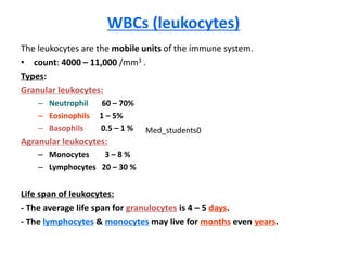
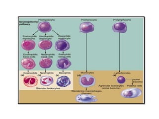
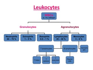
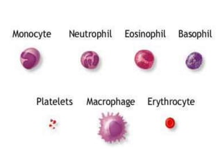
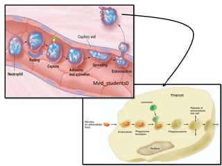
SlideShare a Scribd company logo
White Blood Cells
(WBCs)
Med_students0
At the end of the session the students should
be able to:
Describe the different types of WBCs.
Explain the development of leucocytes
(leucopoiesis).
Discuss the function of different types of
WBCs.
Describe the mechanism of phagocytosis.
WBCs (leukocytes)
The leukocytes are the mobile units of the immune system.
• count: 4000 – 11,000 /mm3 .
Types:
Granular leukocytes:
– Neutrophil 60 – 70%
– Eosinophils 1 – 5%
– Basophils 0.5 – 1 %
Agranular leukocytes:
– Monocytes 3 – 8 %
– Lymphocytes 20 – 30 %
Life span of leukocytes:
- The average life span for granulocytes is 4 – 5 days.
- The lymphocytes & monocytes may live for months even years.
Med_students0
Pathologic variation in leukocytic count:
1- Leukopenia:
• -  wBCs count below 4000 cell /mm3
• - Found in enteric fever.
2- Leukocytosis:
• - Means ↑WBCs count above 11,000 cell / mm3.
3- Agranulocytosis:
• - Acondition in which bone marrow stops WBCs production.
• - It's due to drug toxicity or irradiation.
4- Leukaemias:
- It's malignant disease of bone marrow → uncontrolled production of WBCs
(very high number).
- The leukaemic cells are non functioning & can't protect the body.
Leukocytes
WBCs
4 – 11 x 103/mm3
Granulocytes Agranulocytes
Monocytes
3 – 8 %
Lymphocytes
20 – 30 %
Neutrophils
60 – 70 %
Basophils
0.5 – 1 %
Esinophils
1 – 5 %
B lymphocytesT lymphocytes
T helper
Plasma
cells
T
suppresso
r
T
cytotoxic
macropha
ges
Functions of leukocytes
WBCs provide the body with a powerful defense
mechanisms against:
– Infections (viral, bacterial, parasitic or fungal).
– Tumours
– Toxins
They act together in different ways to prevent diseases
as by:
– Phagocytosis
– Formation of Sensitized lymphocytes
– Formation of antibodies
Neutrophils
Count: 60 – 70 % of TLC
Histology:
– Diameter:
• 10 – 12 µm
– Cytoplasm:
• contain small granules stained with both acidic and basic dyes (so called
neutrophils)
– Nucleus:
• variable in shape (polymorphnuclear leukocytes)
• Consist of 2 – 5 lobes connected by chromatin filaments
– Cell membrane:
• contain receptors for Ig G and complement (C3b).
½ the total number of neutrophils are circulating free in the
blood and the other ½ are marginated along the blood vessel
walls.
a) Function of neutrophils
- Neutrophils are the first line defense against infection.
- When infection occurs, large number of neutrophils invade the infected area,
then phagocytose & destroy the organism.
Mechanism:
1- Margination: is sticking of the neutrophils to the capillary wall.
2- Diapedesis : the neutrophils squeeze themselves through the capillary
pores .
3- Amaeboid movement : to reach the site of infection .
4- Chemotaxis: the neutrophils are chemically attracted to the site of
infection by a group of substances called chemotactic factors:, they
include :
a) The bacterial toxins.
b) The products of tissue destruction.
c) Components of the complement system (C5a)
d) leukotriens..
a) Function of neutrophils
5- Phagocytosis:
- Once the neutrophils reached the site of infection they ingest the bacteria
by endocytosis. Phagocytosis is facilitated by antibodies & C3 component
of the complement.
1) - They adhere to the bacterial membrane, then C3 binds to specific
receptors on the neutrophil membrane initiating phagocytosis, this
process of fascilitation is called opsonization
2) - Then the neutrophils project their pseudopodia to phagocytose the
organism, which forms phagocytic vesicle.
3) - The organism inside the phagocytic vesicle is then killed by:
1) the lysosomal proteolytic enzymes
2) bactricidal agents formed inside the neutrophil as:
• a - oxidizing agents as hydrogen peroxide & hydroxyl ions.
• b- hypochlorite that results from the reaction between H2O2 & chloride.
• - Neutrophils phagocytose up to 20 organism then die.
Med_students0
Eosinophils
Count: 1 – 5 % of TLC
Structure:
– Diameter: 10 – 12 µm
– Cytoplasm:
• contain coarse granules stained red with acidic dye (so called
eosinophils)
– Nucleus:
• Consist of 2 lobes bilobed
– Cell membrane:
• contain receptors for Ig E and Ig G, Ig M and complement
b) Functions of eosinophils
1- They are weak phagocytes.
2- Protect the body against parasitic infection e.g schistosomiasis:
• - They migrate to the infected area & kill the juvenile forms of the organism
by:
a- Hydrolytic enzymes.
b- Larvicidal polypeptides called major basic proteins.
c- highly reactive forms of oxygen..
3- Prevent spread of the inflammation resulting from allergic reaction
because they:
a) Phagocytose the antigen- antibody complex.
b) Detoxify the inflammation producing substances released from mast cells
and basophils
Basophils
Count: 0.5 – 1 % of TLC
Structure:
– Diameter: 8 – 10 µm
– Cytoplasm: contain coarse granules stained blue with
basic dye (so called basophils)
– Nucleus: segmented of 2 – 3 lobes
– Cell membrane: contain receptors for Ig E
Med_students0
c) Functions of basophils
1- Synthesize & liberate heparin into circulation to prevent blood coagulation.
2- Has a role in allergy:
– the antigen- antibody reaction occurs on their surface (Ig E) leading to
their degranulation & release of:
• histamine,
• bradykinin &
• serotonin,
• slow reacting substance of anaphylaxis and
• a number of lysosomal enzymes
– which are responsible for allergic manifestations.
3) Share in inflammatory reactions: by the released substances
II) Function of non-granular leukocytes
a) Function of lymphocytes:
• - The lymphocytes represent the immune system
b) Function of monocytes:
• Monocytes enter the blood (from bone marrow) then circulate for 72
hours.
• Then they enter the tissues, within 8 hours they  in size & develop
lysosomes & becomes tissue macrophages.
• Tissue macrophages phagocytose & kill bacteria by similar mechanisms to
neutrophils but more powerfully as :
– a) They can phagocytose up to 100 bacteria.
– b) They can engulf much large particles as whole RBCs or a parasite
– c) After destroying the particle, macrophage extrude them outside & survive
for many months.
Tissue macrophage system
(reticulo-endothelial system)
• RES consists of:
– Monocytes
– Mobile macrophages & Fixed tissue macrophages
– Endothelial cells in BM, spleen & lymph nodes
The tissue macrophages are 2 types :
1) Mobile macrophages:
• - wander through the tissues all the time.
2) Fixed macrophages:
• They are attached to the tissues & remain attached for months or even years.
• May become mobile macrophages when stimulated in response to chemotaxis.
• The tissue macrophages are known by different names in various tissues:
– kupffer cells in the liver
– Alveolar macrophages in the lung
– Microglia cells in the brain
– mesangial cells in the kidney
– Histocytes in the skin
– Reticular cells in lymphoid tissue (LN, BM, spleen)
Med_students0
Functions of tissue macrophage system
1- Phagocytosis of micro-organisms e.g bacteria & viruses & dead tissues and
foreign particles.
2- Engulfing of old blood cells.
3- Breakdown of HB & formation of bile pigment (e.g bilirubin).
4- Repair of damaged tissues after inflammation by engulfing the necrotic
tissues & release tissue growth factors.
5 - Production of 10 % of erythropoietin by von kupffer cells in liver.
6- Help in production of antibodies by B-lympholytes:
– a) Upon entry of the foreign antigen to the body, macrophages phagocytize it
then digest it, then present its antigenic structure to B lymphocyte which
begin to form the antibodies .
– b) macrophage then secrete IL1 that activate T helper lymphocyte which by
its turn activates B lymphocytes to form antibodies.
* It represents the difference between the filtered and the reabsorbed
fluids across the capillary membrane.
* Normally, lymph flow rate is: 120 ml./hour (i.e., 2-4 L/day) during
rest.
Lymphatic Circulation
Definition:
Lymph is the fluid that returns to the blood stream from tissue spaces
by lymphatic vessels.
Rate of lymph flow:
Med_students0
Thorathic duct Right lymphatic duct
* drains lymph from:
lower limbs – abdomen – left half of
thorax – left side of head and neck –
left upper limb.
* lymph flow rate: 100 ml./hour.
* drains in the left subclavian.
* drains lymph from:
right half of thorax – right side of head
and neck – right upper limb.
* lymph flow rate: 20 ml./hour.
* drains in the right subclavian.
lymphatic capillaries which unit together large lymphatic vessels (have
unidirectional valves) 2 lymphatic ducts:
Lymph is drained by lymphatic system:
1- Almost all the body have lymphatic drainage except:
Brain - deep parts of nerves – superficial parts of the skin –
bone.
2- Lymphatic system is formed of:
Lymph is a part of the interstitial fluid (I.S.F.) characterized by the following:
Physical:
- colourless. – isotonic. - pH 7.4.
- transparent except during fat absorption where It looks milky due to fat globules.
Chemical: It is similar to plasma except in:
* Less proteins (average 2 gm%) but protein concentration varies according to
the site of drainage form 0-6 gm% .
* A/G ratio is greater in lymph than in plasma because albumin has smaller
molecular weight.
* Less number of lymphocytes than plasma.
* Less amount of fibrinogen, prothrombin, Ca++ and no platelets, so it clots but
slower than plasma.
Composition of lymph:
1. Drainage of excess I.S.F. from capillaries back to the blood --> prevent
accumulation of fluid and edema. This also maintains blood volume.
2. Removal of substances having large molecular weight (proteins and
metabolites), because lymphatic capillaries are more permeable than blood
capillaries. The amount of proteins returns to blood by lymph is 25 - 50% of
total circulating plasma proteins, so lymph flow maintains plasma proteins.
3. Absorption of fat and fat - soluble vitamins from the intestine.
4. Lymph nodes functions are:
- formation of lymphocytes.
- removal of bacteria (protective mechanism).
Functions of lymphatic system:
Immune
organs
Immune defenses can be classified into 2 types which usually
interacts:
Nonspecific immune
defenses
(Innate Immunity)
 Protect against microbes
or F.B. (invaders) without
having to recognize their
specific identity.
 The mechanisms used are
not specific to any invader.
Specific immune
defenses
(Acquired Immunity)
 Protect against microbes to
which the body is previously
exposed (recognized before)
either through infection or
immunization.
 The mechanisms used are
specific for each invader.
Acquired ImmunityInnate Immunity
Specific
i.e., Reacts to a specific
invader each time and can
not react to another.
Non-specific
i.e., Reacts to invaders from
different type
Specificity
Very high
i.e., Although the cells are of
the same type, each group
shows totally different
surface molecules.
LimitedDiversity
Yes
Remembers the invader on
re-exposure.
NoMemory
YesYesNon-reaction to self
Components
LymphocytesPhagocytes & natural killer
cells
Cells
AntibodiesComplementMolecules
Med_students0
Mechanisms of Innate Immunity
I - Defenses at Body Surfaces
Very few microorganisms can penetrate the intact skin,
Various skin glands & tears contain anti-microbial agents
Mucus secreted by epithelial lining contain antimicrobial agents and sticky
to which microbes adhere.
Hairs at the entrance of nose
Cough and sneezing reflex
Acid secretion by the stomach and uterus
II- Inflammation
1. Initial entry of bacteria into tissue
2. Vasodilation in the infected area, leading to increased blood flow
3. Marked increase in protein permeability of the capillaries and venules in
the infected area, with resulting diffusion of protein and filtration of fluid
into the interstitial fluid.
4. Chemotaxis: exit of leukocytes from the venules into the interstitial fluid
of the infected area
5. Destruction of bacteria in the tissue either through phagocytosis or by
mechanisms not requiring phagocytosis.
6. Tissue repair
family of plasma proteins which is involved in:
1) killing of microbes without prior phagocytosis.
2) Opsonization: making phagocytosis easier.
3) Chemotaxis: Direction of phagocytes toward the source of infection.
III- Complement
opsonization
IV- Interferons
are a family of cytokines that nonspecifically inhibit viral replication inside host
cells
Med_students0
Antigen (immune stimulant)
Is any foreign substance which when introduced into the body ,is
capable to stimulates specific immune response.
Antigen nature:
either proteins or high molecular weight polysaccharides (8000
or greater) that are specific for each type organism.
Basic types of acquired immunity
2) Cell-mediated immunity
= T cell immunity
1) Humoral immunity
= B cell immunity
1. Performed by T-lymphocytes
2. It's a major defense against
viral & fungal infection
3. The T-cell them selves can
kill the abnormal cells.
4. It is also responsible for
allergic reactions and
rejection of transplanted
organs.
1. Performed by B-lymphocytes
2. It's a major defense against
bacterial infection
3. The B-lymphocytes produce
antibodies that kill bacteria
Acquired Immunity involves the following steps:
1) Antigen recognition and presentation by “antigen presenting
cells”.
2) Lymphocyte activation and differentiation.
3) Elimination of antigen by:
a) Antibodies secreted by plasma cells (differentiated from B-
lymphocytes).
b) Direct attack by activated T-cytotoxic cells.
Functions of different types of T-lymphocytes
1) Helper T-lymphocytes (TH cells):
- They are the most numerous of the T-cells (75%).
- They serve as the major regulator of all immune functions. They do this by
forming a series of protein mediators, called lymphokines, that act on other cells
of the immune system as well as on the bone marrow cells.
2) Cytotoxic-T cells (Tc) or killer cells
-Tc is a direct attack cell that is capable of killing micro-organisms and some of
the body’s own cells specially when they are cancerous or invaded with viruses.
They are also responsible for rejection of transplants of foreign tissues.
3) Suppressor T-lymphocytes (Ts cells)
-Ts cells are capable of suppressing the functions of both cytotoxic and helper T
cells.
Med_students0
Med_students0

More Related Content

What's hot

Rbc Structure and Physiology
Rbc Structure and PhysiologyRbc Structure and Physiology
Rbc Structure and Physiology
Sourav Chowdhury
 
Plasma proteins
Plasma proteinsPlasma proteins
All About Blood From Rtibloodinfo
All About Blood From RtibloodinfoAll About Blood From Rtibloodinfo
All About Blood From Rtibloodinfo
rtibloodinfo
 
Erythropoiesis
ErythropoiesisErythropoiesis
Erythropoiesis
Farhanaz Irani
 
PLATELETS
PLATELETSPLATELETS
PLATELETS
Dr Nilesh Kate
 
Platelet cell
Platelet cell Platelet cell
Platelet cell
Tagore medical College
 
Hemoglobin
HemoglobinHemoglobin
Hemoglobin
vigyanashram
 
Plasma proteins
Plasma proteins Plasma proteins
Plasma proteins
Prof Viyatprajna Acharya
 
Metabolism of Vitamin B12 and Folic Acid
Metabolism of Vitamin B12 and Folic Acid Metabolism of Vitamin B12 and Folic Acid
Metabolism of Vitamin B12 and Folic Acid
Dr Christa Maria Joel MBBS MPH MRSPH
 
RBC
RBCRBC
ERYTHROPOIESIS
ERYTHROPOIESISERYTHROPOIESIS
ERYTHROPOIESIS
Dr Nilesh Kate
 
Body fluid compartments slide share
Body fluid compartments slide shareBody fluid compartments slide share
Body fluid compartments slide share
Anu Priya
 
Blood physiology
Blood physiologyBlood physiology
Blood physiology
Raniagaye Mansibang
 
Local control of blood flow
Local control of blood flow Local control of blood flow
Local control of blood flow
mediciti institute of medical sciences
 
Structure and function of hemoglobin
Structure and function of hemoglobinStructure and function of hemoglobin
Structure and function of hemoglobin
Asif Zeb
 
White blood cells & Immunity (The Guyton and Hall Physiology)
White blood cells & Immunity  (The Guyton and Hall Physiology)White blood cells & Immunity  (The Guyton and Hall Physiology)
White blood cells & Immunity (The Guyton and Hall Physiology)
Maryam Fida
 

What's hot (20)

Rbc Structure and Physiology
Rbc Structure and PhysiologyRbc Structure and Physiology
Rbc Structure and Physiology
 
Plasma proteins
Plasma proteinsPlasma proteins
Plasma proteins
 
All About Blood From Rtibloodinfo
All About Blood From RtibloodinfoAll About Blood From Rtibloodinfo
All About Blood From Rtibloodinfo
 
Erythropoiesis
ErythropoiesisErythropoiesis
Erythropoiesis
 
PLATELETS
PLATELETSPLATELETS
PLATELETS
 
Platelet cell
Platelet cell Platelet cell
Platelet cell
 
WBC
WBCWBC
WBC
 
Hemoglobin
HemoglobinHemoglobin
Hemoglobin
 
Plasma proteins
Plasma proteins Plasma proteins
Plasma proteins
 
Metabolism of Vitamin B12 and Folic Acid
Metabolism of Vitamin B12 and Folic Acid Metabolism of Vitamin B12 and Folic Acid
Metabolism of Vitamin B12 and Folic Acid
 
Biochemistry of blood, heme biosynthesis and degradation
Biochemistry of blood, heme biosynthesis and degradationBiochemistry of blood, heme biosynthesis and degradation
Biochemistry of blood, heme biosynthesis and degradation
 
RBC
RBCRBC
RBC
 
Haemopoiesis
HaemopoiesisHaemopoiesis
Haemopoiesis
 
ERYTHROPOIESIS
ERYTHROPOIESISERYTHROPOIESIS
ERYTHROPOIESIS
 
Body fluid compartments slide share
Body fluid compartments slide shareBody fluid compartments slide share
Body fluid compartments slide share
 
Blood physiology
Blood physiologyBlood physiology
Blood physiology
 
Local control of blood flow
Local control of blood flow Local control of blood flow
Local control of blood flow
 
White blood cells
White blood cells White blood cells
White blood cells
 
Structure and function of hemoglobin
Structure and function of hemoglobinStructure and function of hemoglobin
Structure and function of hemoglobin
 
White blood cells & Immunity (The Guyton and Hall Physiology)
White blood cells & Immunity  (The Guyton and Hall Physiology)White blood cells & Immunity  (The Guyton and Hall Physiology)
White blood cells & Immunity (The Guyton and Hall Physiology)
 

Similar to White Blood Cells

Hematology lecture 1
Hematology lecture 1Hematology lecture 1
Hematology lecture 1
apazmraz
 
8-Reticuloendothelial System (RES).pptx
8-Reticuloendothelial System (RES).pptx8-Reticuloendothelial System (RES).pptx
8-Reticuloendothelial System (RES).pptx
KavithaAnandhan2
 
⑧ 혈액
⑧ 혈액⑧ 혈액
⑧ 혈액
Gloomy Marvin
 
Blood/White Blood Cells/Fluid Connective tissue
Blood/White Blood Cells/Fluid Connective tissue Blood/White Blood Cells/Fluid Connective tissue
Blood/White Blood Cells/Fluid Connective tissue
Pharmacy Universe
 
Wbc ppt
Wbc pptWbc ppt
Cell of the immune system by narayan singh rajpoot
Cell of the immune system by narayan singh rajpootCell of the immune system by narayan singh rajpoot
Cell of the immune system by narayan singh rajpoot
Narayan Singh Rajpoot
 
section 1, chapter 14: blood
section 1, chapter 14: bloodsection 1, chapter 14: blood
section 1, chapter 14: blood
Michael Walls
 
Cells of immune system notes1 @.docx
Cells of immune system notes1 @.docxCells of immune system notes1 @.docx
Cells of immune system notes1 @.docx
PriyankLashkari2
 
Lecture 9 (white blood cells)
Lecture 9 (white blood cells)Lecture 9 (white blood cells)
Lecture 9 (white blood cells)
Ayub Abdi
 
Chap 1 transport form 5
Chap 1  transport form 5Chap 1  transport form 5
Chap 1 transport form 5SITI NORMAIDAH
 
White blood cells 1.ppt
White blood cells 1.pptWhite blood cells 1.ppt
White blood cells 1.ppt
UMAMAHISHAQ
 
Reticuloendothelial system
Reticuloendothelial systemReticuloendothelial system
Reticuloendothelial system
Abdelwahab Khalid
 
Blood and its components.pptx
Blood and its components.pptxBlood and its components.pptx
Blood and its components.pptx
lekhakuriakose1
 
24. blood 2-07-08
24. blood 2-07-0824. blood 2-07-08
24. blood 2-07-08Nasir Koko
 
white blood cell (wbc).pptx
white blood cell (wbc).pptxwhite blood cell (wbc).pptx
white blood cell (wbc).pptx
siyum Alebachew Mekonen
 
Cells of immune system by pranzly.ppt
Cells of immune system by pranzly.pptCells of immune system by pranzly.ppt
Cells of immune system by pranzly.ppt
Pranzly Rajput
 
Functions of spleen and lymph nodes
Functions of spleen and lymph nodesFunctions of spleen and lymph nodes
Functions of spleen and lymph nodesRajesh Goit
 
whitebloodcells-200419065400 (1).pdf
whitebloodcells-200419065400 (1).pdfwhitebloodcells-200419065400 (1).pdf
whitebloodcells-200419065400 (1).pdf
MariumNSiddiqui
 
White blood cells.
White blood cells.White blood cells.
White blood cells.
EXCELRA
 
CHAPTER 2.pdf
CHAPTER 2.pdfCHAPTER 2.pdf
CHAPTER 2.pdf
AmanuelMerga
 

Similar to White Blood Cells (20)

Hematology lecture 1
Hematology lecture 1Hematology lecture 1
Hematology lecture 1
 
8-Reticuloendothelial System (RES).pptx
8-Reticuloendothelial System (RES).pptx8-Reticuloendothelial System (RES).pptx
8-Reticuloendothelial System (RES).pptx
 
⑧ 혈액
⑧ 혈액⑧ 혈액
⑧ 혈액
 
Blood/White Blood Cells/Fluid Connective tissue
Blood/White Blood Cells/Fluid Connective tissue Blood/White Blood Cells/Fluid Connective tissue
Blood/White Blood Cells/Fluid Connective tissue
 
Wbc ppt
Wbc pptWbc ppt
Wbc ppt
 
Cell of the immune system by narayan singh rajpoot
Cell of the immune system by narayan singh rajpootCell of the immune system by narayan singh rajpoot
Cell of the immune system by narayan singh rajpoot
 
section 1, chapter 14: blood
section 1, chapter 14: bloodsection 1, chapter 14: blood
section 1, chapter 14: blood
 
Cells of immune system notes1 @.docx
Cells of immune system notes1 @.docxCells of immune system notes1 @.docx
Cells of immune system notes1 @.docx
 
Lecture 9 (white blood cells)
Lecture 9 (white blood cells)Lecture 9 (white blood cells)
Lecture 9 (white blood cells)
 
Chap 1 transport form 5
Chap 1  transport form 5Chap 1  transport form 5
Chap 1 transport form 5
 
White blood cells 1.ppt
White blood cells 1.pptWhite blood cells 1.ppt
White blood cells 1.ppt
 
Reticuloendothelial system
Reticuloendothelial systemReticuloendothelial system
Reticuloendothelial system
 
Blood and its components.pptx
Blood and its components.pptxBlood and its components.pptx
Blood and its components.pptx
 
24. blood 2-07-08
24. blood 2-07-0824. blood 2-07-08
24. blood 2-07-08
 
white blood cell (wbc).pptx
white blood cell (wbc).pptxwhite blood cell (wbc).pptx
white blood cell (wbc).pptx
 
Cells of immune system by pranzly.ppt
Cells of immune system by pranzly.pptCells of immune system by pranzly.ppt
Cells of immune system by pranzly.ppt
 
Functions of spleen and lymph nodes
Functions of spleen and lymph nodesFunctions of spleen and lymph nodes
Functions of spleen and lymph nodes
 
whitebloodcells-200419065400 (1).pdf
whitebloodcells-200419065400 (1).pdfwhitebloodcells-200419065400 (1).pdf
whitebloodcells-200419065400 (1).pdf
 
White blood cells.
White blood cells.White blood cells.
White blood cells.
 
CHAPTER 2.pdf
CHAPTER 2.pdfCHAPTER 2.pdf
CHAPTER 2.pdf
 

More from med_students0

Anti diabetic medications
Anti diabetic medicationsAnti diabetic medications
Anti diabetic medications
med_students0
 
Renal pathology iii
Renal pathology iiiRenal pathology iii
Renal pathology iii
med_students0
 
Renal pathology iv
Renal pathology ivRenal pathology iv
Renal pathology iv
med_students0
 
Renal pathology ii
Renal pathology iiRenal pathology ii
Renal pathology ii
med_students0
 
Renal pathology i
Renal pathology iRenal pathology i
Renal pathology i
med_students0
 
Cervical ca screening ..
Cervical ca screening ..Cervical ca screening ..
Cervical ca screening ..
med_students0
 
01 cardiac pathology
01 cardiac pathology01 cardiac pathology
01 cardiac pathology
med_students0
 
05 cardiac pathology
05 cardiac pathology05 cardiac pathology
05 cardiac pathology
med_students0
 
04 cardiac pathology
04 cardiac pathology04 cardiac pathology
04 cardiac pathology
med_students0
 
03 cardiac pathology
03 cardiac pathology03 cardiac pathology
03 cardiac pathology
med_students0
 
02 cardiac pathology
02 cardiac pathology02 cardiac pathology
02 cardiac pathology
med_students0
 
09 respiratory pleura
09 respiratory   pleura09 respiratory   pleura
09 respiratory pleura
med_students0
 
08 respiratory restrictive
08 respiratory   restrictive08 respiratory   restrictive
08 respiratory restrictive
med_students0
 
07 respiratory obstructive2
07 respiratory   obstructive207 respiratory   obstructive2
07 respiratory obstructive2
med_students0
 
06 respiratory obstructive1
06 respiratory   obstructive106 respiratory   obstructive1
06 respiratory obstructive1
med_students0
 
05 respiratory tumors
05 respiratory   tumors05 respiratory   tumors
05 respiratory tumors
med_students0
 
04 respiratory infection2
04 respiratory   infection204 respiratory   infection2
04 respiratory infection2
med_students0
 
03 Respiratory infection1
03 Respiratory   infection103 Respiratory   infection1
03 Respiratory infection1
med_students0
 
02 respiratory vascular
02 respiratory   vascular02 respiratory   vascular
02 respiratory vascular
med_students0
 
01 respiratory RDS
01 respiratory   RDS01 respiratory   RDS
01 respiratory RDS
med_students0
 

More from med_students0 (20)

Anti diabetic medications
Anti diabetic medicationsAnti diabetic medications
Anti diabetic medications
 
Renal pathology iii
Renal pathology iiiRenal pathology iii
Renal pathology iii
 
Renal pathology iv
Renal pathology ivRenal pathology iv
Renal pathology iv
 
Renal pathology ii
Renal pathology iiRenal pathology ii
Renal pathology ii
 
Renal pathology i
Renal pathology iRenal pathology i
Renal pathology i
 
Cervical ca screening ..
Cervical ca screening ..Cervical ca screening ..
Cervical ca screening ..
 
01 cardiac pathology
01 cardiac pathology01 cardiac pathology
01 cardiac pathology
 
05 cardiac pathology
05 cardiac pathology05 cardiac pathology
05 cardiac pathology
 
04 cardiac pathology
04 cardiac pathology04 cardiac pathology
04 cardiac pathology
 
03 cardiac pathology
03 cardiac pathology03 cardiac pathology
03 cardiac pathology
 
02 cardiac pathology
02 cardiac pathology02 cardiac pathology
02 cardiac pathology
 
09 respiratory pleura
09 respiratory   pleura09 respiratory   pleura
09 respiratory pleura
 
08 respiratory restrictive
08 respiratory   restrictive08 respiratory   restrictive
08 respiratory restrictive
 
07 respiratory obstructive2
07 respiratory   obstructive207 respiratory   obstructive2
07 respiratory obstructive2
 
06 respiratory obstructive1
06 respiratory   obstructive106 respiratory   obstructive1
06 respiratory obstructive1
 
05 respiratory tumors
05 respiratory   tumors05 respiratory   tumors
05 respiratory tumors
 
04 respiratory infection2
04 respiratory   infection204 respiratory   infection2
04 respiratory infection2
 
03 Respiratory infection1
03 Respiratory   infection103 Respiratory   infection1
03 Respiratory infection1
 
02 respiratory vascular
02 respiratory   vascular02 respiratory   vascular
02 respiratory vascular
 
01 respiratory RDS
01 respiratory   RDS01 respiratory   RDS
01 respiratory RDS
 

Recently uploaded

Ocular injury ppt Upendra pal optometrist upums saifai etawah
Ocular injury  ppt  Upendra pal  optometrist upums saifai etawahOcular injury  ppt  Upendra pal  optometrist upums saifai etawah
Ocular injury ppt Upendra pal optometrist upums saifai etawah
pal078100
 
Report Back from SGO 2024: What’s the Latest in Cervical Cancer?
Report Back from SGO 2024: What’s the Latest in Cervical Cancer?Report Back from SGO 2024: What’s the Latest in Cervical Cancer?
Report Back from SGO 2024: What’s the Latest in Cervical Cancer?
bkling
 
Evaluation of antidepressant activity of clitoris ternatea in animals
Evaluation of antidepressant activity of clitoris ternatea in animalsEvaluation of antidepressant activity of clitoris ternatea in animals
Evaluation of antidepressant activity of clitoris ternatea in animals
Shweta
 
Cervical & Brachial Plexus By Dr. RIG.pptx
Cervical & Brachial Plexus By Dr. RIG.pptxCervical & Brachial Plexus By Dr. RIG.pptx
Cervical & Brachial Plexus By Dr. RIG.pptx
Dr. Rabia Inam Gandapore
 
Thyroid Gland- Gross Anatomy by Dr. Rabia Inam Gandapore.pptx
Thyroid Gland- Gross Anatomy by Dr. Rabia Inam Gandapore.pptxThyroid Gland- Gross Anatomy by Dr. Rabia Inam Gandapore.pptx
Thyroid Gland- Gross Anatomy by Dr. Rabia Inam Gandapore.pptx
Dr. Rabia Inam Gandapore
 
263778731218 Abortion Clinic /Pills In Harare ,
263778731218 Abortion Clinic /Pills In Harare ,263778731218 Abortion Clinic /Pills In Harare ,
263778731218 Abortion Clinic /Pills In Harare ,
sisternakatoto
 
Basavarajeeyam - Ayurvedic heritage book of Andhra pradesh
Basavarajeeyam - Ayurvedic heritage book of Andhra pradeshBasavarajeeyam - Ayurvedic heritage book of Andhra pradesh
Basavarajeeyam - Ayurvedic heritage book of Andhra pradesh
Dr. Madduru Muni Haritha
 
KDIGO 2024 guidelines for diabetologists
KDIGO 2024 guidelines for diabetologistsKDIGO 2024 guidelines for diabetologists
KDIGO 2024 guidelines for diabetologists
د.محمود نجيب
 
Light House Retreats: Plant Medicine Retreat Europe
Light House Retreats: Plant Medicine Retreat EuropeLight House Retreats: Plant Medicine Retreat Europe
Light House Retreats: Plant Medicine Retreat Europe
Lighthouse Retreat
 
heat stroke and heat exhaustion in children
heat stroke and heat exhaustion in childrenheat stroke and heat exhaustion in children
heat stroke and heat exhaustion in children
SumeraAhmad5
 
ANATOMY AND PHYSIOLOGY OF URINARY SYSTEM.pptx
ANATOMY AND PHYSIOLOGY OF URINARY SYSTEM.pptxANATOMY AND PHYSIOLOGY OF URINARY SYSTEM.pptx
ANATOMY AND PHYSIOLOGY OF URINARY SYSTEM.pptx
Swetaba Besh
 
Pictures of Superficial & Deep Fascia.ppt.pdf
Pictures of Superficial & Deep Fascia.ppt.pdfPictures of Superficial & Deep Fascia.ppt.pdf
Pictures of Superficial & Deep Fascia.ppt.pdf
Dr. Rabia Inam Gandapore
 
New Directions in Targeted Therapeutic Approaches for Older Adults With Mantl...
New Directions in Targeted Therapeutic Approaches for Older Adults With Mantl...New Directions in Targeted Therapeutic Approaches for Older Adults With Mantl...
New Directions in Targeted Therapeutic Approaches for Older Adults With Mantl...
i3 Health
 
Sex determination from mandible pelvis and skull
Sex determination from mandible pelvis and skullSex determination from mandible pelvis and skull
Sex determination from mandible pelvis and skull
ShashankRoodkee
 
Maxilla, Mandible & Hyoid Bone & Clinical Correlations by Dr. RIG.pptx
Maxilla, Mandible & Hyoid Bone & Clinical Correlations by Dr. RIG.pptxMaxilla, Mandible & Hyoid Bone & Clinical Correlations by Dr. RIG.pptx
Maxilla, Mandible & Hyoid Bone & Clinical Correlations by Dr. RIG.pptx
Dr. Rabia Inam Gandapore
 
basicmodesofventilation2022-220313203758.pdf
basicmodesofventilation2022-220313203758.pdfbasicmodesofventilation2022-220313203758.pdf
basicmodesofventilation2022-220313203758.pdf
aljamhori teaching hospital
 
Ophthalmology Clinical Tests for OSCE exam
Ophthalmology Clinical Tests for OSCE examOphthalmology Clinical Tests for OSCE exam
Ophthalmology Clinical Tests for OSCE exam
KafrELShiekh University
 
Superficial & Deep Fascia of the NECK.pptx
Superficial & Deep Fascia of the NECK.pptxSuperficial & Deep Fascia of the NECK.pptx
Superficial & Deep Fascia of the NECK.pptx
Dr. Rabia Inam Gandapore
 
Non-respiratory Functions of the Lungs.pdf
Non-respiratory Functions of the Lungs.pdfNon-respiratory Functions of the Lungs.pdf
Non-respiratory Functions of the Lungs.pdf
MedicoseAcademics
 
Physiology of Special Chemical Sensation of Taste
Physiology of Special Chemical Sensation of TastePhysiology of Special Chemical Sensation of Taste
Physiology of Special Chemical Sensation of Taste
MedicoseAcademics
 

Recently uploaded (20)

Ocular injury ppt Upendra pal optometrist upums saifai etawah
Ocular injury  ppt  Upendra pal  optometrist upums saifai etawahOcular injury  ppt  Upendra pal  optometrist upums saifai etawah
Ocular injury ppt Upendra pal optometrist upums saifai etawah
 
Report Back from SGO 2024: What’s the Latest in Cervical Cancer?
Report Back from SGO 2024: What’s the Latest in Cervical Cancer?Report Back from SGO 2024: What’s the Latest in Cervical Cancer?
Report Back from SGO 2024: What’s the Latest in Cervical Cancer?
 
Evaluation of antidepressant activity of clitoris ternatea in animals
Evaluation of antidepressant activity of clitoris ternatea in animalsEvaluation of antidepressant activity of clitoris ternatea in animals
Evaluation of antidepressant activity of clitoris ternatea in animals
 
Cervical & Brachial Plexus By Dr. RIG.pptx
Cervical & Brachial Plexus By Dr. RIG.pptxCervical & Brachial Plexus By Dr. RIG.pptx
Cervical & Brachial Plexus By Dr. RIG.pptx
 
Thyroid Gland- Gross Anatomy by Dr. Rabia Inam Gandapore.pptx
Thyroid Gland- Gross Anatomy by Dr. Rabia Inam Gandapore.pptxThyroid Gland- Gross Anatomy by Dr. Rabia Inam Gandapore.pptx
Thyroid Gland- Gross Anatomy by Dr. Rabia Inam Gandapore.pptx
 
263778731218 Abortion Clinic /Pills In Harare ,
263778731218 Abortion Clinic /Pills In Harare ,263778731218 Abortion Clinic /Pills In Harare ,
263778731218 Abortion Clinic /Pills In Harare ,
 
Basavarajeeyam - Ayurvedic heritage book of Andhra pradesh
Basavarajeeyam - Ayurvedic heritage book of Andhra pradeshBasavarajeeyam - Ayurvedic heritage book of Andhra pradesh
Basavarajeeyam - Ayurvedic heritage book of Andhra pradesh
 
KDIGO 2024 guidelines for diabetologists
KDIGO 2024 guidelines for diabetologistsKDIGO 2024 guidelines for diabetologists
KDIGO 2024 guidelines for diabetologists
 
Light House Retreats: Plant Medicine Retreat Europe
Light House Retreats: Plant Medicine Retreat EuropeLight House Retreats: Plant Medicine Retreat Europe
Light House Retreats: Plant Medicine Retreat Europe
 
heat stroke and heat exhaustion in children
heat stroke and heat exhaustion in childrenheat stroke and heat exhaustion in children
heat stroke and heat exhaustion in children
 
ANATOMY AND PHYSIOLOGY OF URINARY SYSTEM.pptx
ANATOMY AND PHYSIOLOGY OF URINARY SYSTEM.pptxANATOMY AND PHYSIOLOGY OF URINARY SYSTEM.pptx
ANATOMY AND PHYSIOLOGY OF URINARY SYSTEM.pptx
 
Pictures of Superficial & Deep Fascia.ppt.pdf
Pictures of Superficial & Deep Fascia.ppt.pdfPictures of Superficial & Deep Fascia.ppt.pdf
Pictures of Superficial & Deep Fascia.ppt.pdf
 
New Directions in Targeted Therapeutic Approaches for Older Adults With Mantl...
New Directions in Targeted Therapeutic Approaches for Older Adults With Mantl...New Directions in Targeted Therapeutic Approaches for Older Adults With Mantl...
New Directions in Targeted Therapeutic Approaches for Older Adults With Mantl...
 
Sex determination from mandible pelvis and skull
Sex determination from mandible pelvis and skullSex determination from mandible pelvis and skull
Sex determination from mandible pelvis and skull
 
Maxilla, Mandible & Hyoid Bone & Clinical Correlations by Dr. RIG.pptx
Maxilla, Mandible & Hyoid Bone & Clinical Correlations by Dr. RIG.pptxMaxilla, Mandible & Hyoid Bone & Clinical Correlations by Dr. RIG.pptx
Maxilla, Mandible & Hyoid Bone & Clinical Correlations by Dr. RIG.pptx
 
basicmodesofventilation2022-220313203758.pdf
basicmodesofventilation2022-220313203758.pdfbasicmodesofventilation2022-220313203758.pdf
basicmodesofventilation2022-220313203758.pdf
 
Ophthalmology Clinical Tests for OSCE exam
Ophthalmology Clinical Tests for OSCE examOphthalmology Clinical Tests for OSCE exam
Ophthalmology Clinical Tests for OSCE exam
 
Superficial & Deep Fascia of the NECK.pptx
Superficial & Deep Fascia of the NECK.pptxSuperficial & Deep Fascia of the NECK.pptx
Superficial & Deep Fascia of the NECK.pptx
 
Non-respiratory Functions of the Lungs.pdf
Non-respiratory Functions of the Lungs.pdfNon-respiratory Functions of the Lungs.pdf
Non-respiratory Functions of the Lungs.pdf
 
Physiology of Special Chemical Sensation of Taste
Physiology of Special Chemical Sensation of TastePhysiology of Special Chemical Sensation of Taste
Physiology of Special Chemical Sensation of Taste
 

White Blood Cells

  • 2. At the end of the session the students should be able to: Describe the different types of WBCs. Explain the development of leucocytes (leucopoiesis). Discuss the function of different types of WBCs. Describe the mechanism of phagocytosis.
  • 3.
  • 4. WBCs (leukocytes) The leukocytes are the mobile units of the immune system. • count: 4000 – 11,000 /mm3 . Types: Granular leukocytes: – Neutrophil 60 – 70% – Eosinophils 1 – 5% – Basophils 0.5 – 1 % Agranular leukocytes: – Monocytes 3 – 8 % – Lymphocytes 20 – 30 % Life span of leukocytes: - The average life span for granulocytes is 4 – 5 days. - The lymphocytes & monocytes may live for months even years. Med_students0
  • 5.
  • 6. Pathologic variation in leukocytic count: 1- Leukopenia: • -  wBCs count below 4000 cell /mm3 • - Found in enteric fever. 2- Leukocytosis: • - Means ↑WBCs count above 11,000 cell / mm3. 3- Agranulocytosis: • - Acondition in which bone marrow stops WBCs production. • - It's due to drug toxicity or irradiation. 4- Leukaemias: - It's malignant disease of bone marrow → uncontrolled production of WBCs (very high number). - The leukaemic cells are non functioning & can't protect the body.
  • 7. Leukocytes WBCs 4 – 11 x 103/mm3 Granulocytes Agranulocytes Monocytes 3 – 8 % Lymphocytes 20 – 30 % Neutrophils 60 – 70 % Basophils 0.5 – 1 % Esinophils 1 – 5 % B lymphocytesT lymphocytes T helper Plasma cells T suppresso r T cytotoxic macropha ges
  • 8. Functions of leukocytes WBCs provide the body with a powerful defense mechanisms against: – Infections (viral, bacterial, parasitic or fungal). – Tumours – Toxins They act together in different ways to prevent diseases as by: – Phagocytosis – Formation of Sensitized lymphocytes – Formation of antibodies
  • 9. Neutrophils Count: 60 – 70 % of TLC Histology: – Diameter: • 10 – 12 µm – Cytoplasm: • contain small granules stained with both acidic and basic dyes (so called neutrophils) – Nucleus: • variable in shape (polymorphnuclear leukocytes) • Consist of 2 – 5 lobes connected by chromatin filaments – Cell membrane: • contain receptors for Ig G and complement (C3b). ½ the total number of neutrophils are circulating free in the blood and the other ½ are marginated along the blood vessel walls.
  • 10.
  • 11. a) Function of neutrophils - Neutrophils are the first line defense against infection. - When infection occurs, large number of neutrophils invade the infected area, then phagocytose & destroy the organism. Mechanism: 1- Margination: is sticking of the neutrophils to the capillary wall. 2- Diapedesis : the neutrophils squeeze themselves through the capillary pores . 3- Amaeboid movement : to reach the site of infection . 4- Chemotaxis: the neutrophils are chemically attracted to the site of infection by a group of substances called chemotactic factors:, they include : a) The bacterial toxins. b) The products of tissue destruction. c) Components of the complement system (C5a) d) leukotriens..
  • 12.
  • 13. a) Function of neutrophils 5- Phagocytosis: - Once the neutrophils reached the site of infection they ingest the bacteria by endocytosis. Phagocytosis is facilitated by antibodies & C3 component of the complement. 1) - They adhere to the bacterial membrane, then C3 binds to specific receptors on the neutrophil membrane initiating phagocytosis, this process of fascilitation is called opsonization 2) - Then the neutrophils project their pseudopodia to phagocytose the organism, which forms phagocytic vesicle. 3) - The organism inside the phagocytic vesicle is then killed by: 1) the lysosomal proteolytic enzymes 2) bactricidal agents formed inside the neutrophil as: • a - oxidizing agents as hydrogen peroxide & hydroxyl ions. • b- hypochlorite that results from the reaction between H2O2 & chloride. • - Neutrophils phagocytose up to 20 organism then die.
  • 15. Eosinophils Count: 1 – 5 % of TLC Structure: – Diameter: 10 – 12 µm – Cytoplasm: • contain coarse granules stained red with acidic dye (so called eosinophils) – Nucleus: • Consist of 2 lobes bilobed – Cell membrane: • contain receptors for Ig E and Ig G, Ig M and complement
  • 16. b) Functions of eosinophils 1- They are weak phagocytes. 2- Protect the body against parasitic infection e.g schistosomiasis: • - They migrate to the infected area & kill the juvenile forms of the organism by: a- Hydrolytic enzymes. b- Larvicidal polypeptides called major basic proteins. c- highly reactive forms of oxygen.. 3- Prevent spread of the inflammation resulting from allergic reaction because they: a) Phagocytose the antigen- antibody complex. b) Detoxify the inflammation producing substances released from mast cells and basophils
  • 17. Basophils Count: 0.5 – 1 % of TLC Structure: – Diameter: 8 – 10 µm – Cytoplasm: contain coarse granules stained blue with basic dye (so called basophils) – Nucleus: segmented of 2 – 3 lobes – Cell membrane: contain receptors for Ig E Med_students0
  • 18. c) Functions of basophils 1- Synthesize & liberate heparin into circulation to prevent blood coagulation. 2- Has a role in allergy: – the antigen- antibody reaction occurs on their surface (Ig E) leading to their degranulation & release of: • histamine, • bradykinin & • serotonin, • slow reacting substance of anaphylaxis and • a number of lysosomal enzymes – which are responsible for allergic manifestations. 3) Share in inflammatory reactions: by the released substances
  • 19. II) Function of non-granular leukocytes a) Function of lymphocytes: • - The lymphocytes represent the immune system b) Function of monocytes: • Monocytes enter the blood (from bone marrow) then circulate for 72 hours. • Then they enter the tissues, within 8 hours they  in size & develop lysosomes & becomes tissue macrophages. • Tissue macrophages phagocytose & kill bacteria by similar mechanisms to neutrophils but more powerfully as : – a) They can phagocytose up to 100 bacteria. – b) They can engulf much large particles as whole RBCs or a parasite – c) After destroying the particle, macrophage extrude them outside & survive for many months.
  • 20. Tissue macrophage system (reticulo-endothelial system) • RES consists of: – Monocytes – Mobile macrophages & Fixed tissue macrophages – Endothelial cells in BM, spleen & lymph nodes The tissue macrophages are 2 types : 1) Mobile macrophages: • - wander through the tissues all the time. 2) Fixed macrophages: • They are attached to the tissues & remain attached for months or even years. • May become mobile macrophages when stimulated in response to chemotaxis. • The tissue macrophages are known by different names in various tissues: – kupffer cells in the liver – Alveolar macrophages in the lung – Microglia cells in the brain – mesangial cells in the kidney – Histocytes in the skin – Reticular cells in lymphoid tissue (LN, BM, spleen) Med_students0
  • 21. Functions of tissue macrophage system 1- Phagocytosis of micro-organisms e.g bacteria & viruses & dead tissues and foreign particles. 2- Engulfing of old blood cells. 3- Breakdown of HB & formation of bile pigment (e.g bilirubin). 4- Repair of damaged tissues after inflammation by engulfing the necrotic tissues & release tissue growth factors. 5 - Production of 10 % of erythropoietin by von kupffer cells in liver. 6- Help in production of antibodies by B-lympholytes: – a) Upon entry of the foreign antigen to the body, macrophages phagocytize it then digest it, then present its antigenic structure to B lymphocyte which begin to form the antibodies . – b) macrophage then secrete IL1 that activate T helper lymphocyte which by its turn activates B lymphocytes to form antibodies.
  • 22. * It represents the difference between the filtered and the reabsorbed fluids across the capillary membrane. * Normally, lymph flow rate is: 120 ml./hour (i.e., 2-4 L/day) during rest. Lymphatic Circulation Definition: Lymph is the fluid that returns to the blood stream from tissue spaces by lymphatic vessels. Rate of lymph flow: Med_students0
  • 23. Thorathic duct Right lymphatic duct * drains lymph from: lower limbs – abdomen – left half of thorax – left side of head and neck – left upper limb. * lymph flow rate: 100 ml./hour. * drains in the left subclavian. * drains lymph from: right half of thorax – right side of head and neck – right upper limb. * lymph flow rate: 20 ml./hour. * drains in the right subclavian. lymphatic capillaries which unit together large lymphatic vessels (have unidirectional valves) 2 lymphatic ducts: Lymph is drained by lymphatic system: 1- Almost all the body have lymphatic drainage except: Brain - deep parts of nerves – superficial parts of the skin – bone. 2- Lymphatic system is formed of:
  • 24. Lymph is a part of the interstitial fluid (I.S.F.) characterized by the following: Physical: - colourless. – isotonic. - pH 7.4. - transparent except during fat absorption where It looks milky due to fat globules. Chemical: It is similar to plasma except in: * Less proteins (average 2 gm%) but protein concentration varies according to the site of drainage form 0-6 gm% . * A/G ratio is greater in lymph than in plasma because albumin has smaller molecular weight. * Less number of lymphocytes than plasma. * Less amount of fibrinogen, prothrombin, Ca++ and no platelets, so it clots but slower than plasma. Composition of lymph:
  • 25. 1. Drainage of excess I.S.F. from capillaries back to the blood --> prevent accumulation of fluid and edema. This also maintains blood volume. 2. Removal of substances having large molecular weight (proteins and metabolites), because lymphatic capillaries are more permeable than blood capillaries. The amount of proteins returns to blood by lymph is 25 - 50% of total circulating plasma proteins, so lymph flow maintains plasma proteins. 3. Absorption of fat and fat - soluble vitamins from the intestine. 4. Lymph nodes functions are: - formation of lymphocytes. - removal of bacteria (protective mechanism). Functions of lymphatic system:
  • 27. Immune defenses can be classified into 2 types which usually interacts: Nonspecific immune defenses (Innate Immunity)  Protect against microbes or F.B. (invaders) without having to recognize their specific identity.  The mechanisms used are not specific to any invader. Specific immune defenses (Acquired Immunity)  Protect against microbes to which the body is previously exposed (recognized before) either through infection or immunization.  The mechanisms used are specific for each invader.
  • 28. Acquired ImmunityInnate Immunity Specific i.e., Reacts to a specific invader each time and can not react to another. Non-specific i.e., Reacts to invaders from different type Specificity Very high i.e., Although the cells are of the same type, each group shows totally different surface molecules. LimitedDiversity Yes Remembers the invader on re-exposure. NoMemory YesYesNon-reaction to self Components LymphocytesPhagocytes & natural killer cells Cells AntibodiesComplementMolecules Med_students0
  • 29. Mechanisms of Innate Immunity I - Defenses at Body Surfaces Very few microorganisms can penetrate the intact skin, Various skin glands & tears contain anti-microbial agents Mucus secreted by epithelial lining contain antimicrobial agents and sticky to which microbes adhere. Hairs at the entrance of nose Cough and sneezing reflex Acid secretion by the stomach and uterus
  • 30. II- Inflammation 1. Initial entry of bacteria into tissue 2. Vasodilation in the infected area, leading to increased blood flow 3. Marked increase in protein permeability of the capillaries and venules in the infected area, with resulting diffusion of protein and filtration of fluid into the interstitial fluid. 4. Chemotaxis: exit of leukocytes from the venules into the interstitial fluid of the infected area 5. Destruction of bacteria in the tissue either through phagocytosis or by mechanisms not requiring phagocytosis. 6. Tissue repair
  • 31. family of plasma proteins which is involved in: 1) killing of microbes without prior phagocytosis. 2) Opsonization: making phagocytosis easier. 3) Chemotaxis: Direction of phagocytes toward the source of infection. III- Complement opsonization
  • 32. IV- Interferons are a family of cytokines that nonspecifically inhibit viral replication inside host cells Med_students0
  • 33. Antigen (immune stimulant) Is any foreign substance which when introduced into the body ,is capable to stimulates specific immune response. Antigen nature: either proteins or high molecular weight polysaccharides (8000 or greater) that are specific for each type organism.
  • 34. Basic types of acquired immunity 2) Cell-mediated immunity = T cell immunity 1) Humoral immunity = B cell immunity 1. Performed by T-lymphocytes 2. It's a major defense against viral & fungal infection 3. The T-cell them selves can kill the abnormal cells. 4. It is also responsible for allergic reactions and rejection of transplanted organs. 1. Performed by B-lymphocytes 2. It's a major defense against bacterial infection 3. The B-lymphocytes produce antibodies that kill bacteria
  • 35. Acquired Immunity involves the following steps: 1) Antigen recognition and presentation by “antigen presenting cells”. 2) Lymphocyte activation and differentiation. 3) Elimination of antigen by: a) Antibodies secreted by plasma cells (differentiated from B- lymphocytes). b) Direct attack by activated T-cytotoxic cells.
  • 36.
  • 37. Functions of different types of T-lymphocytes 1) Helper T-lymphocytes (TH cells): - They are the most numerous of the T-cells (75%). - They serve as the major regulator of all immune functions. They do this by forming a series of protein mediators, called lymphokines, that act on other cells of the immune system as well as on the bone marrow cells. 2) Cytotoxic-T cells (Tc) or killer cells -Tc is a direct attack cell that is capable of killing micro-organisms and some of the body’s own cells specially when they are cancerous or invaded with viruses. They are also responsible for rejection of transplants of foreign tissues. 3) Suppressor T-lymphocytes (Ts cells) -Ts cells are capable of suppressing the functions of both cytotoxic and helper T cells.

Editor's Notes

  1. Med_students0