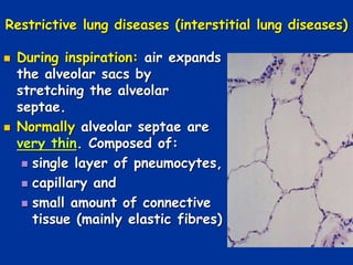
Spr 09 1
Restrictive lung diseases
(interstitial lung diseases)
(ILD)
Restrictive lung diseases (interstitial lung diseases)
 During inspiration: air expands
the alveolar sacs by
stretching the alveolar
septae.
 Normally alveolar septae are
very thin. Composed of:
 single layer of pneumocytes,
 capillary and
 small amount of connective
tissue (mainly elastic fibres)
Spr 09 3
The alveolar wall= Alveolar septa
 Interstitial lung diseases (ILD's)
add cells and fibrous tissue to the
interstitium.
 This:
 Thickens and stiffens the
septae and
 Restricts stretching,
 Making it harder to breath in.
 Therefore, ILD associated with:
 Reduced lung compliance (
decreased filling of lungs on
inspiration)
 Increased elasticity (increased
recoil of lungs on expiration)
Restrictive lung diseases (interstitial lung diseases)
 The PFT reveal the following:
 Decreased FEV 1sec (3L vs 4L)
 Decreased FVC (3L vs 5L)
 Increased FEV 1sec / FVC ratio ~ 3/3 =
100%.
 Compare with
 obstructive lung diseases:
 FEV 1sec / FVC ratio = 33%.
Findings in all interstitial lung diseases
 Clinical:
 Stiff lung increased effort of breathing 
DYSPNEA
 DRY COUGH
 Ventilation perfusion abnormalities  Hypoxemia 
pulmonary hypertension  cor pulmonale 
Respiratory failure
 All lung volumes and capacities are decreased
 Increased FEV 1sec / FVC ratio
 Pathologic:
 Diffuse fibrosis of alveolar interstitium
 X-Ray:
 Diffuse infiltrate (ground glass shadows)
Interstitial fibrosis
Ground glass opacities
Causes of Restrictive lung diseases
1. Chest wall and other disorders in the presence
of normal lungs:
 Kyphoscoliosis, pleural disease (mesothelioma)
2. Acute or chronic interstitial lung disease:
1. Acute
 Acute respiratory distress syndrome
(ARDS)
2. Chronic
 Based on etiology: Classified in to two
major groups:
 ILD with known cause
 ILD with unknown cause
ILD with known cause
1. Occupational and environmental exposures
a) Inorganic:
1. Pneumoconiosis
1. Asbestosis**
2. Silicosis**
3. Coal worker’s pneumoconiosis**
4. Berylliosis*
b) Organic
 Hypersensitivity pneumonitis **
2. Drug or treatment related
 Chemotherapeutic agents:
 Busulfan, bleomycin, methotrexate
 Ionizing radiation
ILD with unknown cause
 Sarcoidosis**
 Idiopathic pulmonary fibrosis*
 Pulmonary hemorrhage syndrome:
1. Goodpasture syndrome*
2. Wegener’s granulomatosis
3. Idiopathic hemosiderosis
 Collagen vascular diseases
 Eosinophilic granuloma
Pneumoconioses
 The most common cause of ILD
 Refers to a group of occupational lung diseases
characterized by
 inhalation of mineral dust particles (coal dust,
silica, asbestos, beryllium) that leads to
interstitial fibrosis.
 Coal Dust : coal workers pneumoconiosis
(CWP)
 Silica : silicosis
 Asbestos : asbestosis
 Beryllium : berylliosis
 Coal dust is least fibrogenic
 Silica, asbestos and beryllium are very fibrogenic.
 Reaction of lung to mineral dust depends on:
 Size, shape, solubility and reactivity of particles
 Most inhaled dust removed by ciliary action.
 Some get impacted at bifurcation of alveolar ducts.
 Macrophages accumulate and engulf the particles.
 Macrophage release a number of factors:
 Fibrogenic factors
 Proinflammatory factors
 Toxic factors
 These factors cause lung injury, inflammatory
reaction, collagen deposition and fibrosis.
Pathogenesis
Spr 09 13
 Caused by inhaling coal dust for prolonged
periods.
 Source of coal dust (anthracotic pigment) :
 coal mines, large urban centers, cigarette
smoke
 Three forms of Coal worker's pneumoconiosis:
1. Asymptomatic anthracosis = pulmonary
anthracosis
2. Simple CWP and
3. Complicated CWP (progressive massive
fibrosis = PMF).
Coal worker's pneumoconiosis(CWP)
 Pulmonary anthracosis:
 Usually asymptomatic
 Carbon pigment (anthracotic pigment) accumulates in
macrophages along lymphatics and in interstitial lung
tissue.
 Alveolar macrophages with anthracotic pigment are
called dust cells.
 Simple CWP
 Characterized by presence of nodular masses in lung
composed of collagen fibers and anthracotic pigment.
 X ray: opacities <1 cm.
 Little pulmonary dysfunction.
 Characterized by : fibrotic opacities >1 cm on X ray.
 Microscopically:
 Opacities show dense collagen and anthracotic
pigment.
 Complications:
 Crippling lung disease (black lung disease)
 Pulmonary hypertension  cor pulmonale
 No increased risk of TB or lung cancer.
 Associations:
 Caplan’s syndrome.
 CWP plus Rheumatoid nodule in the lungs***.
Complicated CWP
or Progressive massive fibrosis (PMF)
"Black lung disease"
Silicosis
 Most common occupational
disease in the world
 Due to inhalation of crystalline
silicone dioxide (found in
quartz)  inflammation and
fibrosis of the lung tissue.
 Occupations associated with
development of silicosis:
1. Foundries
2. Sandblasting
3. Mining, Drilling
4. Tunneling ,Stone cutting
 Silica is highly fibrogenic.
 Chronic exposure:
 nodular opacities in lungs
 Concentric layers of collagen.
 Birefringent silica particles can be seen with
Polarized light.
 Complications:
 Cor pulmonale, Caplan syndrome
 Increased risk of developing lung cancer and
TB
Silicosis
Silicotic nodule
Lungs, silicosis - Radiograph
Asbestos related disease
 Due to inhalation of
asbestos fibers.
 Occupations:
 Pipe fitting in
shipyards,
 roofing,
 insulation and
construction
industries,
 demolition of building
containing asbestos..
Spr 09 23
Pathogenesis
 Asbestos exposure causes fibrosis of lung by
interacting with lung macrophages.
 Asbestos a crystalline silicate with two
subfamilies:
 Serpentine (Chrysotile)
 Curley and flexible fibers
 Tend to be in upper airway
 Cause interstitial fibrosis and lung cancer.
 Amphibole (Crocidolite)
 Straight, stiff, needle like
 Delivered deep
 Cause lung cancer and mesotheliomas.
Asbestos related diseases
1. Benign pleural plaques: Most common lesion***.
• Calcified plaques on pleura and domes of diaphragm.
• Not a precursor for mesothelioma
2. Diffuse Interstitial Fibrosis .
3. Primary bronchogenic carcinoma***:
• Risk further increases if person smokes
• Occurs ~20 years after first exposure.
4. Malignant mesothelioma of pleura***:
• No etiologic relationship with smoking***
• Arises from cells lining the pleura
• Occurs ~25-40 years after 1st exposure.
5. Complications: Cor pulmonale and Caplan’s
syndrome.
Interstitial fibrosis
Asbestos: appearance in tissue
1. Asbestos body
2. Asbestos fibers coated by iron and protein are
called
 Ferruginous bodies (Look like a dumbbell)
 Macrophages phagocytose and coat fibers
with ferritin***
 Golden beaded appearance in sputum or in
airways of lung.
Asbestos body: high power****
A
B
“Ferruginous body"
Berylliosis
 Exposure in nuclear and aerospace industry.
 Lesion:
 Diffuse interstitial fibrosis with noncaseating
granulomas
 Increased incidence of cor pulmonale and
primary lung cancer.
Sarcoidosis
 Multisystem granulomatous diseases of unknown
etiology characterized by
 Noncaseating granulomas in many tissues and
organs**.
 Most commonly targets the lungs, face,
salivary glands and skin***.
 Epidemiology:
 Second most common ILD
 More common in African American women**.
 Occurs in adults of 20-40 yrs age**.
Sarcoidosis
 Pathogenesis:
 An unknown antigen interacts with CD4 helper
T cells , leading to formation of non-caseating
granulomas.
 Sarcoidosis is a diagnosis of exclusion
 Must rule out other granulomatous diseases
Clinical and pathological findings:
 Primarily targets the lungs
 Noncaseating granulomas in the lung
interstitium and hilar lymphnodes (potato
nodes)***.
 Granulomas contain multinucleated giant cells
with laminated calcium concretions (Schaumann
bodies) and stellate inclusions (Asteroid
bodies)***
 Diffuse interstitial fibrosis
 Cough and shortness of breath is the most
common CF
Laminated calcium
Concretions
= Schaumann bodies
Stellate inclusions
=asteroid bodies
 Skin lesions : 2 types
 Erythema nodosum: raised, red, tender
nodules (anterior aspect of legs), no
granulomas
 Nodular, painless lesions containing granulomas
 Eye lesion ; produces uveitis  blurry vision**.
 Mikulicz syndrome: involvement of lacrimal gland
and parotid (enlargement).
 Liver: granulomatous hepatitis  hepatomegaly
 Diabetes insipidus ( hypothalamic and or
posterior pituitary disease)
 Other organs: granulomas in spleen, CNS , heart
and bone marrow.
 Major clinical findings: dyspnea, cough ,fever
and night sweats.
 Laboratory findings:
 Hypercalcemia:** due to synthesis of 1 alpha
hydroxylase by macrophages in granulomas
(increases vit D)
 Elevated serum angiotensin converting enzyme
(ACE)***
 Chest X ray:
 Enlarged hilar lymphnodes – potato nodes***
 Prognosis:
 Majority of patients recover with
corticosteroid therapy.
Giant cell
Granuloma
Hypersensitivity pneumonitis
 Immunologically mediated disorder caused by
repetitive exposure to a known inhaled antigen.
 Does not involve IgE ab or have eosinophilia***
 Primarily affects the alveoli
 Also known as allergic alveolitis.
 Microscopy:
 Mononuclear cell infiltrate in the interstitium.
 Interstitial noncaseating granulomas (in >2/3rd of
cases)***
 Advanced cases show diffuse interstitial fibrosis.
 Clinical presentation:
 Acute reaction: fever, cough, dyspnea ( hours
after exposure)***
 Chronic disease: cough, dyspnea, weight loss.
Noncaseating
granuloma
Selected hypersensitivity pneumonitis
 Farmer’s lung**
 associated with
exposure to moldy
hay containing
thermophilic
actinomycetes
 Silo filler’s disease**
 Inhalation of wheat
weevil protein ( in
fumes originating
from fermenting
corn)
Other hypersensitivity pneumonitis
1. Byssinosis:* *
1. Occurs in workers in textile factories
2. Associated with exposure to cotton, hemp or
linen (“Monday morning blues”)
2. Bagassosis: lung disease that develops after
repeated exposure to moldy sugar cane.
3. Pigeon breeders lung: proteins from bird
feathers or excreta.
4. Humidifier or air conditioner lung: thermophilic
bacteria.
Diffuse alveolar hemorrhage syndromes
 A group of immune mediated diseases that present as
the triad of:
 Hemoptysis, Anemia and diffuse pulmonary
infiltrates. Examples:
1. Goodpasture syndrome
2. Wegener Granulomatosis
 GOODPASTURE SYNDROME:***
 Male predominant disease characterized by:
 Glumerulonephritis and
 Hemorrhagic interstitial pneumonitis.
 Both lesions caused by anti-basement membrane
antibodies directed against BM of pulmonary and
glomerular capillaries.
 Patients first present with hemoptysis and then
acute glomerulonephritis.***
 Micro:
 Focal necrosis of alveolar walls
 Intra-alveolar hemorrhage
 Intra or extracellular hemosiderin
 Fibrous thickening of septa
 Immunofluorescence:
 Linear pattern of immunoglobulin deposition**
 Kidney and lungs
 Treatment:
 Plasmapheresis and immunosuppressive therapy.
Goodpasture’s Syndrome, Hemorrhage, Necrosis
and IgG Deposits
Idiopathic pulmonary fibrosis
 A disorder of unknown cause.
 Pathogenesis:
 Repeated cycles of alveolitis triggered by an
unknown ag.
 Release of cytokines produces interstitial
fibrosis.
 Alveolar fibrosis leads to dilatation of airways
 Lung has a honeycomb appearance
 Course:
 Progression  pulmonary insufficiency, cor
pulmonale and cardiac failure.
Honeycomb lung.
Spr 09 47
 Drugs producing ILD include:
 Nitrofurantoin, amiodarone, bleomycin,
busulfan and cyclophosphamide.
 Collagen vascular diseases producing restrictive
lung disease: SLE, RA, systemic sclerosis.
Other Restrictive lung diseases

08 respiratory restrictive

  • 1.
    Spr 09 1 Restrictivelung diseases (interstitial lung diseases) (ILD)
  • 2.
    Restrictive lung diseases(interstitial lung diseases)  During inspiration: air expands the alveolar sacs by stretching the alveolar septae.  Normally alveolar septae are very thin. Composed of:  single layer of pneumocytes,  capillary and  small amount of connective tissue (mainly elastic fibres)
  • 3.
    Spr 09 3 Thealveolar wall= Alveolar septa
  • 4.
     Interstitial lungdiseases (ILD's) add cells and fibrous tissue to the interstitium.  This:  Thickens and stiffens the septae and  Restricts stretching,  Making it harder to breath in.  Therefore, ILD associated with:  Reduced lung compliance ( decreased filling of lungs on inspiration)  Increased elasticity (increased recoil of lungs on expiration)
  • 5.
    Restrictive lung diseases(interstitial lung diseases)  The PFT reveal the following:  Decreased FEV 1sec (3L vs 4L)  Decreased FVC (3L vs 5L)  Increased FEV 1sec / FVC ratio ~ 3/3 = 100%.  Compare with  obstructive lung diseases:  FEV 1sec / FVC ratio = 33%.
  • 6.
    Findings in allinterstitial lung diseases  Clinical:  Stiff lung increased effort of breathing  DYSPNEA  DRY COUGH  Ventilation perfusion abnormalities  Hypoxemia  pulmonary hypertension  cor pulmonale  Respiratory failure  All lung volumes and capacities are decreased  Increased FEV 1sec / FVC ratio  Pathologic:  Diffuse fibrosis of alveolar interstitium  X-Ray:  Diffuse infiltrate (ground glass shadows)
  • 7.
  • 8.
    Causes of Restrictivelung diseases 1. Chest wall and other disorders in the presence of normal lungs:  Kyphoscoliosis, pleural disease (mesothelioma) 2. Acute or chronic interstitial lung disease: 1. Acute  Acute respiratory distress syndrome (ARDS) 2. Chronic  Based on etiology: Classified in to two major groups:  ILD with known cause  ILD with unknown cause
  • 9.
    ILD with knowncause 1. Occupational and environmental exposures a) Inorganic: 1. Pneumoconiosis 1. Asbestosis** 2. Silicosis** 3. Coal worker’s pneumoconiosis** 4. Berylliosis* b) Organic  Hypersensitivity pneumonitis ** 2. Drug or treatment related  Chemotherapeutic agents:  Busulfan, bleomycin, methotrexate  Ionizing radiation
  • 10.
    ILD with unknowncause  Sarcoidosis**  Idiopathic pulmonary fibrosis*  Pulmonary hemorrhage syndrome: 1. Goodpasture syndrome* 2. Wegener’s granulomatosis 3. Idiopathic hemosiderosis  Collagen vascular diseases  Eosinophilic granuloma
  • 11.
    Pneumoconioses  The mostcommon cause of ILD  Refers to a group of occupational lung diseases characterized by  inhalation of mineral dust particles (coal dust, silica, asbestos, beryllium) that leads to interstitial fibrosis.  Coal Dust : coal workers pneumoconiosis (CWP)  Silica : silicosis  Asbestos : asbestosis  Beryllium : berylliosis  Coal dust is least fibrogenic  Silica, asbestos and beryllium are very fibrogenic.
  • 12.
     Reaction oflung to mineral dust depends on:  Size, shape, solubility and reactivity of particles  Most inhaled dust removed by ciliary action.  Some get impacted at bifurcation of alveolar ducts.  Macrophages accumulate and engulf the particles.  Macrophage release a number of factors:  Fibrogenic factors  Proinflammatory factors  Toxic factors  These factors cause lung injury, inflammatory reaction, collagen deposition and fibrosis. Pathogenesis
  • 13.
  • 14.
     Caused byinhaling coal dust for prolonged periods.  Source of coal dust (anthracotic pigment) :  coal mines, large urban centers, cigarette smoke  Three forms of Coal worker's pneumoconiosis: 1. Asymptomatic anthracosis = pulmonary anthracosis 2. Simple CWP and 3. Complicated CWP (progressive massive fibrosis = PMF). Coal worker's pneumoconiosis(CWP)
  • 15.
    Coal worker's pneumoconiosis(CWP) Pulmonary anthracosis:  Usually asymptomatic  Carbon pigment (anthracotic pigment) accumulates in macrophages along lymphatics and in interstitial lung tissue.  Alveolar macrophages with anthracotic pigment are called dust cells.  Simple CWP  Characterized by presence of nodular masses in lung composed of collagen fibers and anthracotic pigment.  X ray: opacities <1 cm.  Little pulmonary dysfunction.
  • 16.
     Characterized by: fibrotic opacities >1 cm on X ray.  Microscopically:  Opacities show dense collagen and anthracotic pigment.  Complications:  Crippling lung disease (black lung disease)  Pulmonary hypertension  cor pulmonale  No increased risk of TB or lung cancer.  Associations:  Caplan’s syndrome.  CWP plus Rheumatoid nodule in the lungs***. Complicated CWP or Progressive massive fibrosis (PMF)
  • 17.
  • 18.
    Silicosis  Most commonoccupational disease in the world  Due to inhalation of crystalline silicone dioxide (found in quartz)  inflammation and fibrosis of the lung tissue.  Occupations associated with development of silicosis: 1. Foundries 2. Sandblasting 3. Mining, Drilling 4. Tunneling ,Stone cutting
  • 19.
     Silica ishighly fibrogenic.  Chronic exposure:  nodular opacities in lungs  Concentric layers of collagen.  Birefringent silica particles can be seen with Polarized light.  Complications:  Cor pulmonale, Caplan syndrome  Increased risk of developing lung cancer and TB Silicosis
  • 20.
  • 21.
  • 22.
    Asbestos related disease Due to inhalation of asbestos fibers.  Occupations:  Pipe fitting in shipyards,  roofing,  insulation and construction industries,  demolition of building containing asbestos..
  • 23.
    Spr 09 23 Pathogenesis Asbestos exposure causes fibrosis of lung by interacting with lung macrophages.  Asbestos a crystalline silicate with two subfamilies:  Serpentine (Chrysotile)  Curley and flexible fibers  Tend to be in upper airway  Cause interstitial fibrosis and lung cancer.  Amphibole (Crocidolite)  Straight, stiff, needle like  Delivered deep  Cause lung cancer and mesotheliomas.
  • 24.
    Asbestos related diseases 1.Benign pleural plaques: Most common lesion***. • Calcified plaques on pleura and domes of diaphragm. • Not a precursor for mesothelioma 2. Diffuse Interstitial Fibrosis . 3. Primary bronchogenic carcinoma***: • Risk further increases if person smokes • Occurs ~20 years after first exposure. 4. Malignant mesothelioma of pleura***: • No etiologic relationship with smoking*** • Arises from cells lining the pleura • Occurs ~25-40 years after 1st exposure. 5. Complications: Cor pulmonale and Caplan’s syndrome.
  • 26.
  • 27.
    Asbestos: appearance intissue 1. Asbestos body 2. Asbestos fibers coated by iron and protein are called  Ferruginous bodies (Look like a dumbbell)  Macrophages phagocytose and coat fibers with ferritin***  Golden beaded appearance in sputum or in airways of lung.
  • 28.
    Asbestos body: highpower**** A B “Ferruginous body"
  • 29.
    Berylliosis  Exposure innuclear and aerospace industry.  Lesion:  Diffuse interstitial fibrosis with noncaseating granulomas  Increased incidence of cor pulmonale and primary lung cancer.
  • 30.
    Sarcoidosis  Multisystem granulomatousdiseases of unknown etiology characterized by  Noncaseating granulomas in many tissues and organs**.  Most commonly targets the lungs, face, salivary glands and skin***.  Epidemiology:  Second most common ILD  More common in African American women**.  Occurs in adults of 20-40 yrs age**.
  • 31.
    Sarcoidosis  Pathogenesis:  Anunknown antigen interacts with CD4 helper T cells , leading to formation of non-caseating granulomas.  Sarcoidosis is a diagnosis of exclusion  Must rule out other granulomatous diseases
  • 32.
    Clinical and pathologicalfindings:  Primarily targets the lungs  Noncaseating granulomas in the lung interstitium and hilar lymphnodes (potato nodes)***.  Granulomas contain multinucleated giant cells with laminated calcium concretions (Schaumann bodies) and stellate inclusions (Asteroid bodies)***  Diffuse interstitial fibrosis  Cough and shortness of breath is the most common CF
  • 33.
    Laminated calcium Concretions = Schaumannbodies Stellate inclusions =asteroid bodies
  • 34.
     Skin lesions: 2 types  Erythema nodosum: raised, red, tender nodules (anterior aspect of legs), no granulomas  Nodular, painless lesions containing granulomas  Eye lesion ; produces uveitis  blurry vision**.  Mikulicz syndrome: involvement of lacrimal gland and parotid (enlargement).  Liver: granulomatous hepatitis  hepatomegaly  Diabetes insipidus ( hypothalamic and or posterior pituitary disease)  Other organs: granulomas in spleen, CNS , heart and bone marrow.
  • 35.
     Major clinicalfindings: dyspnea, cough ,fever and night sweats.  Laboratory findings:  Hypercalcemia:** due to synthesis of 1 alpha hydroxylase by macrophages in granulomas (increases vit D)  Elevated serum angiotensin converting enzyme (ACE)***  Chest X ray:  Enlarged hilar lymphnodes – potato nodes***  Prognosis:  Majority of patients recover with corticosteroid therapy.
  • 37.
  • 38.
    Hypersensitivity pneumonitis  Immunologicallymediated disorder caused by repetitive exposure to a known inhaled antigen.  Does not involve IgE ab or have eosinophilia***  Primarily affects the alveoli  Also known as allergic alveolitis.  Microscopy:  Mononuclear cell infiltrate in the interstitium.  Interstitial noncaseating granulomas (in >2/3rd of cases)***  Advanced cases show diffuse interstitial fibrosis.  Clinical presentation:  Acute reaction: fever, cough, dyspnea ( hours after exposure)***  Chronic disease: cough, dyspnea, weight loss.
  • 39.
  • 40.
    Selected hypersensitivity pneumonitis Farmer’s lung**  associated with exposure to moldy hay containing thermophilic actinomycetes  Silo filler’s disease**  Inhalation of wheat weevil protein ( in fumes originating from fermenting corn)
  • 41.
    Other hypersensitivity pneumonitis 1.Byssinosis:* * 1. Occurs in workers in textile factories 2. Associated with exposure to cotton, hemp or linen (“Monday morning blues”) 2. Bagassosis: lung disease that develops after repeated exposure to moldy sugar cane. 3. Pigeon breeders lung: proteins from bird feathers or excreta. 4. Humidifier or air conditioner lung: thermophilic bacteria.
  • 42.
    Diffuse alveolar hemorrhagesyndromes  A group of immune mediated diseases that present as the triad of:  Hemoptysis, Anemia and diffuse pulmonary infiltrates. Examples: 1. Goodpasture syndrome 2. Wegener Granulomatosis  GOODPASTURE SYNDROME:***  Male predominant disease characterized by:  Glumerulonephritis and  Hemorrhagic interstitial pneumonitis.  Both lesions caused by anti-basement membrane antibodies directed against BM of pulmonary and glomerular capillaries.
  • 43.
     Patients firstpresent with hemoptysis and then acute glomerulonephritis.***  Micro:  Focal necrosis of alveolar walls  Intra-alveolar hemorrhage  Intra or extracellular hemosiderin  Fibrous thickening of septa  Immunofluorescence:  Linear pattern of immunoglobulin deposition**  Kidney and lungs  Treatment:  Plasmapheresis and immunosuppressive therapy.
  • 44.
    Goodpasture’s Syndrome, Hemorrhage,Necrosis and IgG Deposits
  • 45.
    Idiopathic pulmonary fibrosis A disorder of unknown cause.  Pathogenesis:  Repeated cycles of alveolitis triggered by an unknown ag.  Release of cytokines produces interstitial fibrosis.  Alveolar fibrosis leads to dilatation of airways  Lung has a honeycomb appearance  Course:  Progression  pulmonary insufficiency, cor pulmonale and cardiac failure.
  • 46.
  • 47.
    Spr 09 47 Drugs producing ILD include:  Nitrofurantoin, amiodarone, bleomycin, busulfan and cyclophosphamide.  Collagen vascular diseases producing restrictive lung disease: SLE, RA, systemic sclerosis. Other Restrictive lung diseases