Downloaded 168 times

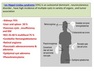
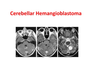
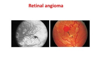
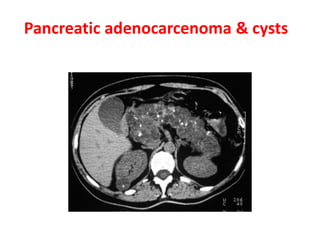
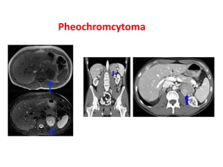
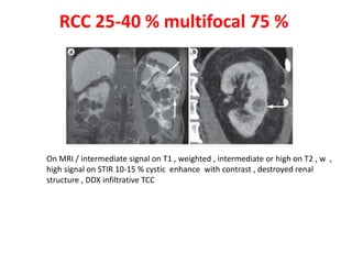
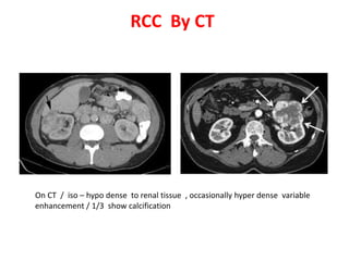

Von Hippel-Lindau disease is an autosomal dominant neurocutaneous disorder characterized by a high incidence of cysts in multiple organs and tumor development. Common manifestations include renal cell carcinoma in 25-40% of cases, cerebellar hemangioblastoma, retinal angioma, pancreatic cysts and tumors, and pheochromocytoma. Imaging findings include lesions that are intermediate to high signal on MRI and variably enhancing on CT and IVU.


![PERI-PROSTHETIC FRACTURE NAIL-PLATE CONSTRUCT [NPC].pptx](https://cdn.slidesharecdn.com/ss_thumbnails/drarunkumardrmohamedashrafperiprostheticfrasturenail-plateconstructnpc-260209164459-7e9d15a1-thumbnail.jpg?width=640&height=640&fit=bounds)


![ONFH[AVN HIP] -TRIPLE REGIME -A NOVAL SURGICAL CONCEPT .pptx](https://cdn.slidesharecdn.com/ss_thumbnails/onfhavnhip2026koaconcalicutdrgokuldevdrmashraf-260210064517-213ec005-thumbnail.jpg?width=640&height=640&fit=bounds)
