Downloaded 106 times


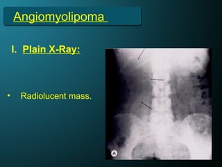

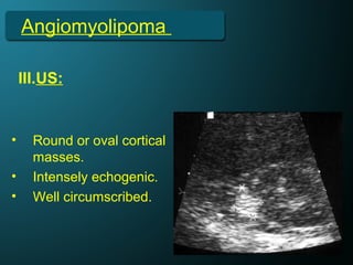


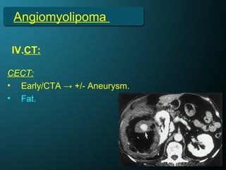
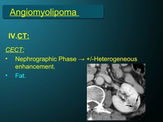
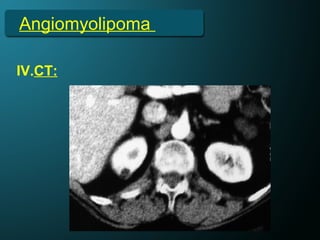
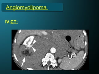
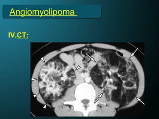


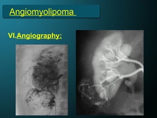
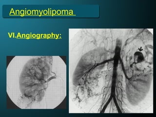
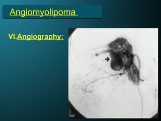


This document discusses angiomyolipomas (AML), a type of benign renal tumor composed of blood vessels, smooth muscle, and fat cells. It describes the typical presentation of isolated and tuberous sclerosis-associated AML, including that isolated AML most often present as solitary lesions in the right kidney in women, while AML associated with tuberous sclerosis are typically bilateral and occur equally in men and women. Imaging findings are also summarized, noting the characteristic fat content visible on CT and MRI that appears echogenic on ultrasound and enhances heterogeneously after contrast. Angiography further demonstrates the hypervascular and tortuous nature of AML vasculature.


