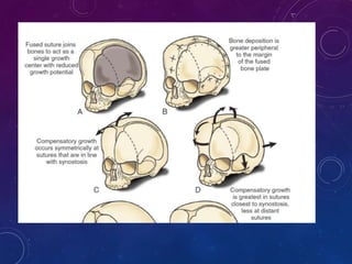
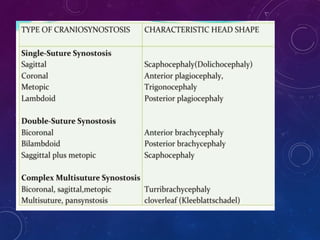
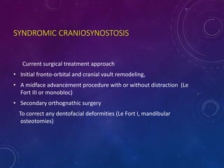
2. ⢠Pathological condition that results from premature fusion
of one or more sutures in the cranial vault.
⢠Associated with a deformity of the vault and cranial base.
3. BRAIN GROWTH
⢠At term has nearly 40 percent of his or her adult brain
volume.
⢠This increases to 80 percent by three
years of age.
⢠Continues to grow until the age of 12 yrs
4. THE CRANIUM
⢠At term is 40 percent of adult size;
⢠By seven years, this increases to 90 percent.
5. SUTURE GROWTH
⢠Sutures allow growth perpendicular to them
⢠Growth at suture lines related to brain growth
⢠In 1851, Virchow described how skull growth is restricted to a
plane perpendicular to the affected, prematurely fused suture
and is enhanced in a plane parallel to it.
6.
7.
8. Mature suture closure occurs by 12 years of age, but
completion continues into the third decade of life and beyond.
Timing of Closure of Sutures
and Fontanelles
Type of suture Time to closure
Metopic suture Nine months to two
years (may persist
into adulthood)
Coronal, sagittal, 3rd decade of life
lambdoid sutures
9. IN HUMANS, THE SEQUENCE OF FONTANELLE
CLOSURE IS AS FOLLOWS:
1) posterior fontanelle generally closes 2-3 months after birth
2) sphenoidal fontanelle is the next to close around 6 months after birth
3) mastoid fontanelle closes next from 6-18 months after birth
4) the anterior fontanelle is generally the last to close between 1-3 years of
age
10. HISTORY AND PATHOGENESIS
⢠Otto ( 1830) coined the term craniosynostosis.
⢠Virchow : Classified the different types of skull deformity.
⢠L.C. Lane: 1st surgical procedure to release stenosed suture
⢠Lannelogue (1890) :- performed B/L strip craniectomies
⢠Paul Tessier - father of modern craniofacial Surgery.
- First to attempt major surgical procedures on the craniofacial
skeleton.
11. THEORIES OF CRANOISYNOSTOSIS
⢠Sommering( 1839)â Noted that bone growth in skull primarily occurs at
suture line and if it prematurely fused, an abnormal skull shape developed
and skull growth restricted.
⢠Virchow(1821) and Otto(1830)- Similar observation were made and they
noted restriction of growth adjacent to suture and compensatory growth
occurred at elsewhere in skull to accommodate growing brain .
12.
13. THEORIES OF CRANIOSYNOSTOSIS
⢠Moss(1959) â Described functional matrix theory. According to this theory
cranial base abnormality was the primary pathological process and cranial
vault suture abnormality was secondary as cranial base mature
embryologically before cranial vault.
⢠Persson (1979) â Cranial vault suture pathology may be primary in the
development of synostosis leading to cranial base and facial deformity.
14. ⢠Animal studies
The cranial vault abnormalities typical of synostosis can be
produced with experimental fusion of developing cranial vault
sutures.
⢠1. Persing JA, Babler WJ, Jane JA, et al. Experimental unilateral coronal synostosis in
rabbits. Plast Reconstr Surg. 1986;77:369â376.
15. INCIDENCE
⢠One per 1,800 to 2,200 live births
⢠Males - Sagittal and metopic stenosis
⢠Females - Coronal
17. PRIMARY VS SECONDARY
17
⢠Primary defect of
ossification
⢠suture growth is altered
⢠head frequently asymmetric
⢠the brain continues to grow
in areas where sutures are
open
⢠most individuals are normal
neurologically
⢠benefit from surgery
⢠Secondary to brain
malformation
⢠Head symmetric
⢠growth of brain is
impaired
⢠neurologically
abnormal
⢠usually No benifit
of surgery
18.
19.
20. CRANIOSYNOSTOSIS
SYNDROMES
⢠10-20 % of cases
⢠Autosomal Dominant
⢠Linked to Chromosome 10
⢠Multi-sutural, complex case
⢠If a suture is fused, check hands, feet, big toe and
thumb
23. CROUZONâS
⢠Autosomal - dominant pattern.
⢠One of every 25,000 live births
⢠5 percent of cases of craniosynostosis.
⢠Numerous missense mutations in the Ig III domain of the FGFR2 gene have
been implicated
26. APERTâS -âCROUZONâS WITH HAND
INVOLVEMENTâ
â˘1 in 55,000
⢠Varying intellect (50 % with MR)
⢠syndactyly
⢠Cervical vertebral anomalies
⢠Rare hydrocephalus
27. PFEIFFER SYNDROME
⢠1 in 2 lakhs
⢠Clover leaf skull in 20%
⢠Broad thumbs, broad great toes
⢠Intelligence is reported to be normal
28.
29. CLOVERLEAF SKULL DEFORMITY
(TRIPHYLLOCEPHALY)
⢠(Derived from the Greek word triphyllos,
meaning trefoil, with 3 leaves),
⢠Multiple suture synostosis
⢠Head shaped like a cloverleaf
⢠Three bulges-two temporal and top
⢠Pronounced constrictions in both sylvian
fissures
30. NON SYNDROMIC -1. SAGITTAL- SCAPHOCEPHALY
⢠most common form of craniosynostosis
⢠occurs at a rate of 1 in 5000 children
⢠male-to-female ratio of 3.5 : 1
31. 2. CORONAL - PLAGIOCEPHALY
⢠Greek word plagios, meaning oblique or sloping, and corresponds to unilateral
coronal synostosis.
⢠10-20% -second most common sutural fusion and occurs at a rate of 1 in
10,000 children
⢠male-to-female ratio of 1 : 2.
⢠Posterior plagiocephaly corresponds to lambdoid synostosis. 1.3%
33. 3.METOPIC - TRIGONOCEPHALY
⢠Derived from the Greek word trigonos, meaning triangular
⢠5 â 10% incidence - 1 out of 10,000 to 15,000 live births
⢠male-to-female ratio of 3.3 : 1
40. ⢠Passage of the head through the birth canal deforms the
head. This shape is retained for 2-3 weeks postnatally.
⢠Early diagnosis is important
The brain grows rapidly during this period
Delay only worsens the deformity of the head shape.
41. ⢠Birth, sleeping position.
⢠Head tilt, torticollis
deformational plagiocephaly
⢠Family history
Abnormal head shape or multiple systemic problems
(eg,cardiac, genitourinary, musculoskeletal)
Detailed history
42. CLINICAL EXAM
⢠Head shape (from above, side)
⢠Ear and facial symmetry
⢠Palpate suture lines & fontanelles
⢠Look for ridging
⢠Look for associated anomalies
⢠If a suture is fused,
check hands, feet, big toe and thumb
48. MENTAL DELAY
⢠Mental outcome varies widely among patients with syndromic
craniosynostosis .
⢠Mental retardation common in Apertâs syndrome & in some patients with
Pfeifferâs syndrome
⢠The proportion of children with developmental and behavioral problems at
first consultation increases with age.
⢠patients who undergo surgery before the age of 1 year have better
developmental scores than do those who undergo surgery after the age of
1 year.
49. PLAIN FILMS
⢠Simple and inexpensive,
⢠Absent or line of increased density
⢠Harlequin appearenceâcoronal
⢠Cannot differentiate
Lambdoid synostosis and deformational plagiocephaly (plagiocephaly
without synostosis).
⢠To visualize all the sutures, special Waters views must be taken.
51. CT SCAN
⢠Standard for the complete visualization of the skull and cranial sutures.
⢠Detailed anatomy of the calvaria and the brain parenchyma
⢠Document effect of corrective surgery
54. ICP MONITORING
⢠Radiological signs inconclusive
⢠Deciding nature and timing of surgery
⢠Features of â ICT
Mean pressure > 15 mm Hg
Raised base line value
Prolonged plateau wave
56. GOAL
⢠Normalization of deviated appearance, growth and function of skull
⢠Keep the suture open till brain growth is complete
57. INDICATIONS
⢠Correction of cosmetic abnormality
⢠Early treatment of intracranial hypertension
⢠Optimizing brain growth
⢠Severe proptosis and impending corneal damage
⢠upward and caudal cerebellar tocillar herniation ,hindbrain compression
and distortion
58. TIMING OF SURGERY
Early operation(3-6 months)
⢠Rapid brain growth reshape bone
⢠Better compliance of brain dura and scalp
⢠Calvarium in an infant aged 3-9 months is much more
malleable, easier to shape and providing a better outcome.
59. ⢠Surgical intervention should be performed during infancy, preferably
in the first 6 months of postnatal life, to prevent the further
progression of the deformity and possible complications associated
with increased intracranial pressure.
60. LATE INTERVENTION
⢠Closer the cranium is to the adult size, the less
overcorrection for reconstruction and the better the
ultimate skull shape.
⢠Higher risk of recurrent deformity
⢠Surgical correction more complex
62. BASIC MECHANISMS
Passive reshapement
1. Generous removal of bone
2. Strip craniectomy
3. Morcellation
Active reshapement
1. Fronto orbital advancement
2. Cranial vault reshapement
63. INCISION
⢠Zigzag bicoronal incision
⢠Prevents parting of the hair along a straight line
⢠Scar tends to spread less - redistribution of the forces.
⢠Incision begins slightly anterior and superior to the helix of the ear.
⢠Electrocautery is used cautiously
64.
65. SAGITTAL CRANIOSYNOSTOSIS
⢠Objectives
⢠Correction of scaphocephaly
⢠Frontal bossing and occipital protrusion
⢠Initial surgical procedures included a narrow-strip craniectomyâ higher
restenosis .
⢠Wider and more extensive craniectomy
Do not address the frontal bossing and occipital bathrocephaly relied on
the growing brain to correct these deformities.
68. BILATERAL CORONAL STENOSIS
⢠Extended bicoronal craniectomies with
reconstruction of the forehead.
⢠The supraorbital bar or brow is
reshaped and advanced forward with
the forehead.
⢠The reconstructed forehead and brow
are rigidly fixed to the nose and lateral
orbits with microplates.
70. SYNDROMIC CRANIOSYNOSTOSIS
Current surgical treatment approach
⢠Initial fronto-orbital and cranial vault remodeling,
⢠A midface advancement procedure with or without distraction (Le
Fort III or monobloc)
⢠Secondary orthognathic surgery
To correct any dentofacial deformities (Le Fort I, mandibular
osteotomies)