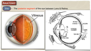
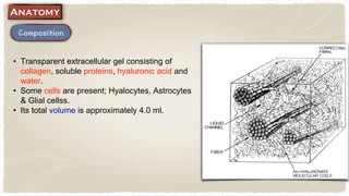
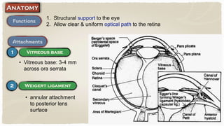
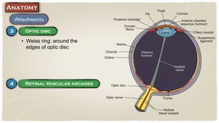
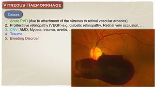
Vitreous
Mohamed Abdelzaher MD, FRCS
• The posterior segment of the eye between Lens & Retina.
• Transparent extracellular gel consisting of
collagen, soluble proteins, hyaluronic acid and
water.
• Some cells are present; Hyalocytes, Astrocytes
& Glial cellss.
• Its total volume is approximately 4.0 ml.
1. Structural support to the eye
2. Allow clear & uniform optical path to the retina
• Vitreous base: 3-4 mm
across ora serrata
• annular attachment
to posterior lens
surface
• Weiss ring: around the
edges of optic disc
• e.g. lattice degeneration
• Fly, cobweb or thread-like lesions best seen against a pale background.
1. Embryological remnants of vitreous cells
2. Vitreous haemorrhage
3. Vitreous degeneration
• If associated with photopsia,
Posterior vitreous detachment
(PVD) or Retinal break is
suspected.
1. Reassurance & observation
2. Vitrectomy: in severe visually disabling cases
3. YAG vitreolysis (Debatable): the laser energy evaporates the collagen
and hyaluronic molecules to form a gas
1. Acute PVD (due to attachment of the vitreous to retinal vascular arcades)
2. Proliferative retinopathy (VEGF) e.g. diabetic retinopathy, Retinal vein occlusion, …
3. CNV: AMD, Myopia, trauma, uveitis, …
4. Trauma
5. Bleeding Disorder
1. Mild haemorrhage; sudden onset of floaters
2. Severe haemorrhage; sudden painless diminution of vision
1. Visual Acuity; diminished according to the
density of haemorrhage
2. Red reflex: dark red or dim
3. Fundus: Vitreous Haemorrhage might
preclude good retinal view
• Retinal Detachment
• Retinal edema
• Central Retinal Artery occlusion
• Central Retinal Vein occlusion
• B scan Ultrasound (to exclude
retinal detachment)
• Proliferative
Vitreoretinopathy
(PVR)
• Tractional Retinal
Detachment
• Treat the cause
• Alpha chymotrypsin
• Vitrectomy, When;
1. Non resolving vitreous
haemorrhage for 3 - 6 months
2. Pre macular haemorrhage
3. Vitreous haemorrhage combined
with retinal detachment
Separation of the posterior hyaloid membrane from the retina anywhere
posterior to the vitreous base.
1. Senility
2. High myopia
3. Trauma
• Photopsia (flashes of light)
• Floaters
• Ring floater to the temporal side of central vision (Weiss ring)
• Retinal break
• Retinal Detachment
• Not required in
non complicated
cases
• Retinal Tear >>>
Argon laser
retinopexy
• Retinal
detachment >>>
Vitrectomy
• A common degenerative process in which calcium pyrophosphate particles collect
within the vitreous gel
• Commonly Unilateral
• Asymptomatic (do not affect the
vision)
• Numerous tiny round yellow–white
opacities of varying size and density
• Moves with vitreous during eye
movement
• Do not sediment when the eye is
immobile
•Occurs as a consequence of chronic vitreous haemorrhage, often in a blind eye
•Crystals are composed of cholesterol and are derived from plasma cells or degraded
products of erythrocytes
• Often blind eye
• Numerous Golden brown refractile
particles of varying size and density
• Anterior chamber may be involved
• Tend to sediment inferiorly when the
eye is immobile
Asteroid Hyalosis Synchysis Scintillans
Vision Not affected Often blind
Nature Calcium Cholesterol
Clinically
tiny round yellow–white
opacities
Golden brown refractile
particles
Anterior chamber Not affected May be affected
Sedimentation No Yes
Vitreous

Vitreous

  • 1.
  • 2.
    • The posteriorsegment of the eye between Lens & Retina.
  • 3.
    • Transparent extracellulargel consisting of collagen, soluble proteins, hyaluronic acid and water. • Some cells are present; Hyalocytes, Astrocytes & Glial cellss. • Its total volume is approximately 4.0 ml.
  • 4.
    1. Structural supportto the eye 2. Allow clear & uniform optical path to the retina • Vitreous base: 3-4 mm across ora serrata • annular attachment to posterior lens surface
  • 5.
    • Weiss ring:around the edges of optic disc
  • 6.
    • e.g. latticedegeneration
  • 7.
    • Fly, cobwebor thread-like lesions best seen against a pale background. 1. Embryological remnants of vitreous cells 2. Vitreous haemorrhage 3. Vitreous degeneration • If associated with photopsia, Posterior vitreous detachment (PVD) or Retinal break is suspected.
  • 9.
    1. Reassurance &observation 2. Vitrectomy: in severe visually disabling cases 3. YAG vitreolysis (Debatable): the laser energy evaporates the collagen and hyaluronic molecules to form a gas
  • 10.
    1. Acute PVD(due to attachment of the vitreous to retinal vascular arcades) 2. Proliferative retinopathy (VEGF) e.g. diabetic retinopathy, Retinal vein occlusion, … 3. CNV: AMD, Myopia, trauma, uveitis, … 4. Trauma 5. Bleeding Disorder
  • 11.
    1. Mild haemorrhage;sudden onset of floaters 2. Severe haemorrhage; sudden painless diminution of vision
  • 12.
    1. Visual Acuity;diminished according to the density of haemorrhage 2. Red reflex: dark red or dim 3. Fundus: Vitreous Haemorrhage might preclude good retinal view
  • 13.
    • Retinal Detachment •Retinal edema • Central Retinal Artery occlusion • Central Retinal Vein occlusion • B scan Ultrasound (to exclude retinal detachment)
  • 14.
  • 15.
    • Treat thecause • Alpha chymotrypsin • Vitrectomy, When; 1. Non resolving vitreous haemorrhage for 3 - 6 months 2. Pre macular haemorrhage 3. Vitreous haemorrhage combined with retinal detachment
  • 16.
    Separation of theposterior hyaloid membrane from the retina anywhere posterior to the vitreous base. 1. Senility 2. High myopia 3. Trauma
  • 17.
    • Photopsia (flashesof light) • Floaters • Ring floater to the temporal side of central vision (Weiss ring)
  • 18.
    • Retinal break •Retinal Detachment
  • 19.
    • Not requiredin non complicated cases • Retinal Tear >>> Argon laser retinopexy • Retinal detachment >>> Vitrectomy
  • 20.
    • A commondegenerative process in which calcium pyrophosphate particles collect within the vitreous gel • Commonly Unilateral • Asymptomatic (do not affect the vision) • Numerous tiny round yellow–white opacities of varying size and density • Moves with vitreous during eye movement • Do not sediment when the eye is immobile
  • 21.
    •Occurs as aconsequence of chronic vitreous haemorrhage, often in a blind eye •Crystals are composed of cholesterol and are derived from plasma cells or degraded products of erythrocytes • Often blind eye • Numerous Golden brown refractile particles of varying size and density • Anterior chamber may be involved • Tend to sediment inferiorly when the eye is immobile
  • 22.
    Asteroid Hyalosis SynchysisScintillans Vision Not affected Often blind Nature Calcium Cholesterol Clinically tiny round yellow–white opacities Golden brown refractile particles Anterior chamber Not affected May be affected Sedimentation No Yes