The document provides a historical overview and summary of virology:
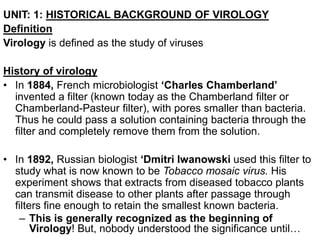
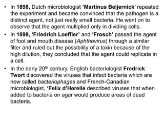
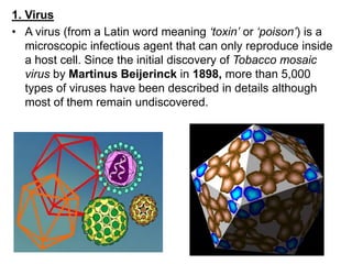
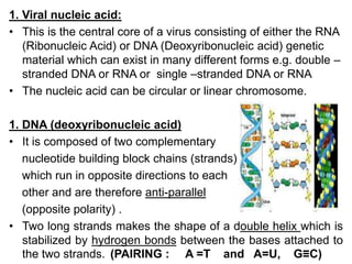
- Virology is the study of viruses, which are microscopic infectious agents that can only reproduce inside host cells. Important early discoveries included identifying that the agents causing tobacco mosaic disease and foot-and-mouth disease could pass through filters, indicating they were not bacteria.
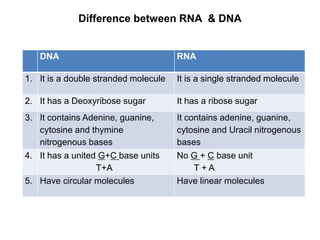
- Major milestones in the 20th century included imaging viruses using electron microscopy, growing viruses in cell cultures and eggs, discovering they contain either DNA or RNA, determining viruses cause diseases like polio, yellow fever, and cancer, and ultimately eradicating smallpox through vaccination. Today over 5,000 virus types have been described though many remain undiscovered.
History, Albert mayor, Stanley, Smith, Crystalline nature, DNA as genetic material, antigenic properties, tmv structure, RNA as genetic material, contribution of Indian scientists to plant virology
It talks about Contribution of Antonie Van Leeuwenhoek to the society. It also talks about his life style like a story. It also explains the contribution about microscope and micro organisms.
Sergei Nikolaievich Winogradsky And Martinus Willem Beijerinck-Discoveries,Nitrogen Fixing Bacteria and the Discovery of Chemosynthesis, Scientific contributions
What is bacteria?(Structures Present in Bacteria And their Functions | Prokar...sehriqayyum
Explains what bacteria is and where it exists.
A key feature of nearly all prokaryotic cells is the cell wall, which maintains cell shape, protects the cell, and prevents it from bursting in a hypotonic environment.
The cell walls of prokaryotes differ in structure from those of eukaryotes. In eukaryotes that have cell walls, such as plants and fungi, the walls are usually made of cellulose or chitin. In contrast, most bacterial cell walls contain peptidoglycan, a polymer composed of modified sugars cross-linked by short polypeptides.
Using a technique called the Gram stain, developed by the 19th-century Danish physician Hans Christian Gram, scientists can categorize many bacterial species according to differences in cell wall composition.
Gram-positive bacteria have simpler walls with a relatively large amount of peptidoglycan. Gram-negative bacteria have less peptidoglycan
and are structurally more complex, with an outer membrane
that contains lipopolysaccharides (carbohydrates bonded
to lipids).
LEARN ABOUT:
- Bacteria
- The number of viruses on earth is staggering
- Pathogenic yeasts
- Helminths
- Harnessing bacteria
- Microbes on the tree of life
- Living and working together
- Archaea
- Protozoa
LEARN ABOUT:
- Bacteria
- The number of viruses on earth is staggering
- Pathogenic yeasts
- Helminths
- Harnessing bacteria
- Microbes on the tree of life
- Living and working together
- Archaea
- Protozoa
The bacterial flagellum has three main parts (the motor, hook, and filament) that are themselves composed of 42 different kinds of proteins.The cells of prokaryotes are simpler than those of eukaryotes
in both their internal structure and the physical arrangement
of their DNA. The genome of a prokaryote is structurally different from
a eukaryotic genome and in most cases has considerably less DNA. Prokaryotes generally have circular chromosomes, whereas eukaryotes have linear chromosomes.
Viruses are infectious intracellular obligate parasites with subcellular level of organisation and without protoplasm, cell, cell organells and the molecular machineries for energy metabolism and protein synthesis,Grouped on the basis of size and shape, chemical composition and structure of the genome and mode of replication
most viruses have a specific shape that is determined by the capsomeres or the envelope.
Capsid symmetry - Three types
(1) HELICAL CAPSID
(2) ICOSAHEDRALCAPSID
(3) COMPLEX CAPSID
Capsid - large macromolecular structures.
Made up of proteins called capsomers.
Chemical unit of capsomers are polypeptide chain.
Capsid - surrounded by lipoprotein layer called envelop
Envelop is made up of proteins and glycoproteins
Presence of lipid -envelope seems flexible and loose.
Envelope is composed of both the host viral components.
projections on the envelope known as spikes/peplomers which are arranged into distinct units.
In Louis Pasteur's scientific career it is striking to note the exponential character
of the research he introduced in all the fields he opened up. He offered fabulous
opportunities to stereochemistry. He is acknowledged as one of the founders of
microbiology. He established the possibility of anaerobic life. He pointed the way
to epidemiology, public health, and the bacteriologic fight. He struggled against
the idea of spontaneous generation of life. He irrevocably substantiated the
microbial theory of infectious diseases. He demonstrated that bacterial virulence
could be attenuated, he evidenced immunity and generalised the vaccination
principle. He also was an incomparable experimenter.
Bacteriophage is the most common and extensively studied virus. The life cycle of bacteriophages. The transfer of their genetic system via the process of transduction (Generalised and Specialised) and studying the gene mapping in phages. This theoretical explanation about viruses and their genetic system will help the learner in the fields of biotechnology, microbiology, basic science, life science, and various other fields of biology.
Virus, infectious agent of small size and simple composition that can multiply only in living cells of animals, plants, or bacteria. The name is from a Latin word meaning “slimy liquid” or “poison.”
Presentation comprises of introductory information on virus, related terminology, its composition and structure, classification, nomenclature and taxonomy for under graduate students.
Biology is a natural science concerned with the study of life and living organisms. Modern biology is a vast and eclectic field composed of many specialized disciplines that study the structure, function, growth, distribution, evolution, or other features of living organisms
A comprehensive illustration about viruses and their genetic system. The life cycle of bacteriophages. The transfer of their genetic system via the process of transduction (Generalised and Specialised) and studying the gene mapping in phages.
Unit 8 - Information and Communication Technology (Paper I).pdfThiyagu K
This slides describes the basic concepts of ICT, basics of Email, Emerging Technology and Digital Initiatives in Education. This presentations aligns with the UGC Paper I syllabus.
The Art Pastor's Guide to Sabbath | Steve ThomasonSteve Thomason
What is the purpose of the Sabbath Law in the Torah. It is interesting to compare how the context of the law shifts from Exodus to Deuteronomy. Who gets to rest, and why?
The French Revolution, which began in 1789, was a period of radical social and political upheaval in France. It marked the decline of absolute monarchies, the rise of secular and democratic republics, and the eventual rise of Napoleon Bonaparte. This revolutionary period is crucial in understanding the transition from feudalism to modernity in Europe.
For more information, visit-www.vavaclasses.com
Welcome to TechSoup New Member Orientation and Q&A (May 2024).pdfTechSoup
In this webinar you will learn how your organization can access TechSoup's wide variety of product discount and donation programs. From hardware to software, we'll give you a tour of the tools available to help your nonprofit with productivity, collaboration, financial management, donor tracking, security, and more.
We all have good and bad thoughts from time to time and situation to situation. We are bombarded daily with spiraling thoughts(both negative and positive) creating all-consuming feel , making us difficult to manage with associated suffering. Good thoughts are like our Mob Signal (Positive thought) amidst noise(negative thought) in the atmosphere. Negative thoughts like noise outweigh positive thoughts. These thoughts often create unwanted confusion, trouble, stress and frustration in our mind as well as chaos in our physical world. Negative thoughts are also known as “distorted thinking”.
Ethnobotany and Ethnopharmacology:
Ethnobotany in herbal drug evaluation,
Impact of Ethnobotany in traditional medicine,
New development in herbals,
Bio-prospecting tools for drug discovery,
Role of Ethnopharmacology in drug evaluation,
Reverse Pharmacology.
Students, digital devices and success - Andreas Schleicher - 27 May 2024..pptxEduSkills OECD
Andreas Schleicher presents at the OECD webinar ‘Digital devices in schools: detrimental distraction or secret to success?’ on 27 May 2024. The presentation was based on findings from PISA 2022 results and the webinar helped launch the PISA in Focus ‘Managing screen time: How to protect and equip students against distraction’ https://www.oecd-ilibrary.org/education/managing-screen-time_7c225af4-en and the OECD Education Policy Perspective ‘Students, digital devices and success’ can be found here - https://oe.cd/il/5yV
2. UNIT: 1: HISTORICAL BACKGROUND OF VIROLOGY
Definition
Virology is defined as the study of viruses
History of virology
• In 1884, French microbiologist ‘Charles Chamberland’
invented a filter (known today as the Chamberland filter or
Chamberland-Pasteur filter), with pores smaller than bacteria.
Thus he could pass a solution containing bacteria through the
filter and completely remove them from the solution.
• In 1892, Russian biologist ‘Dmitri Iwanowski used this filter to
study what is now known to be Tobacco mosaic virus. His
experiment shows that extracts from diseased tobacco plants
can transmit disease to other plants after passage through
filters fine enough to retain the smallest known bacteria.
– This is generally recognized as the beginning of
Virology! But, nobody understood the significance until…
3. • In 1898, Dutch microbiologist ‘Martinus Beijernick’ repeated
the experiment and became convinced that the pathogen is a
distinct agent, not just really small bacteria. He went on to
observe that the agent multiplied only in dividing cells.
• In 1899, ‘Friedrich Loeffler’ and ‘Frosch’ passed the agent
of foot and mouth disease (Aphthovirus) through a similar
filter and ruled out the possibility of a toxin because of the
high dilution, they concluded that the agent could replicate in
a cell.
• In the early 20th century, English bacteriologist Fredrick
Twort discovered the viruses that infect bacteria which are
now called bacteriophages and French-Canadian
microbiologist, ‘Felix d’Herelle described viruses that when
added to bacteria on agar would produce areas of dead
bacteria.
4. • In 1906, ‘Harrison invented a method for growing tissue in
lymph and in 1913, ‘E. Steinhardt, C. Israeli and R. A.
Lambert used this method to grow Vacinia virus in
suspension in fragments of guinea pigs corneal tissue.
• 1911 - Francis Peyton Rous demonstrated that a virus
(Rous sarcoma virus) can cause cancer in chickens. Rous
is the first person to show that a virus could cause cancer in
animals.
• In 1928, H. B. Maltland and M.C. Maltland grew Vacinia
virus in suspension of minced hens’ kidneys. Their method
was not widely adopted until 1950s, when Poliovirus was
grown on a large scale for vaccine production.
• In 1931, an American pathologist Ernest William
Goodpasture grew Influenza and other several viruses in
fertilized chicken eggs.
• In 1931, German engineers Ernest Ruska and Max Knoll
came up with the first images of viruses using Electron
microscope.
5. • In 1935 - Wendell Stanley crystallized Tobacco mosaic virus
and showed that it remains infectious.
• In 1935, American biochemist and virologist Wendell Stanley
examined the Tobacco mosaic virus and found it to be mostly
made from proteins which was latter separated into protein
and RNA parts
• In 1937- Max Theiler was the first to propagate yellow fever
virus in chick embryos and successfully produced an
attenuated vaccine.
• In 1940 - Helmuth Ruska used an electron microscope to
take the first pictures of virus particles.
• In 1949 - John Enders, Thomas Weller and Frederick
Robbins were able to grow poliovirus in vitro using human
tissue culture.
6. • In 1955, Rosalind Franklin discovered the full structure of
virus.
• In 1955 -The Salk vaccine against polio was introduced into
general use.
• In 1957, Equine arterivirus and Pestivirus (cause of bovine
virus diarrhea) were discovered.
• In 1963, the Hepatitis B virus was discovered by Baruch
Blumberg.
• In 1965, Howard Temin described the first Retrovirus.
• In 1970, reverse transcriptase enzyme which is the key
enzyme in Retroviruses in translation of RNA into DNA was
discovered by Howard Temin and David Baltimore.
• In 1979 - The W.H.O. officially declared smallpox to be
completely eradicated!
– First microbial disease ever to be completely eliminated
7. • In 1981 – First recorded cases of AIDS, mostly in
homosexual populations
• In 1983, Luc Montagnier’s team at the Pasteur Institute in
France first isolated the Retrovirus now called HIV the
causative agent of AIDS.
8. 1. Virus
• A virus (from a Latin word meaning ‘toxin’ or ‘poison’) is a
microscopic infectious agent that can only reproduce inside
a host cell. Since the initial discovery of Tobacco mosaic
virus by Martinus Beijerinck in 1898, more than 5,000
types of viruses have been described in details although
most of them remain undiscovered.
9. 2. Virion is a Complete infectious particle of a virus. It is
composed of nucleic acid, protein capsid (coat), which may
be surrounded by an envelope
3. Viral Genome: Viral genome contains EITHER RNA or DNA
genome surrounded by a protective virus-coded protein coat
(Capsid)
4. Bacteriophage: This is a Virus that infects
prokaryotic (bacterial) cells.
5. Prion: It is a proteinacious infectious agent that
do not have nucleic acid but are thought by many to consist
solely of protein and perhaps lipids. It was discovered in1982
and are disease agents of:
Scrapie (sheep)
Kuru (Tribes in New Guinea)
Creutzfeldt-Jacob disease (CJD)
Bovine Spongiform Encephalopathy (BSE) Mad Cow
Disease.
10. 6. Defective virus: A virus particle that is functionally
deficient in some aspect of replication. Defective virus may
interfere with the replication of normal virus.
7. Pseudovirus: During viral replication the capsid
sometimes encloses host nucleic acid rather than viral
nucleic acid. Such particles look like ordinary virus,
particles when observed by electron microscopy, but they
do not replicate. Pseudo-virions contain the “wrong”
nucleic acid.
11. UNIT: 2: GENERAL PROPERTIES OF VIRUSES.
1. They are obligate intra-cellular parasites that reproduce
within a living cell.
2. They are acellular organisms (organisms without cell wall).
3. They multiply within a cell by means of replication but not
binary fission or mitosis like bacteria.
4. A virus usually has only a single type of nucleic acid
serving as its genetic material. This can be single or double
stranded DNA or RNA, linear or circular
• It can be double-stranded
DNA or RNA.
• It can be single-stranded
DNA or RNA
13. 5. Viruses infect animals, insects, bacteria, fungi, plants, human
beings and protozoa
6. They are sensitive to interferons (anti-retro-viral drugs) but
resistant to antibiotic.
7. They lack enzymes necessary for protein and nucleic acid
synthesis and other structures necessary for reproduction
but depend on or upon synthesized foods from the host cell.
8. They are either oval, round shaped or complex.
9. Cannot grow on ordinary laboratory cultures but in
embryonated eggs and susceptible animals.
10. They are extremely small in size ranging
(20-400nm in diameter)
11. Some viruses have an outer lipo-protein
membrane called an Envelope.
12. Viruses do not have a nucleus, cytoplasm, mitochondria or
ribosomes.
14. DIFFERENCES BETWEEN
VIRUSES CELLS
1 Have either DNA or RNA but
not both
1 Have both DNA and RNA
2 They have few proteins 2 They have numerous proteins
3 Some viruses have an
envelope called Lipo-protein
membrane
3 They have cell wall
4 They lack ribosome’s 4 They have ribosome’s
5 They lack mitochondria 5 They have mitochondria
6 They have none or few
enzymes
6 They have many enzymes
7 They multiply by means of
replication
7 They multiply by means of
binary fission or mitosis
15. UNIT:3: GENERAL STRUCTURE OF A VIRUS: 2HRS
Introduction
A virus is an obligate intra-cellular parasite (can reproduce
within a living cell). An isolated virus cannot replicate itself or
carry on metabolic activities because it lacks many of the
enzymes and structures necessary for reproduction, protein
synthesis and energy generation. Therefore, it must invade and
take control of host metabolic machinery in order to multiply.
Viruses are structured in a way to lead a parasitic life and are
composed of mostly nucleic acid surrounded by a protein coat.
Size and shape
• Viruses range from 20-400 nm in diameter.
• They are complex structures which are determined by the
arrangement of the repeating sub-units that forms protein
coat called CAPSID of the virus.
• Symmetry (Icosahedral or Helical or Complex).
16. 1. Helical symmetry
• The virus is composed of a single type of capsomere around
a central axis to form a helical structure which may have a
central cavity or hollow tube. The arrangement results in rod-
shaped or filamentous virions which can be short and highly
rigid or long and very flexible. The genetic materials are
generally single-stranded RNA (ssRNA) but (ssDNA) in some
cases. It is bound into protein helix. Examples: Tobacco
mosaic virus.
Viral Genome
(DNA or RNA)
Capsid
Proteins
Repeating structure
Of proteins
surrounding viral
genome
17. 2. Icosahedral symmetry
• Most animal viruses are icosahedral or spherical forming
closed shell forms from identical sub-units. It requires at least
twelve capsomeres each consisting of five identical sub-
units. Many viruses such as Rotavirus have more than
twelve capsomeres and therefore appear spherical but they
retain the symmetry. Examples: Rotavirus.
Capsid Proteins
(Genome in the
center)
An Icosahedron is a
20-sided Structure.
Viral genome at the
core
18. 3. Complex symmetry
• Viruses contain capsid that is neither purely helical nor purely
icosahedral and may contain extra structures such as protein
tails or a complex outer wall. Some Bacteriophages, such as
Enterobacteria phage T4 have a complex structure consisting
of an icosahedral head bound to a helical tail which may have
a hexagonal base plate with protruding protein tail fibers. The
protein tail acts as a molecular syringe attaching to the
bacterial host and then injecting the viral genome into the
cell. Examples: Poxvirus, Adenovirus.
19. :
VIRAL STRUCTURE – OVERVIEW
Nucleic acid
(RNA or DNA)
Capsid
Nucleo-capsid
Envelope protein
Membrane protein
(Peplomer)
Viral envelope**
20.
21. 1. Viral nucleic acid:
• This is the central core of a virus consisting of either the RNA
(Ribonucleic Acid) or DNA (Deoxyribonucleic acid) genetic
material which can exist in many different forms e.g. double –
stranded DNA or RNA or single –stranded DNA or RNA
• The nucleic acid can be circular or linear chromosome.
1. DNA (deoxyribonucleic acid)
• It is composed of two complementary
nucleotide building block chains (strands)
which run in opposite directions to each
other and are therefore anti-parallel
(opposite polarity) .
• Two long strands makes the shape of a double helix which is
stabilized by hydrogen bonds between the bases attached to
the two strands. (PAIRING : A =T and A=U, G≡C)
22. BASES
• Chemically, DNA consists of two long polymers of simple
units called nucleotides, with backbones made of base,
sugars and phosphate groups.
Sugar +Base = nucleoside
Phosphate+ sugar + Base = nucleotide
Sugar + Base = nucleoside
23. • The Nitrogen bases include : Guanine (G), Thymine (T) and
Cytosine and Adenine (A).
• Purines (adenine and guanine) are fused five- and six-
membered heterocyclic compounds.
• Pyrimidine (cytosine & thymine) are six-membered rings.
• A fifth pyrimidine base, called uracil (U), usually takes the
place of thymine in RNA and differs from thymine by lacking
a methyl group on its ring.
24. DNA belongs to a class of macro-molecule which has a
Deoxyribose pentose sugar which lacks hydroxyl group
(OH) on the 2’-carbon atom.
25. 2. RNA (ribonucleic acid)
• RNA belongs to a class of macro-molecules called nucleic
acid made up of a ribose sugar. This means that, the
pentose sugar has a hydroxyl (OH) group in the 2I-carbon
atom, Phosphate group, Nitrogen base containing:
Adenine (A), Guanine (G), Uracil (U), and Cytosine (C) and it
is a single-stranded molecule.
Ribose sugar
26. • Most RNA are:
single-stranded.
Positive-sense RNA: The genome is ready for
immediate translation into proteins.
Negative-sense RNA: The genome has to be converted
into the proper form to be made
into proteins.
Segmented: The individual genes exist on separate
pieces of RNA
27. • Double-stranded RNA
Double-stranded RNA (dsRNA) is RNA with two complementary
strands, similar to the DNA found in all cells.
It forms the genetic material of some viruses (double-stranded
RNA viruses).
28. Types of RNA
1. Messenger RNA (mRNA)
• mRNA carries information about a protein sequence to the
ribosomes, the protein synthesis factories in the cell.
• It is coded so that every three nucleotides (a codon)
correspond to one amino acid.
• The mRNA is then exported from the nucleus to the
cytoplasm, where it is bound to ribosomes and translated
into its corresponding protein form with the help of tRNA.
29. 2. Transfer RNA (tRNA)
• Transfer RNA (tRNA) is a small RNA chain of about 80
nucleotides that transfers a specific amino acid to a growing
polypeptide chain at the ribosomal site of protein synthesis
during translation.
• It has sites for amino acid attachment and an anticodon
region for codon recognition
• that site binds to a specific sequence on the messenger RNA
chain through hydrogen bonding.
30. 3. Ribosomal RNA
• Ribosomal RNA (rRNA) is the catalytic component of the
ribosomes.
• Eukaryotic ribosomes contain four different rRNA molecules:
18S, 5.8S, 28S and 5S rRNA.
• rRNA molecules are synthesized in the nucleolus.
• In the cytoplasm, ribosomal RNA and protein combine to form
a nucleoprotein called a ribosome.
• The ribosome binds mRNA and carries
out protein synthesis. Several ribosomes
may be attached to a single mRNA at
any time.
31. Difference between RNA & DNA
DNA RNA
1. It is a double stranded molecule It is a single stranded molecule
2. It has a Deoxyribose sugar It has a ribose sugar
3. It contains Adenine, guanine,
cytosine and thymine
nitrogenous bases
It contains adenine, guanine,
cytosine and Uracil nitrogenous
bases
4. It has a united G+C base units
T+A
No G + C base unit
T + A
5. Have circular molecules Have linear molecules
32. 2. The protein coat or capsid
The nucleic acid is surrounded by a protein coat called the
CAPSID which is made up of a number of sub-units called
capsomers each capsomer consists of one or several
proteins. The work of the capsid is:
Protects the nucleic acid of the viruses from being
destroyed by enzymes in the surrounding environment.
Provides the viruses with nutrients from the host cell.
It facilitates attachment to the susceptible host cell.
It confers or gives the virus structural or morphological
characteristics.
It confers antigenicity of the virus.
It exhibits viral receptor binding sites.
Nucleo-capsid: This is the capsid together with the
enclosed nucleic acid.
33. Types of Capsid
Helical: It has Rod-shaped capsomers which Assembles in
to helical nucleo-capsid.
Icosahedral: It has three-dimensional, 20-sided figure with
12 evenly spaced corners.
Complex: Contains both
Helical and Icosahedral
capsid.
34. 3. Capsomers
• These are protein structural units or Morphologic units
(repeating units) seen in the electron microscope developing
from the capsid or protein coat of a virus particles.
Capsomeres represent clusters of polypeptides, which when
completely assembled form the capsid.
4. Peplomer
• These are protein structured units (Glycoprotein e.g.gp120)
seen as projecting spikes from the surface of the envelope.
Their work is to enable the virus to recognize and attack the
next host cell. Presence or absence of an envelope or spike
is determined by the nucleic acid
Structural units: The basic protein building blocks of the
capsid.
35. 5. Envelope
• This is a lipid bi-layer (a membrane and not a cell wall) that is
derived from the host cell membrane before or as the virus
leaves the host cell after maturation during replication. The
envelope is composed of lipids, proteins and carbohydrates
(lipoprotein membrane). It is a glycoprotein in form of spike-
like projections (peplomers) on the surface and attaches to
the host cell receptors during entry of the virus into the host
cell. It provides stability of the virus.
36. 6. Matrix Protein (Protein layer between Capsid and Envelope)
Proteins are macromolecules that play many functions in the
cell. They are used for support, storage, transport of other
substances, defense against invaders and catalytic enzymes.
A protein is made up of repeating units called amino acids.
An amino acid consist of an amino group (-NH2), a carboxyl
group (-COOH) and a specific side- chain (R- group or
radical group) attached to the same carbon atom.
Amino acids are enzymatically linked or joined together by
covalent bonds forming between the carboxyl group (-COOH)
of one Amino acid and the amino group (-NH2) of another
forming a peptide bond and releases a molecule of water.
Many Amino acids joined together forms polypeptide chains.
Protein molecules consist of one or more of these
polypeptides chains folded and coiled together.
37. Drawing
R
Matrix proteins: These are external virion proteins whose
functions are to link the internal nucleo-capsid assembly.
Glyco-proteins: Trans-membrane proteins anchored to the
membrane of the virus. They can be sub-divided into 2-types
based on functions i.e.
External glyco-proteins(Gp120)
• They are anchored in the envelope by a single trans-
membrane domain (short tail) on the surface of many
enveloped viruses as seen in Electron microscope e.g.
Influenza virus enzyme: Neuraminidase.
Transport channels(Gp41)
• Proteins that contain multiple trans-membrane domains
which enables the virus to alter the permeability of the
membrane forming channels e.g. Ion channel.
COOH
C
+H3N
H