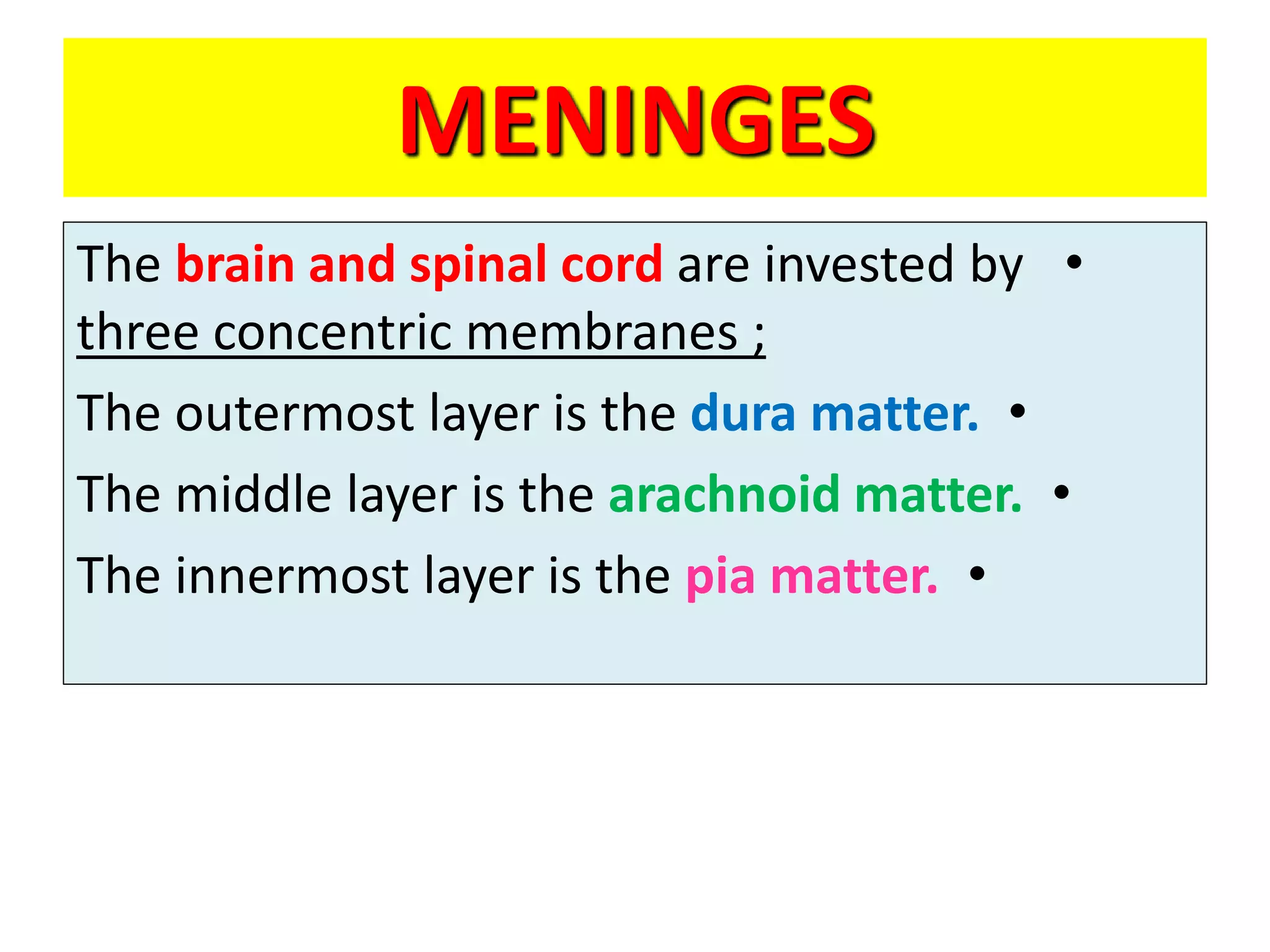
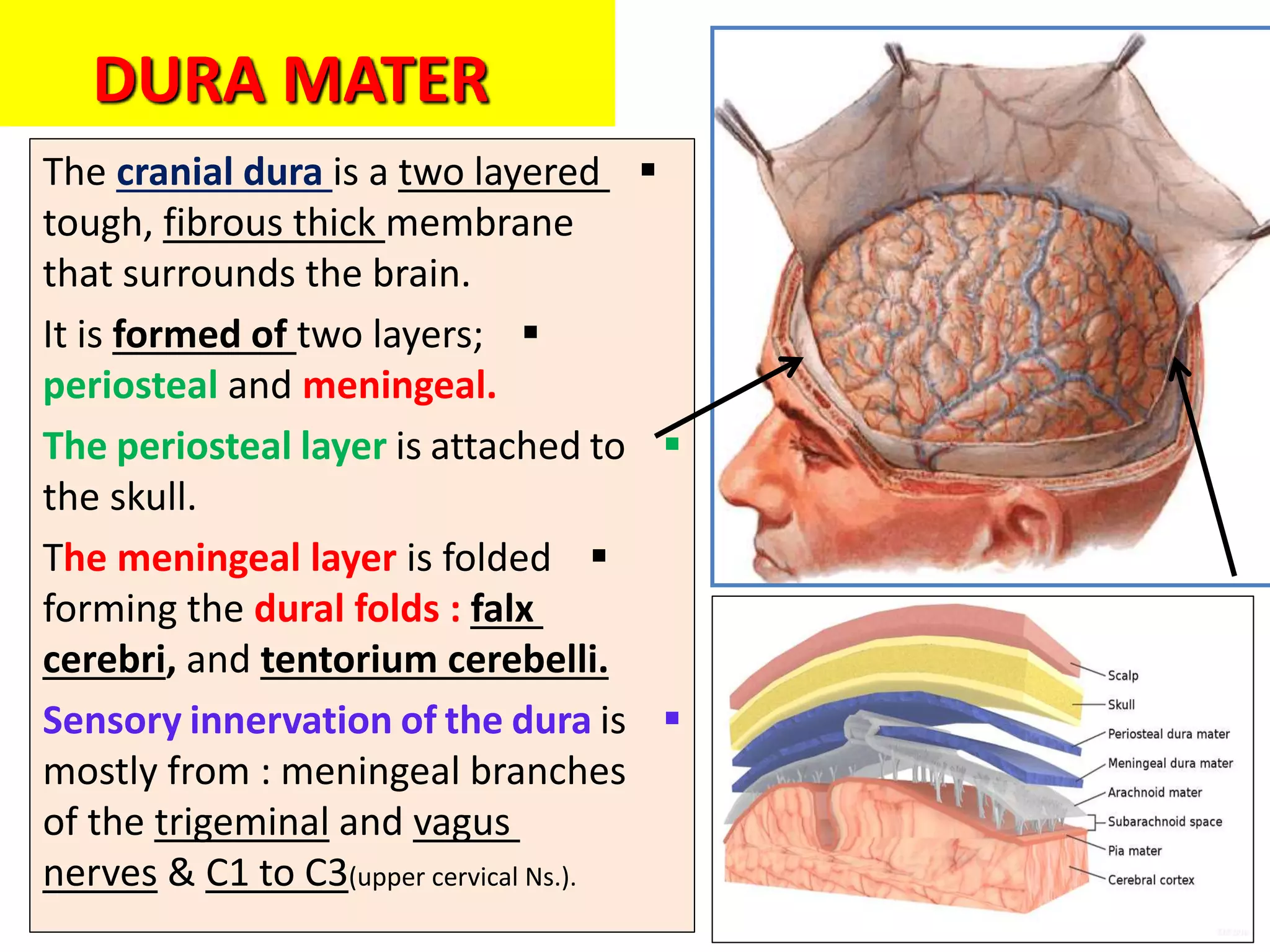
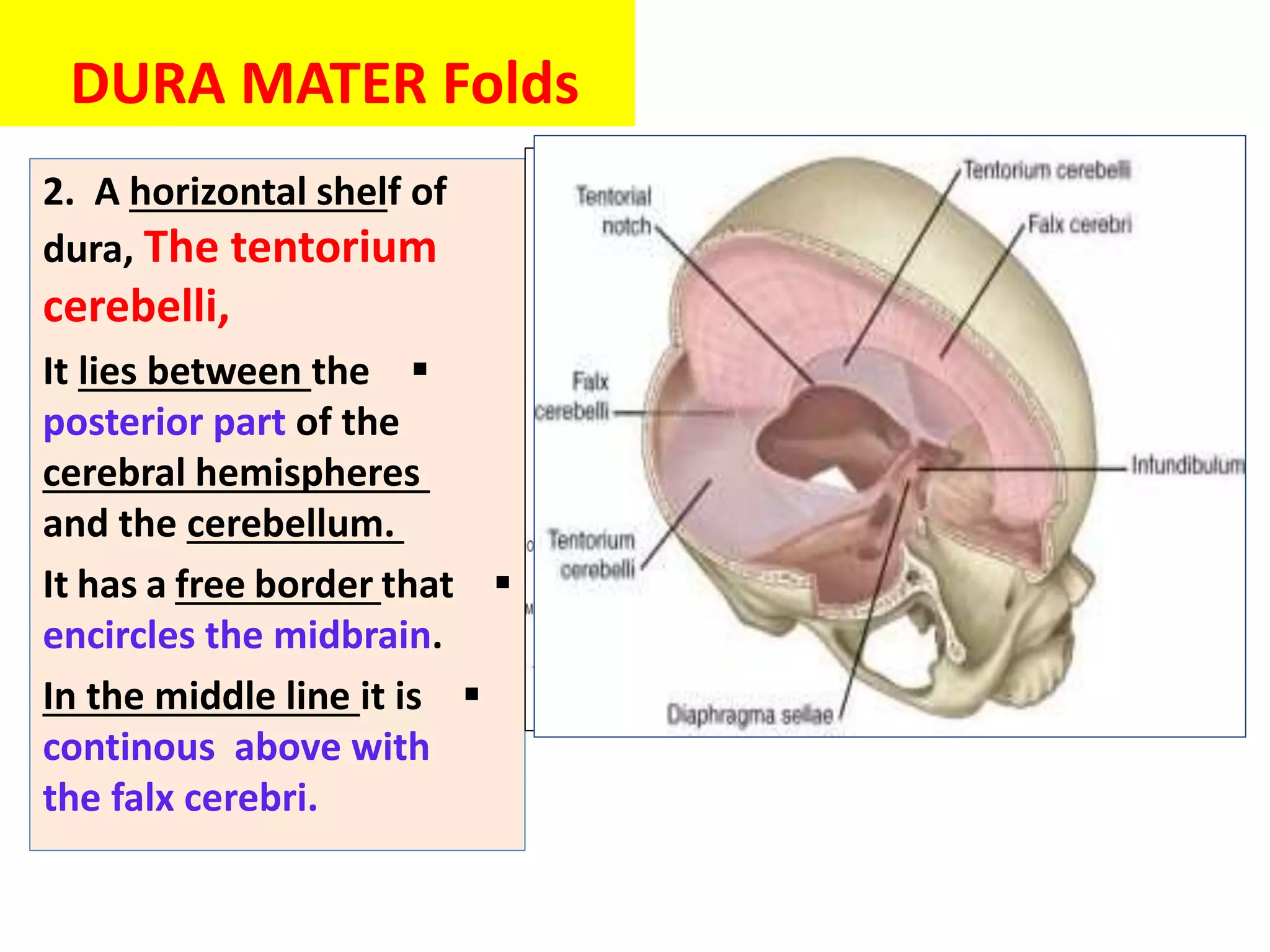
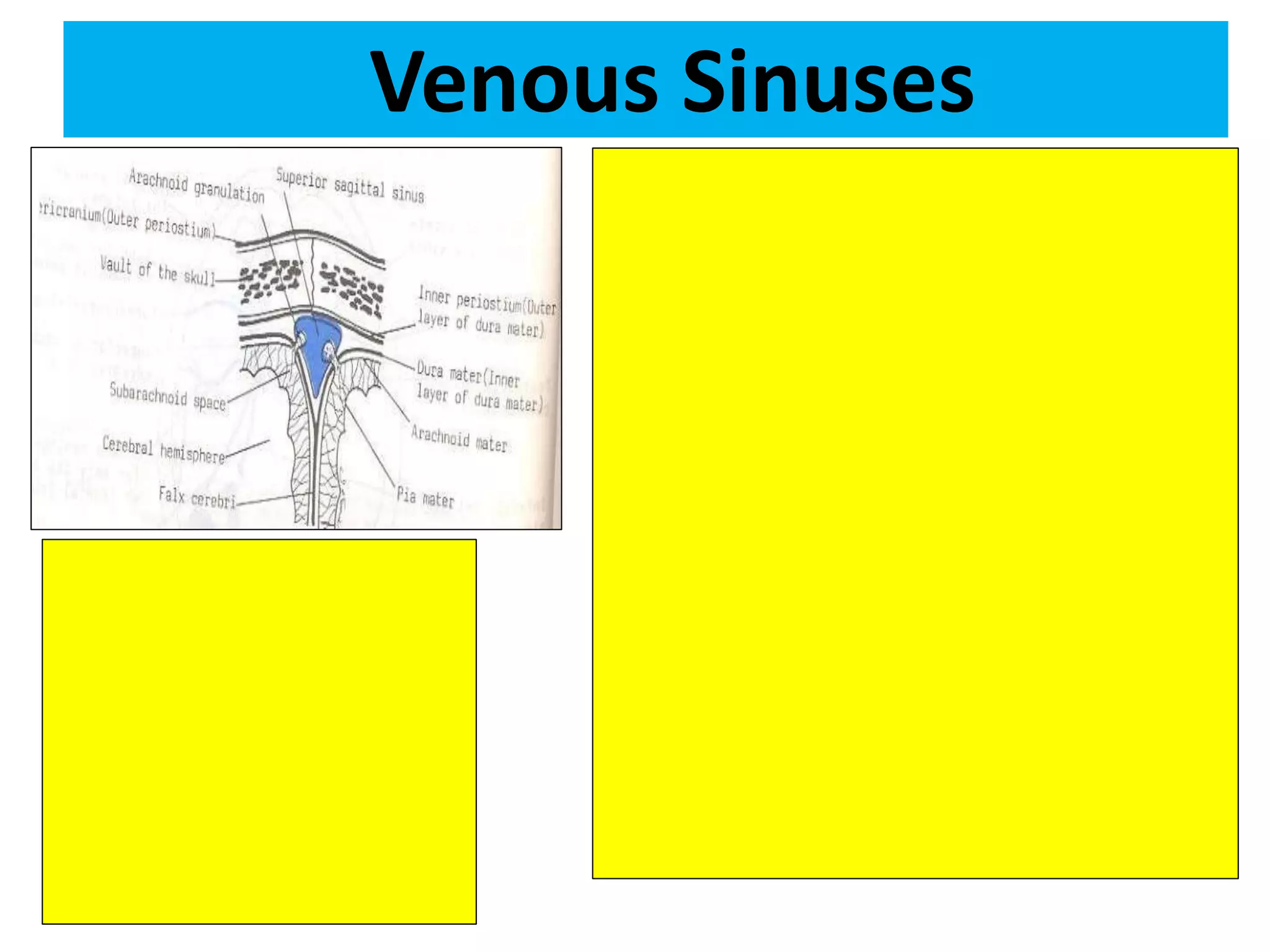
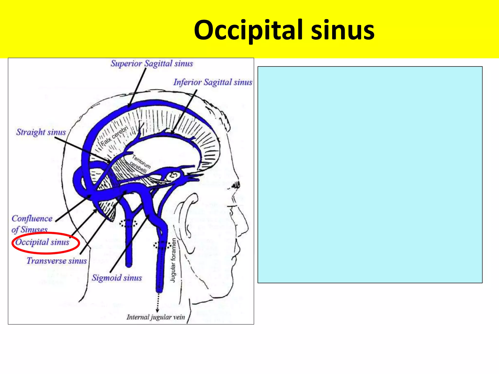
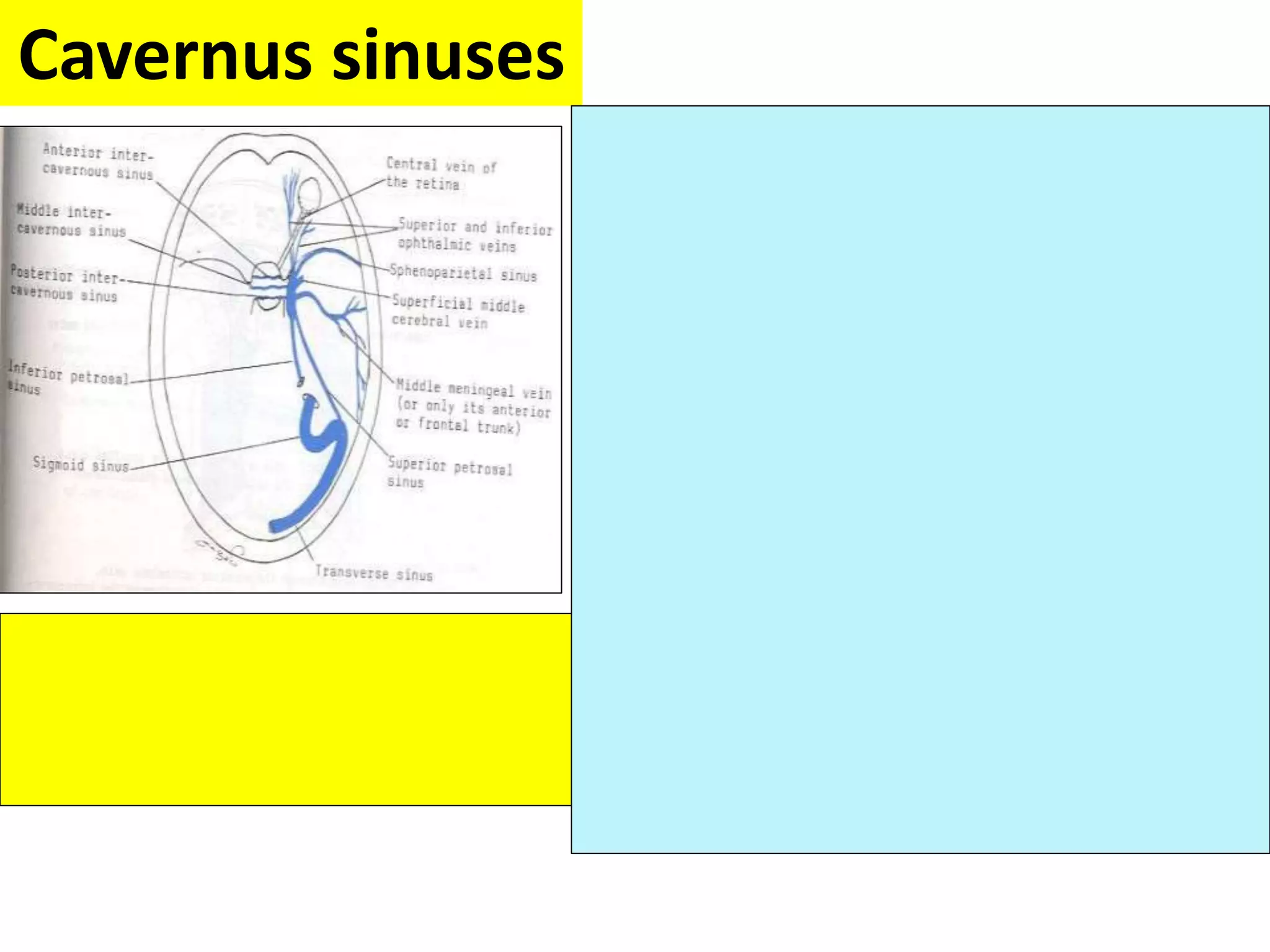
SlideShare a Scribd company logo
1 of 30
Meninges ,ventricles &
CSF
OBJECTIVES
•
By the end of the lecture the student
should be able to:
•
Describe the cerebral meninges & list the main dural
folds.
•
Describe the spinal meninges & locate the level of the
termination of each of them.
•
Describe the importance of the subarachnoid space.
•
List the Ventricular system of the CNS and locate the
site of each of them.
•
Describe the formation, circulation, drainage, and
functions of the CSF.
•
Know some clinical point about the CSF
MENINGES
•
The brain and spinal cord are invested by
three concentric membranes ;
•
The outermost layer is the dura matter.
•
The middle layer is the arachnoid matter.
•
The innermost layer is the pia matter.
DURA MATER

The cranial dura is a two layered
tough, fibrous thick membrane
that surrounds the brain.

It is formed of two layers;
periosteal and meningeal.

The periosteal layer is attached to
the skull.

The meningeal layer is folded
forming the dural folds : falx
cerebri, and tentorium cerebelli.

Sensory innervation of the dura is
mostly from : meningeal branches
of the trigeminal and vagus
nerves & C1 to C3(upper cervical Ns.).
DURA MATER Folds

Two large reflection of dura
extend into the cranial cavity
:
1.The falx cerebri,
In the midline,

It is a vertical sickle-shaped
sheet of dura, extends from
the cranial roof into the great
longitudinal fissure between
the two cerebral
hemispheres.

It has an attached border
adherent to the skull.

And a free border lies above
the corpus callosum.
2. A horizontal shelf of
dura, The tentorium
cerebelli,

It lies between the
posterior part of the
cerebral hemispheres
and the cerebellum.

It has a free border that
encircles the midbrain.

In the middle line it is
continous above with
the falx cerebri.
DURA MATER Folds
Arachnoid Mater& Pia Mater

The arachnoid mater is a soft,
translucent membrane loosely
envelops the brain.

The arachnoid mater is separated
from the dura by a narrow subdural
space.

The pia mater is the innermost,
thin, delicate & highly vascular
membrane that is closely adherent
to the gyri and fitted into the sulci.

Between the pia and arachnoid
mater lies the subarachnoid
space which contains; fibrous
trabechulae, main blood vessels
and CSF.
Subarachnoid
Space

It is varied in depth forming;
subarachnoid cisterns ;
.1
The cisterna magna, or
cerebllomedullary cistern
which lies between the inferior
surface of the cerebellum and
the back of the medulla.
•
At this cistern CSF flows
out of the 4th ventricle via
the 2 lateral aperatures
and median aperature.
Subarachnoid
Space
2. The interpeduncular
cistern, which is located
at the base of the brain,
where the arachnoid
spans between the two
cerebral peduncles of
midbrain.
•
This cistern
contains the optic
chiasma & circulus
arteriosus of Wills.
Spinal meninges

The spinal cord, is invested by three
meningeal coverings: pia mater,
arachnoid mater and dura mater.

The dura matter; The outer covering;
is a thick, tough fibrous membrane.

It envelopes the cord loosely.

It is separated from arachnoid matter
by the subdural space, and from the
bony wall of the vertebral canal by the
epidural space.

The arachnoid matter is a translucent
membrane lies between the pia and
dura,

Between arachnoid and pia lies the
subarachnoid space contains CSF.

The pia matter; The innermost
covering, is a delicate fibrous
membrane closely envelops the cord
and nerve roots.

It is attached through the arachnoid to
the dura by the denticulate ligament.
Spinal meninges

The spinal cord terminates
at level L1-L2, while

The dura and arachnoid
and, subarachnoid space,
continue caudally to S2.

The pia extends
downwards forming the
filum terminale which
pierces the arachnoid and
dural sacs and passes
through the sacral hiatus to
be attached to the back of
the coccyx.
VENTRICULAR SYSTEM

Interconnecting channels within
the CNS.

In the spinal cord; represented
by the central canal.

Within the brain; a system of
ventricles is found.

The central canal of the spinal
cord is continuous upwards to
the forth ventricle.

On each side of the forth
ventricle laterally, lateral recess
extend to open into lateral
aperture (foramen of Luscka),
central defect in its roof
VENTRICULAR SYSTEM

The forth ventricle
is continuous up
with the cerebral
aqueduct, that
opens in the third
ventricle.

The third ventricle
is continuous with
the lateral ventricle
through the
interventricular
foramen (foramen
of Monro).
CEREBROSPINAL FLUID

Present in the ventricular
system, together with the
cranial and spinal
subarachnoid spaces.

It is colourless clear fluid
containing little protein
and few cells.

It is about 150 ml.

It acts as a cushion for
the brain from sudden
movements of the head.

It is produced by the
choroid plexus, which is
located in the lateral,
third & fourth ventricles.

From lateral ventricle it
flows: through the
interventricular foramen
into the 3rd ventricle and,
by way of the cerebral
aqueduct, into the 4th
ventricle.
CEREBROSPINAL FLUID

It leaves the
ventricular system
through the three
apertures of the 4th
ventricle (median
foramen of Magindi &
2 lateral foraminae of
Leushka), to enters the
subarachnoid space.
CEREBROSPINAL FLUID

reabsorbed finally
into the venous
system along

arachnoid villi (small
microscopic herniation of
arachnoid mater), and

arachnoid
granulation
(Macroscopic
multilobulated structures)
that project into the
dural venous
sinuses , mainly
superior saggital
CEREBROSPINAL FLUID
CEREBROSPINAL FLUID
clinical point

The obstruction of the
flow of CSF leads to a
rise in fluid pressure
causing swelling of the
ventricles
(hydrocephalus).

Causes :

Congenital : ( Arnold-Chiari
malformation).

Acquired :

Stenosis of the cerebral aqueduct
by tumor of pineal region.

Obstruction of the interventricular
foramina secondary to tumors,
hemorrhages or infections such as
meningitis

Decompression of
the dilated ventricles
is achieved by
inserting a shunt
connecting the
ventricles to the
jugular vein or the
abdominal
peritoneum.
CEREBROSPINAL FLUID
clinical point
Summary
•
The brain & spinal cord are covered by 3 layers of
meninges : dura, arachnoid & pia mater.
•
The important dural folds inside the brain are the
falax cerebri & tentorium cerebelli.
•
CSF is produced by the choroid plexuses of the
ventricles of the brain : lateral ,3rd & 4th ventricles.
•
CSF circulates in the subarachnoid space.
•
CSF is drained into the dural venous sinuses
principally superior saggital sinus.
•
The subarachnoid space in the spinal cord
terminates at the 2nd sacral vertebra.
•
Obstruction of the flow of CSF as in tumors of the
brain leads to hydrocephalus.
Thank U & Good Luck
Venous Sinuses
Superior Sagittal Sinus
It lies in the upper convex fixed
border of falx cerebri.
It begins at frontal crest &
foramen cecum.
Ends as right transverse sinus.
Confluences of the sinuses :
it is formed by junction of
superior sagittal, straight +
2 transverse sinuses.
Inferior Sagittal Sinus
It lies : in the free lower border
of falx cerebri.
It terminates : by joining great
cerebral vein to form straight
sinus.
Transverse Sinuses
Right sinus is continuous with
the superior sagittal sinus, and
 Left sinus is continuous with the
straight sinus.
Each sinus occupies the attached
margin of tentorium cerebelli,
grooving the occipital bone forming
groove for transverse sinus.
They receive the superior petrosal
sinuses.
They end by becoming sigmoid
sinuses.
Sigmoid Sinuses
Occipital sinus
Cavernus sinuses
It lies in middle cranial fossa on either sides
of hypophyseal fossa.
Relations :
Superiorly : optic chiasma.
Inferiorly : sphenoidal air sinus.
Medially : hypophysis cerebri.
Laterally : temporal bone.
Inside the sinus (in the floor) :
1- Internal carotid artery .
2- Abducent nerve.
In the lateral wall of cavernus sinus :
1-Oculomotor nerve.
2-Trochlear nerve.
3-Ophthalmic nerve of trigeminal N.
4-Maxillary nerve of trigeminal N.
Cavernus sinuses
Superior & inferior Petrosal sinuses

More Related Content

What's hot

Arteries of head and neck; subclavian vessels
Arteries of head and neck; subclavian vesselsArteries of head and neck; subclavian vessels
Arteries of head and neck; subclavian vesselsDr. Mohammad Mahmoud
 
Posterior triangle of the neck
Posterior triangle of the neckPosterior triangle of the neck
Posterior triangle of the neckMan B Paudyal
 
Anatomy of medulla oblangata
Anatomy of  medulla oblangataAnatomy of  medulla oblangata
Anatomy of medulla oblangataravijangid39
 
15 dural venous sinuses
15 dural venous sinuses15 dural venous sinuses
15 dural venous sinusesMed Study
 
Suboccipital triangle
Suboccipital triangleSuboccipital triangle
Suboccipital trianglemgmcri1234
 
14.triangles of the neck and root of the
14.triangles of  the neck and root of the14.triangles of  the neck and root of the
14.triangles of the neck and root of theJenn Ryse
 
Anatomy of brainstem
Anatomy of brainstemAnatomy of brainstem
Anatomy of brainstemMBBS IMS MSU
 
Meninges of brain and spinal cord
Meninges of brain and spinal cordMeninges of brain and spinal cord
Meninges of brain and spinal cordmunnam37
 
Anatomy of the cranial nerves
Anatomy of the cranial nervesAnatomy of the cranial nerves
Anatomy of the cranial nervesMukhtar Khan
 
Cranial nerves xi and xii
Cranial nerves xi and xiiCranial nerves xi and xii
Cranial nerves xi and xiiNeurologyKota
 
Carotid Arteries (Anatomy of the Neck)
Carotid Arteries (Anatomy of the Neck)Carotid Arteries (Anatomy of the Neck)
Carotid Arteries (Anatomy of the Neck)Dr. Sherif Fahmy
 
Mandibular nerve and otic ganglion
Mandibular nerve and otic ganglionMandibular nerve and otic ganglion
Mandibular nerve and otic ganglionmgmcri1234
 
Segments of internal carotid artery
Segments of internal carotid arterySegments of internal carotid artery
Segments of internal carotid arteryANDREA TITUS
 
Neck triangles anatomy
Neck triangles anatomyNeck triangles anatomy
Neck triangles anatomyJamil Anwar
 

What's hot (20)

Arteries of head and neck; subclavian vessels
Arteries of head and neck; subclavian vesselsArteries of head and neck; subclavian vessels
Arteries of head and neck; subclavian vessels
 
Posterior triangle of the neck
Posterior triangle of the neckPosterior triangle of the neck
Posterior triangle of the neck
 
Anatomy of medulla oblangata
Anatomy of  medulla oblangataAnatomy of  medulla oblangata
Anatomy of medulla oblangata
 
Corpus callosum
Corpus callosumCorpus callosum
Corpus callosum
 
15 dural venous sinuses
15 dural venous sinuses15 dural venous sinuses
15 dural venous sinuses
 
ICA anatomy
ICA anatomyICA anatomy
ICA anatomy
 
Lateral Ventricle
Lateral VentricleLateral Ventricle
Lateral Ventricle
 
Suboccipital triangle
Suboccipital triangleSuboccipital triangle
Suboccipital triangle
 
14.triangles of the neck and root of the
14.triangles of  the neck and root of the14.triangles of  the neck and root of the
14.triangles of the neck and root of the
 
The carotid triangle
The carotid triangleThe carotid triangle
The carotid triangle
 
Anatomy of brainstem
Anatomy of brainstemAnatomy of brainstem
Anatomy of brainstem
 
Meninges of brain and spinal cord
Meninges of brain and spinal cordMeninges of brain and spinal cord
Meninges of brain and spinal cord
 
Anatomy of the cranial nerves
Anatomy of the cranial nervesAnatomy of the cranial nerves
Anatomy of the cranial nerves
 
Cranial nerves xi and xii
Cranial nerves xi and xiiCranial nerves xi and xii
Cranial nerves xi and xii
 
Carotid Arteries (Anatomy of the Neck)
Carotid Arteries (Anatomy of the Neck)Carotid Arteries (Anatomy of the Neck)
Carotid Arteries (Anatomy of the Neck)
 
Arteries of the head and neck
Arteries of the head and neckArteries of the head and neck
Arteries of the head and neck
 
Mandibular nerve and otic ganglion
Mandibular nerve and otic ganglionMandibular nerve and otic ganglion
Mandibular nerve and otic ganglion
 
Segments of internal carotid artery
Segments of internal carotid arterySegments of internal carotid artery
Segments of internal carotid artery
 
Neck triangles anatomy
Neck triangles anatomyNeck triangles anatomy
Neck triangles anatomy
 
Ventricles of the brain
Ventricles of the brainVentricles of the brain
Ventricles of the brain
 

Similar to Meninges, Ventricles & CSF: A Concise Overview

19 meninges .pptx
19 meninges .pptx19 meninges .pptx
19 meninges .pptxDrPinkiRai
 
anatomyofmeningesventriclescerebrospinalfluid-100604195832-phpapp02.pdf
anatomyofmeningesventriclescerebrospinalfluid-100604195832-phpapp02.pdfanatomyofmeningesventriclescerebrospinalfluid-100604195832-phpapp02.pdf
anatomyofmeningesventriclescerebrospinalfluid-100604195832-phpapp02.pdfDipaliTalaviya1
 
Meninges, ventricles and cerebro spinal fluid
Meninges, ventricles and cerebro spinal fluidMeninges, ventricles and cerebro spinal fluid
Meninges, ventricles and cerebro spinal fluidSahroz Khan
 
Ventricles of brain
Ventricles of brainVentricles of brain
Ventricles of brainMehdiHayat
 
Meninges covering of the CNS.pdf
Meninges covering of the CNS.pdfMeninges covering of the CNS.pdf
Meninges covering of the CNS.pdfAdebayoAbayomi3
 
Ventricular system of brain & the CSF
Ventricular system of brain & the CSFVentricular system of brain & the CSF
Ventricular system of brain & the CSFABDUL QADEER MEMON
 
2 the central nervous system
2 the central nervous system2 the central nervous system
2 the central nervous systemDAWN V TOMY
 
Contents of vertebral canal .pdf
Contents of vertebral canal .pdfContents of vertebral canal .pdf
Contents of vertebral canal .pdfÖmer aslankan
 
Brain & centers controlling tongue movement
Brain & centers controlling tongue movementBrain & centers controlling tongue movement
Brain & centers controlling tongue movementIndian dental academy
 
Vetricles of brain final.pptx
Vetricles of brain final.pptxVetricles of brain final.pptx
Vetricles of brain final.pptxShilpaNaik50
 
BRAINSTEM-ANATOMICAL DETAILS WITH EXPLANATIOON.pptx
BRAINSTEM-ANATOMICAL DETAILS WITH EXPLANATIOON.pptxBRAINSTEM-ANATOMICAL DETAILS WITH EXPLANATIOON.pptx
BRAINSTEM-ANATOMICAL DETAILS WITH EXPLANATIOON.pptxDrRashidKaziMahbubur
 
envelopes-of-the-brain
envelopes-of-the-brainenvelopes-of-the-brain
envelopes-of-the-brainYoAmoNYC
 
7938186 envelopes-of-the-brain
7938186 envelopes-of-the-brain7938186 envelopes-of-the-brain
7938186 envelopes-of-the-brainabctutor
 

Similar to Meninges, Ventricles & CSF: A Concise Overview (20)

19 meninges .pptx
19 meninges .pptx19 meninges .pptx
19 meninges .pptx
 
Meninges, ventricle and CSF
Meninges, ventricle and CSFMeninges, ventricle and CSF
Meninges, ventricle and CSF
 
anatomyofmeningesventriclescerebrospinalfluid-100604195832-phpapp02.pdf
anatomyofmeningesventriclescerebrospinalfluid-100604195832-phpapp02.pdfanatomyofmeningesventriclescerebrospinalfluid-100604195832-phpapp02.pdf
anatomyofmeningesventriclescerebrospinalfluid-100604195832-phpapp02.pdf
 
Meninges, ventricles and cerebro spinal fluid
Meninges, ventricles and cerebro spinal fluidMeninges, ventricles and cerebro spinal fluid
Meninges, ventricles and cerebro spinal fluid
 
Cerebrospinal fluid.pptx
Cerebrospinal  fluid.pptxCerebrospinal  fluid.pptx
Cerebrospinal fluid.pptx
 
ventricle
ventricleventricle
ventricle
 
Ventricles
VentriclesVentricles
Ventricles
 
Ventricles of brain
Ventricles of brainVentricles of brain
Ventricles of brain
 
Meninges covering of the CNS.pdf
Meninges covering of the CNS.pdfMeninges covering of the CNS.pdf
Meninges covering of the CNS.pdf
 
Ventricular system of brain & the CSF
Ventricular system of brain & the CSFVentricular system of brain & the CSF
Ventricular system of brain & the CSF
 
2 the central nervous system
2 the central nervous system2 the central nervous system
2 the central nervous system
 
Contents of vertebral canal .pdf
Contents of vertebral canal .pdfContents of vertebral canal .pdf
Contents of vertebral canal .pdf
 
Brain & centers controlling tongue movement
Brain & centers controlling tongue movementBrain & centers controlling tongue movement
Brain & centers controlling tongue movement
 
Vetricles of brain final.pptx
Vetricles of brain final.pptxVetricles of brain final.pptx
Vetricles of brain final.pptx
 
Meninges - Anatomy of the Meninges & CSF
Meninges - Anatomy of the Meninges & CSFMeninges - Anatomy of the Meninges & CSF
Meninges - Anatomy of the Meninges & CSF
 
Meninges.pptx
Meninges.pptxMeninges.pptx
Meninges.pptx
 
The meninges
The meningesThe meninges
The meninges
 
BRAINSTEM-ANATOMICAL DETAILS WITH EXPLANATIOON.pptx
BRAINSTEM-ANATOMICAL DETAILS WITH EXPLANATIOON.pptxBRAINSTEM-ANATOMICAL DETAILS WITH EXPLANATIOON.pptx
BRAINSTEM-ANATOMICAL DETAILS WITH EXPLANATIOON.pptx
 
envelopes-of-the-brain
envelopes-of-the-brainenvelopes-of-the-brain
envelopes-of-the-brain
 
7938186 envelopes-of-the-brain
7938186 envelopes-of-the-brain7938186 envelopes-of-the-brain
7938186 envelopes-of-the-brain
 

More from RupaSingh83

Skeletal System and division of axial and appendicular
Skeletal System and division of axial and appendicularSkeletal System and division of axial and appendicular
Skeletal System and division of axial and appendicularRupaSingh83
 
Special Senses (Eye -vision & Ear-hearing).
Special Senses (Eye -vision & Ear-hearing).Special Senses (Eye -vision & Ear-hearing).
Special Senses (Eye -vision & Ear-hearing).RupaSingh83
 
Special Senses eye, ear , nose, tongue ppt
Special Senses eye, ear , nose, tongue pptSpecial Senses eye, ear , nose, tongue ppt
Special Senses eye, ear , nose, tongue pptRupaSingh83
 
Peripheral nervous system,Central nervous system
Peripheral nervous system,Central nervous systemPeripheral nervous system,Central nervous system
Peripheral nervous system,Central nervous systemRupaSingh83
 
THE INTEGUMENTARY SYSTEM.pptx
THE INTEGUMENTARY SYSTEM.pptxTHE INTEGUMENTARY SYSTEM.pptx
THE INTEGUMENTARY SYSTEM.pptxRupaSingh83
 
Introduction to HAP.pptx
Introduction to HAP.pptxIntroduction to HAP.pptx
Introduction to HAP.pptxRupaSingh83
 
RHEUMATOID ARTHRITIS.pptx
RHEUMATOID ARTHRITIS.pptxRHEUMATOID ARTHRITIS.pptx
RHEUMATOID ARTHRITIS.pptxRupaSingh83
 
ANTI GOUT DRUGS.....pptx
ANTI GOUT DRUGS.....pptxANTI GOUT DRUGS.....pptx
ANTI GOUT DRUGS.....pptxRupaSingh83
 
HAEMATINICS.pptx
HAEMATINICS.pptxHAEMATINICS.pptx
HAEMATINICS.pptxRupaSingh83
 
Coagulants & Anti- Coagulants.pptx
Coagulants & Anti- Coagulants.pptxCoagulants & Anti- Coagulants.pptx
Coagulants & Anti- Coagulants.pptxRupaSingh83
 
REPRODUCTIVE SYSTEM.pptx
REPRODUCTIVE SYSTEM.pptxREPRODUCTIVE SYSTEM.pptx
REPRODUCTIVE SYSTEM.pptxRupaSingh83
 
178-Anatomy-Reproductive-System.ppt
178-Anatomy-Reproductive-System.ppt178-Anatomy-Reproductive-System.ppt
178-Anatomy-Reproductive-System.pptRupaSingh83
 
haematinics-160713185907.pptx
haematinics-160713185907.pptxhaematinics-160713185907.pptx
haematinics-160713185907.pptxRupaSingh83
 
lecture-4 [Autosaved].pptx
lecture-4 [Autosaved].pptxlecture-4 [Autosaved].pptx
lecture-4 [Autosaved].pptxRupaSingh83
 
Reproduction.pptx
Reproduction.pptxReproduction.pptx
Reproduction.pptxRupaSingh83
 
The Digestive System.pptx
The Digestive System.pptxThe Digestive System.pptx
The Digestive System.pptxRupaSingh83
 
The Digestive System.pptx
The Digestive System.pptxThe Digestive System.pptx
The Digestive System.pptxRupaSingh83
 

More from RupaSingh83 (20)

Skeletal System and division of axial and appendicular
Skeletal System and division of axial and appendicularSkeletal System and division of axial and appendicular
Skeletal System and division of axial and appendicular
 
Special Senses (Eye -vision & Ear-hearing).
Special Senses (Eye -vision & Ear-hearing).Special Senses (Eye -vision & Ear-hearing).
Special Senses (Eye -vision & Ear-hearing).
 
Special Senses eye, ear , nose, tongue ppt
Special Senses eye, ear , nose, tongue pptSpecial Senses eye, ear , nose, tongue ppt
Special Senses eye, ear , nose, tongue ppt
 
Peripheral nervous system,Central nervous system
Peripheral nervous system,Central nervous systemPeripheral nervous system,Central nervous system
Peripheral nervous system,Central nervous system
 
THE INTEGUMENTARY SYSTEM.pptx
THE INTEGUMENTARY SYSTEM.pptxTHE INTEGUMENTARY SYSTEM.pptx
THE INTEGUMENTARY SYSTEM.pptx
 
Introduction to HAP.pptx
Introduction to HAP.pptxIntroduction to HAP.pptx
Introduction to HAP.pptx
 
RHEUMATOID ARTHRITIS.pptx
RHEUMATOID ARTHRITIS.pptxRHEUMATOID ARTHRITIS.pptx
RHEUMATOID ARTHRITIS.pptx
 
NSAID.....pptx
NSAID.....pptxNSAID.....pptx
NSAID.....pptx
 
ANTI GOUT DRUGS.....pptx
ANTI GOUT DRUGS.....pptxANTI GOUT DRUGS.....pptx
ANTI GOUT DRUGS.....pptx
 
HAEMATINICS.pptx
HAEMATINICS.pptxHAEMATINICS.pptx
HAEMATINICS.pptx
 
Coagulants & Anti- Coagulants.pptx
Coagulants & Anti- Coagulants.pptxCoagulants & Anti- Coagulants.pptx
Coagulants & Anti- Coagulants.pptx
 
REPRODUCTIVE SYSTEM.pptx
REPRODUCTIVE SYSTEM.pptxREPRODUCTIVE SYSTEM.pptx
REPRODUCTIVE SYSTEM.pptx
 
178-Anatomy-Reproductive-System.ppt
178-Anatomy-Reproductive-System.ppt178-Anatomy-Reproductive-System.ppt
178-Anatomy-Reproductive-System.ppt
 
haematinics-160713185907.pptx
haematinics-160713185907.pptxhaematinics-160713185907.pptx
haematinics-160713185907.pptx
 
genetics-.pptx
genetics-.pptxgenetics-.pptx
genetics-.pptx
 
lecture-4.pptx
lecture-4.pptxlecture-4.pptx
lecture-4.pptx
 
lecture-4 [Autosaved].pptx
lecture-4 [Autosaved].pptxlecture-4 [Autosaved].pptx
lecture-4 [Autosaved].pptx
 
Reproduction.pptx
Reproduction.pptxReproduction.pptx
Reproduction.pptx
 
The Digestive System.pptx
The Digestive System.pptxThe Digestive System.pptx
The Digestive System.pptx
 
The Digestive System.pptx
The Digestive System.pptxThe Digestive System.pptx
The Digestive System.pptx
 

Recently uploaded

A Critique of the Proposed National Education Policy Reform
A Critique of the Proposed National Education Policy ReformA Critique of the Proposed National Education Policy Reform
A Critique of the Proposed National Education Policy ReformChameera Dedduwage
 
1029 - Danh muc Sach Giao Khoa 10 . pdf
1029 -  Danh muc Sach Giao Khoa 10 . pdf1029 -  Danh muc Sach Giao Khoa 10 . pdf
1029 - Danh muc Sach Giao Khoa 10 . pdfQucHHunhnh
 
Russian Call Girls in Andheri Airport Mumbai WhatsApp 9167673311 💞 Full Nigh...
Russian Call Girls in Andheri Airport Mumbai WhatsApp  9167673311 💞 Full Nigh...Russian Call Girls in Andheri Airport Mumbai WhatsApp  9167673311 💞 Full Nigh...
Russian Call Girls in Andheri Airport Mumbai WhatsApp 9167673311 💞 Full Nigh...Pooja Nehwal
 
Advanced Views - Calendar View in Odoo 17
Advanced Views - Calendar View in Odoo 17Advanced Views - Calendar View in Odoo 17
Advanced Views - Calendar View in Odoo 17Celine George
 
BASLIQ CURRENT LOOKBOOK LOOKBOOK(1) (1).pdf
BASLIQ CURRENT LOOKBOOK  LOOKBOOK(1) (1).pdfBASLIQ CURRENT LOOKBOOK  LOOKBOOK(1) (1).pdf
BASLIQ CURRENT LOOKBOOK LOOKBOOK(1) (1).pdfSoniaTolstoy
 
Measures of Central Tendency: Mean, Median and Mode
Measures of Central Tendency: Mean, Median and ModeMeasures of Central Tendency: Mean, Median and Mode
Measures of Central Tendency: Mean, Median and ModeThiyagu K
 
The basics of sentences session 2pptx copy.pptx
The basics of sentences session 2pptx copy.pptxThe basics of sentences session 2pptx copy.pptx
The basics of sentences session 2pptx copy.pptxheathfieldcps1
 
Student login on Anyboli platform.helpin
Student login on Anyboli platform.helpinStudent login on Anyboli platform.helpin
Student login on Anyboli platform.helpinRaunakKeshri1
 
Separation of Lanthanides/ Lanthanides and Actinides
Separation of Lanthanides/ Lanthanides and ActinidesSeparation of Lanthanides/ Lanthanides and Actinides
Separation of Lanthanides/ Lanthanides and ActinidesFatimaKhan178732
 
Measures of Dispersion and Variability: Range, QD, AD and SD
Measures of Dispersion and Variability: Range, QD, AD and SDMeasures of Dispersion and Variability: Range, QD, AD and SD
Measures of Dispersion and Variability: Range, QD, AD and SDThiyagu K
 
Arihant handbook biology for class 11 .pdf
Arihant handbook biology for class 11 .pdfArihant handbook biology for class 11 .pdf
Arihant handbook biology for class 11 .pdfchloefrazer622
 
Q4-W6-Restating Informational Text Grade 3
Q4-W6-Restating Informational Text Grade 3Q4-W6-Restating Informational Text Grade 3
Q4-W6-Restating Informational Text Grade 3JemimahLaneBuaron
 
The Most Excellent Way | 1 Corinthians 13
The Most Excellent Way | 1 Corinthians 13The Most Excellent Way | 1 Corinthians 13
The Most Excellent Way | 1 Corinthians 13Steve Thomason
 
Ecosystem Interactions Class Discussion Presentation in Blue Green Lined Styl...
Ecosystem Interactions Class Discussion Presentation in Blue Green Lined Styl...Ecosystem Interactions Class Discussion Presentation in Blue Green Lined Styl...
Ecosystem Interactions Class Discussion Presentation in Blue Green Lined Styl...fonyou31
 
9548086042 for call girls in Indira Nagar with room service
9548086042  for call girls in Indira Nagar  with room service9548086042  for call girls in Indira Nagar  with room service
9548086042 for call girls in Indira Nagar with room servicediscovermytutordmt
 
Web & Social Media Analytics Previous Year Question Paper.pdf
Web & Social Media Analytics Previous Year Question Paper.pdfWeb & Social Media Analytics Previous Year Question Paper.pdf
Web & Social Media Analytics Previous Year Question Paper.pdfJayanti Pande
 
Beyond the EU: DORA and NIS 2 Directive's Global Impact
Beyond the EU: DORA and NIS 2 Directive's Global ImpactBeyond the EU: DORA and NIS 2 Directive's Global Impact
Beyond the EU: DORA and NIS 2 Directive's Global ImpactPECB
 

Recently uploaded (20)

A Critique of the Proposed National Education Policy Reform
A Critique of the Proposed National Education Policy ReformA Critique of the Proposed National Education Policy Reform
A Critique of the Proposed National Education Policy Reform
 
1029 - Danh muc Sach Giao Khoa 10 . pdf
1029 -  Danh muc Sach Giao Khoa 10 . pdf1029 -  Danh muc Sach Giao Khoa 10 . pdf
1029 - Danh muc Sach Giao Khoa 10 . pdf
 
Russian Call Girls in Andheri Airport Mumbai WhatsApp 9167673311 💞 Full Nigh...
Russian Call Girls in Andheri Airport Mumbai WhatsApp  9167673311 💞 Full Nigh...Russian Call Girls in Andheri Airport Mumbai WhatsApp  9167673311 💞 Full Nigh...
Russian Call Girls in Andheri Airport Mumbai WhatsApp 9167673311 💞 Full Nigh...
 
Advanced Views - Calendar View in Odoo 17
Advanced Views - Calendar View in Odoo 17Advanced Views - Calendar View in Odoo 17
Advanced Views - Calendar View in Odoo 17
 
BASLIQ CURRENT LOOKBOOK LOOKBOOK(1) (1).pdf
BASLIQ CURRENT LOOKBOOK  LOOKBOOK(1) (1).pdfBASLIQ CURRENT LOOKBOOK  LOOKBOOK(1) (1).pdf
BASLIQ CURRENT LOOKBOOK LOOKBOOK(1) (1).pdf
 
Measures of Central Tendency: Mean, Median and Mode
Measures of Central Tendency: Mean, Median and ModeMeasures of Central Tendency: Mean, Median and Mode
Measures of Central Tendency: Mean, Median and Mode
 
The basics of sentences session 2pptx copy.pptx
The basics of sentences session 2pptx copy.pptxThe basics of sentences session 2pptx copy.pptx
The basics of sentences session 2pptx copy.pptx
 
Student login on Anyboli platform.helpin
Student login on Anyboli platform.helpinStudent login on Anyboli platform.helpin
Student login on Anyboli platform.helpin
 
Separation of Lanthanides/ Lanthanides and Actinides
Separation of Lanthanides/ Lanthanides and ActinidesSeparation of Lanthanides/ Lanthanides and Actinides
Separation of Lanthanides/ Lanthanides and Actinides
 
Measures of Dispersion and Variability: Range, QD, AD and SD
Measures of Dispersion and Variability: Range, QD, AD and SDMeasures of Dispersion and Variability: Range, QD, AD and SD
Measures of Dispersion and Variability: Range, QD, AD and SD
 
Arihant handbook biology for class 11 .pdf
Arihant handbook biology for class 11 .pdfArihant handbook biology for class 11 .pdf
Arihant handbook biology for class 11 .pdf
 
Advance Mobile Application Development class 07
Advance Mobile Application Development class 07Advance Mobile Application Development class 07
Advance Mobile Application Development class 07
 
Q4-W6-Restating Informational Text Grade 3
Q4-W6-Restating Informational Text Grade 3Q4-W6-Restating Informational Text Grade 3
Q4-W6-Restating Informational Text Grade 3
 
The Most Excellent Way | 1 Corinthians 13
The Most Excellent Way | 1 Corinthians 13The Most Excellent Way | 1 Corinthians 13
The Most Excellent Way | 1 Corinthians 13
 
Ecosystem Interactions Class Discussion Presentation in Blue Green Lined Styl...
Ecosystem Interactions Class Discussion Presentation in Blue Green Lined Styl...Ecosystem Interactions Class Discussion Presentation in Blue Green Lined Styl...
Ecosystem Interactions Class Discussion Presentation in Blue Green Lined Styl...
 
9548086042 for call girls in Indira Nagar with room service
9548086042  for call girls in Indira Nagar  with room service9548086042  for call girls in Indira Nagar  with room service
9548086042 for call girls in Indira Nagar with room service
 
Mattingly "AI & Prompt Design: Structured Data, Assistants, & RAG"
Mattingly "AI & Prompt Design: Structured Data, Assistants, & RAG"Mattingly "AI & Prompt Design: Structured Data, Assistants, & RAG"
Mattingly "AI & Prompt Design: Structured Data, Assistants, & RAG"
 
INDIA QUIZ 2024 RLAC DELHI UNIVERSITY.pptx
INDIA QUIZ 2024 RLAC DELHI UNIVERSITY.pptxINDIA QUIZ 2024 RLAC DELHI UNIVERSITY.pptx
INDIA QUIZ 2024 RLAC DELHI UNIVERSITY.pptx
 
Web & Social Media Analytics Previous Year Question Paper.pdf
Web & Social Media Analytics Previous Year Question Paper.pdfWeb & Social Media Analytics Previous Year Question Paper.pdf
Web & Social Media Analytics Previous Year Question Paper.pdf
 
Beyond the EU: DORA and NIS 2 Directive's Global Impact
Beyond the EU: DORA and NIS 2 Directive's Global ImpactBeyond the EU: DORA and NIS 2 Directive's Global Impact
Beyond the EU: DORA and NIS 2 Directive's Global Impact
 

Meninges, Ventricles & CSF: A Concise Overview

  • 2. OBJECTIVES • By the end of the lecture the student should be able to: • Describe the cerebral meninges & list the main dural folds. • Describe the spinal meninges & locate the level of the termination of each of them. • Describe the importance of the subarachnoid space. • List the Ventricular system of the CNS and locate the site of each of them. • Describe the formation, circulation, drainage, and functions of the CSF. • Know some clinical point about the CSF
  • 3. MENINGES • The brain and spinal cord are invested by three concentric membranes ; • The outermost layer is the dura matter. • The middle layer is the arachnoid matter. • The innermost layer is the pia matter.
  • 4. DURA MATER  The cranial dura is a two layered tough, fibrous thick membrane that surrounds the brain.  It is formed of two layers; periosteal and meningeal.  The periosteal layer is attached to the skull.  The meningeal layer is folded forming the dural folds : falx cerebri, and tentorium cerebelli.  Sensory innervation of the dura is mostly from : meningeal branches of the trigeminal and vagus nerves & C1 to C3(upper cervical Ns.).
  • 5. DURA MATER Folds  Two large reflection of dura extend into the cranial cavity : 1.The falx cerebri, In the midline,  It is a vertical sickle-shaped sheet of dura, extends from the cranial roof into the great longitudinal fissure between the two cerebral hemispheres.  It has an attached border adherent to the skull.  And a free border lies above the corpus callosum.
  • 6. 2. A horizontal shelf of dura, The tentorium cerebelli,  It lies between the posterior part of the cerebral hemispheres and the cerebellum.  It has a free border that encircles the midbrain.  In the middle line it is continous above with the falx cerebri. DURA MATER Folds
  • 7. Arachnoid Mater& Pia Mater  The arachnoid mater is a soft, translucent membrane loosely envelops the brain.  The arachnoid mater is separated from the dura by a narrow subdural space.  The pia mater is the innermost, thin, delicate & highly vascular membrane that is closely adherent to the gyri and fitted into the sulci.  Between the pia and arachnoid mater lies the subarachnoid space which contains; fibrous trabechulae, main blood vessels and CSF.
  • 8. Subarachnoid Space  It is varied in depth forming; subarachnoid cisterns ; .1 The cisterna magna, or cerebllomedullary cistern which lies between the inferior surface of the cerebellum and the back of the medulla. • At this cistern CSF flows out of the 4th ventricle via the 2 lateral aperatures and median aperature.
  • 9. Subarachnoid Space 2. The interpeduncular cistern, which is located at the base of the brain, where the arachnoid spans between the two cerebral peduncles of midbrain. • This cistern contains the optic chiasma & circulus arteriosus of Wills.
  • 10. Spinal meninges  The spinal cord, is invested by three meningeal coverings: pia mater, arachnoid mater and dura mater.  The dura matter; The outer covering; is a thick, tough fibrous membrane.  It envelopes the cord loosely.  It is separated from arachnoid matter by the subdural space, and from the bony wall of the vertebral canal by the epidural space.  The arachnoid matter is a translucent membrane lies between the pia and dura,  Between arachnoid and pia lies the subarachnoid space contains CSF.  The pia matter; The innermost covering, is a delicate fibrous membrane closely envelops the cord and nerve roots.  It is attached through the arachnoid to the dura by the denticulate ligament.
  • 11. Spinal meninges  The spinal cord terminates at level L1-L2, while  The dura and arachnoid and, subarachnoid space, continue caudally to S2.  The pia extends downwards forming the filum terminale which pierces the arachnoid and dural sacs and passes through the sacral hiatus to be attached to the back of the coccyx.
  • 12. VENTRICULAR SYSTEM  Interconnecting channels within the CNS.  In the spinal cord; represented by the central canal.  Within the brain; a system of ventricles is found.  The central canal of the spinal cord is continuous upwards to the forth ventricle.  On each side of the forth ventricle laterally, lateral recess extend to open into lateral aperture (foramen of Luscka), central defect in its roof
  • 13. VENTRICULAR SYSTEM  The forth ventricle is continuous up with the cerebral aqueduct, that opens in the third ventricle.  The third ventricle is continuous with the lateral ventricle through the interventricular foramen (foramen of Monro).
  • 14. CEREBROSPINAL FLUID  Present in the ventricular system, together with the cranial and spinal subarachnoid spaces.  It is colourless clear fluid containing little protein and few cells.  It is about 150 ml.  It acts as a cushion for the brain from sudden movements of the head.
  • 15.  It is produced by the choroid plexus, which is located in the lateral, third & fourth ventricles.  From lateral ventricle it flows: through the interventricular foramen into the 3rd ventricle and, by way of the cerebral aqueduct, into the 4th ventricle. CEREBROSPINAL FLUID
  • 16.  It leaves the ventricular system through the three apertures of the 4th ventricle (median foramen of Magindi & 2 lateral foraminae of Leushka), to enters the subarachnoid space. CEREBROSPINAL FLUID
  • 17.  reabsorbed finally into the venous system along  arachnoid villi (small microscopic herniation of arachnoid mater), and  arachnoid granulation (Macroscopic multilobulated structures) that project into the dural venous sinuses , mainly superior saggital CEREBROSPINAL FLUID
  • 18. CEREBROSPINAL FLUID clinical point  The obstruction of the flow of CSF leads to a rise in fluid pressure causing swelling of the ventricles (hydrocephalus).  Causes :  Congenital : ( Arnold-Chiari malformation).  Acquired :  Stenosis of the cerebral aqueduct by tumor of pineal region.  Obstruction of the interventricular foramina secondary to tumors, hemorrhages or infections such as meningitis
  • 19.  Decompression of the dilated ventricles is achieved by inserting a shunt connecting the ventricles to the jugular vein or the abdominal peritoneum. CEREBROSPINAL FLUID clinical point
  • 20. Summary • The brain & spinal cord are covered by 3 layers of meninges : dura, arachnoid & pia mater. • The important dural folds inside the brain are the falax cerebri & tentorium cerebelli. • CSF is produced by the choroid plexuses of the ventricles of the brain : lateral ,3rd & 4th ventricles. • CSF circulates in the subarachnoid space. • CSF is drained into the dural venous sinuses principally superior saggital sinus. • The subarachnoid space in the spinal cord terminates at the 2nd sacral vertebra. • Obstruction of the flow of CSF as in tumors of the brain leads to hydrocephalus.
  • 21. Thank U & Good Luck
  • 23. Superior Sagittal Sinus It lies in the upper convex fixed border of falx cerebri. It begins at frontal crest & foramen cecum. Ends as right transverse sinus. Confluences of the sinuses : it is formed by junction of superior sagittal, straight + 2 transverse sinuses.
  • 24. Inferior Sagittal Sinus It lies : in the free lower border of falx cerebri. It terminates : by joining great cerebral vein to form straight sinus.
  • 25. Transverse Sinuses Right sinus is continuous with the superior sagittal sinus, and  Left sinus is continuous with the straight sinus. Each sinus occupies the attached margin of tentorium cerebelli, grooving the occipital bone forming groove for transverse sinus. They receive the superior petrosal sinuses. They end by becoming sigmoid sinuses.
  • 28. Cavernus sinuses It lies in middle cranial fossa on either sides of hypophyseal fossa. Relations : Superiorly : optic chiasma. Inferiorly : sphenoidal air sinus. Medially : hypophysis cerebri. Laterally : temporal bone. Inside the sinus (in the floor) : 1- Internal carotid artery . 2- Abducent nerve. In the lateral wall of cavernus sinus : 1-Oculomotor nerve. 2-Trochlear nerve. 3-Ophthalmic nerve of trigeminal N. 4-Maxillary nerve of trigeminal N.
  • 30. Superior & inferior Petrosal sinuses