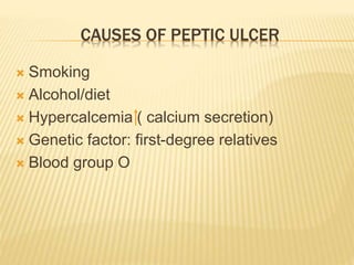
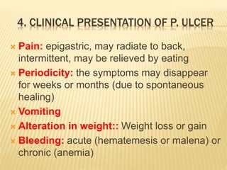
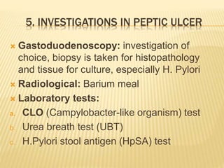
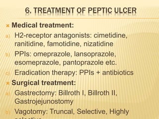
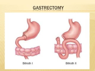
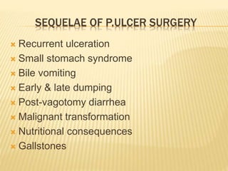
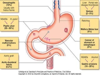
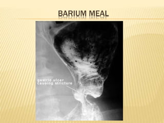
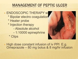
Russian Call Girls in Bangalore Manisha 7001305949 Independent Escort Service...
Peptic ulcer & upper gi bleeding
1. PEPTIC ULCER
&
UPPER GI BLEEDING
By
Dr. Abdul Qadeer
MBBS; FCPS; FICS
Assistant Professor in General Surgery
King Faisal University College of Medicine
Kingdom of Saudi Arabia
2. OBJECTIVES
1. Definition of peptic ulcer
2. Epidemiology of peptic ulcer
3. Causes of peptic ulcer
4. Clinical presentation
5. Investigations
6. Treatment
7. Definition of upper GI bleeding
8. Epidemiology of upper GI bleeding
9. Causes
10. Clinical presentation
11. Investigations
12. Treatment
3. 1. DEFINITION OF PEPTIC ULCER
A lesion in the lining (mucosa) of the digestive
tract, typically in the stomach or duodenum,
caused by the digestive action of pepsin and
stomach acid.
4. 2. EPIDEMIOLOGY OF PEPTIC ULCER
10% of the population has ulcers
Annual incidence of symptomatic peptic ulcer
is about 0.3%
Duodenal ulcers are 4 times as common as
gastric ulcers and occur at the duodenal cap
Gastric ulcers mostly occur in the lesser
curvature. Usually benign. 5% are malignant
May occur on the stoma following gastric
surgery, esophagus & Meckel’s diverticulum
having ectopic gastric tissue
5. In general, the ulcer occurs at a junction
between different types of epithelia, the ulcer
occurring in the epithelium least resistant to
acid damage
Gastric malignancy is common in Japan,
Chile, Finland & Iceland due to
environmental & diet factors.
6. 3. CAUSES OF PEPTIC ULCER
Higher pepsin/gastric acid levels, though the
ulcers have been seen in patients having
normal levels
Gastrinoma (Zollinger-Ellison syndrome)
Helicobacter pylori in 80-95% cases
Consumption of NSAIDs
Stress i.e. emotional, trauma, surgical
Injury or death of mucus-producing cells
8. CAUSES OF PEPTIC ULCER
Smoking
Alcohol/diet
Hypercalcemia ( calcium secretion)
Genetic factor: first-degree relatives
Blood group O
9. 4. CLINICAL PRESENTATION OF P. ULCER
Pain: epigastric, may radiate to back,
intermittent, may be relieved by eating
Periodicity: the symptoms may disappear
for weeks or months (due to spontaneous
healing)
Vomiting
Alteration in weight:: Weight loss or gain
Bleeding: acute (hematemesis or malena) or
chronic (anemia)
10. CLINICAL PRESENTATION OF P. ULCER
O/E:
may be normal or epigastric tenderness
Perforation
GOO (Gastric outlet obstruction)
11. 5. INVESTIGATIONS IN PEPTIC ULCER
Gastoduodenoscopy: investigation of
choice, biopsy is taken for histopathology
and tissue for culture, especially H. Pylori
Radiological: Barium meal
Laboratory tests:
a. CLO (Campylobacter-like organism) test
b. Urea breath test (UBT)
c. H.Pylori stool antigen (HpSA) test
14. 6. TREATMENT OF PEPTIC ULCER
Medical treatment:
a) H2-receptor antagonists: cimetidine,
ranitidine, famotidine, nizatidine
b) PPIs: omeprazole, lansoprazole,
esomeprazole, pantoprazole etc.
c) Eradication therapy: PPIs + antibiotics
Surgical treatment:
a) Gastrectomy: Billroth I, Billroth II,
Gastrojejunostomy
b) Vagotomy: Truncal, Selective, Highly
30. 12. TREATMENT OF UPPER GI BLEEDING
Resuscitation
Treat the cause
31. EMERGENCY MANAGEMENT OF ACUTE NON-VARICEAL
UPPER GIT HAEMORRHAGE
I.V access with large bore cannula
Basic investigations - blood count, routine
biochemistry, cross match blood
Hourly measurements of BP, pulse and urine
output
I.V colloids or crystalloids –pt with hypotension
and tachycardia
Transfuse with blood
Endoscopy for diagnosis & Rx
I.V PPI therapy for bleeding peptic ulcer
33. MANAGEMENT OF PEPTIC ULCER
ENDOSCOPIC THERAPY with
* Bipolar electro coagulation
* Heater probe
* Injection therapy
- Absolute alcohol
- 1:10000 epinephrine
* Clips
High dose constant infusion of iv PPI E.g.
Omeprazole – 80 mg bolus & 8 mg/hr infusion
34. PREVENTION OF RECURRENT BLEEDING
Eradication of H.Pylori infection
Discontinue NSAIDS & acids
If NSAIDS have to be used, use along with
PPI
Use selective COX-2 inhibitors like Coxib or
traditional NSAIDS + Coxib
Coxib + PPI : further significant decrease in
ulcers and recurrent bleeding.
35. MALLORY-WEISS TEARS
Mostly bleeding stops
spontaneously
(Recurrence is only 0-7%)
Endoscopic therapy is only
for actively bleeding
Mallory-Weiss tear.
Angiographic therapy with embolization &
operative therapy with over sewing of tear can
be done
37. ESOPHAGEAL VARICES
I. Vasoconstrictors (somatostatin, octreotide,
terlipressin) i.v terlipressin infusion at 2 mg
6 hourly, generalized vasoconstriction
leading to decreased blood flow to venous
system.
II. Baloon tamponade (Sengastaken–
Blakemore tube): Triple lumen or Four
lumen tube with esophageal and gastric
balloons.
III. Endoscopic variceal ligation (Band ligation)
IV. Sclerotherapy
39. Quinolones – for patients with cirrhosis
decreases the bacterial infection & mortality.
Non selective Beta blockers – Propranolol,
Nadolol
For recurrent esophageal bleeding –
continue therapy with beta blocker +
endoscopic ligation
40. If not subsided with medical therapy, Go for
INVASIVE THERAPY:
TIPSS (Transjugular intrahepatic
portosystemic shunt)
Other shunts e.g. Danver
41. GASTRITIS
Avoiding the long-term use
of alcohol, NSAIDs, coffee,
high-fat foods and drugs
Reducing stress through
relaxation techniques
Antacids, H2 blockers, PPIs
Triple therapy: 2 antibiotics + a PPI is commonly
used to treat H. Pylori related gastritis