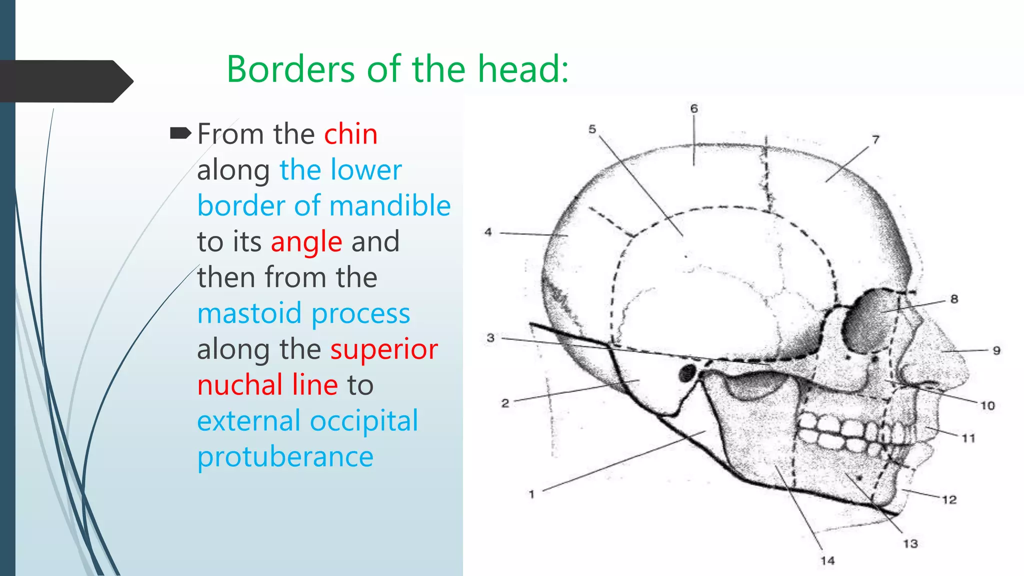
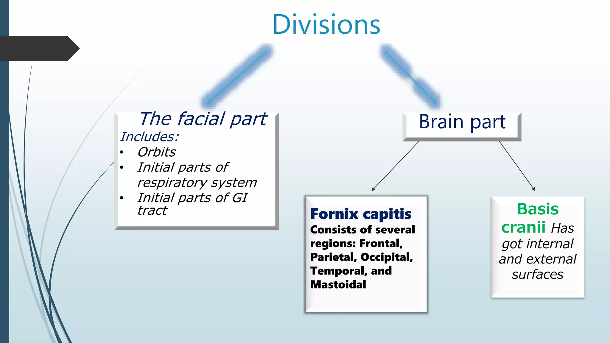
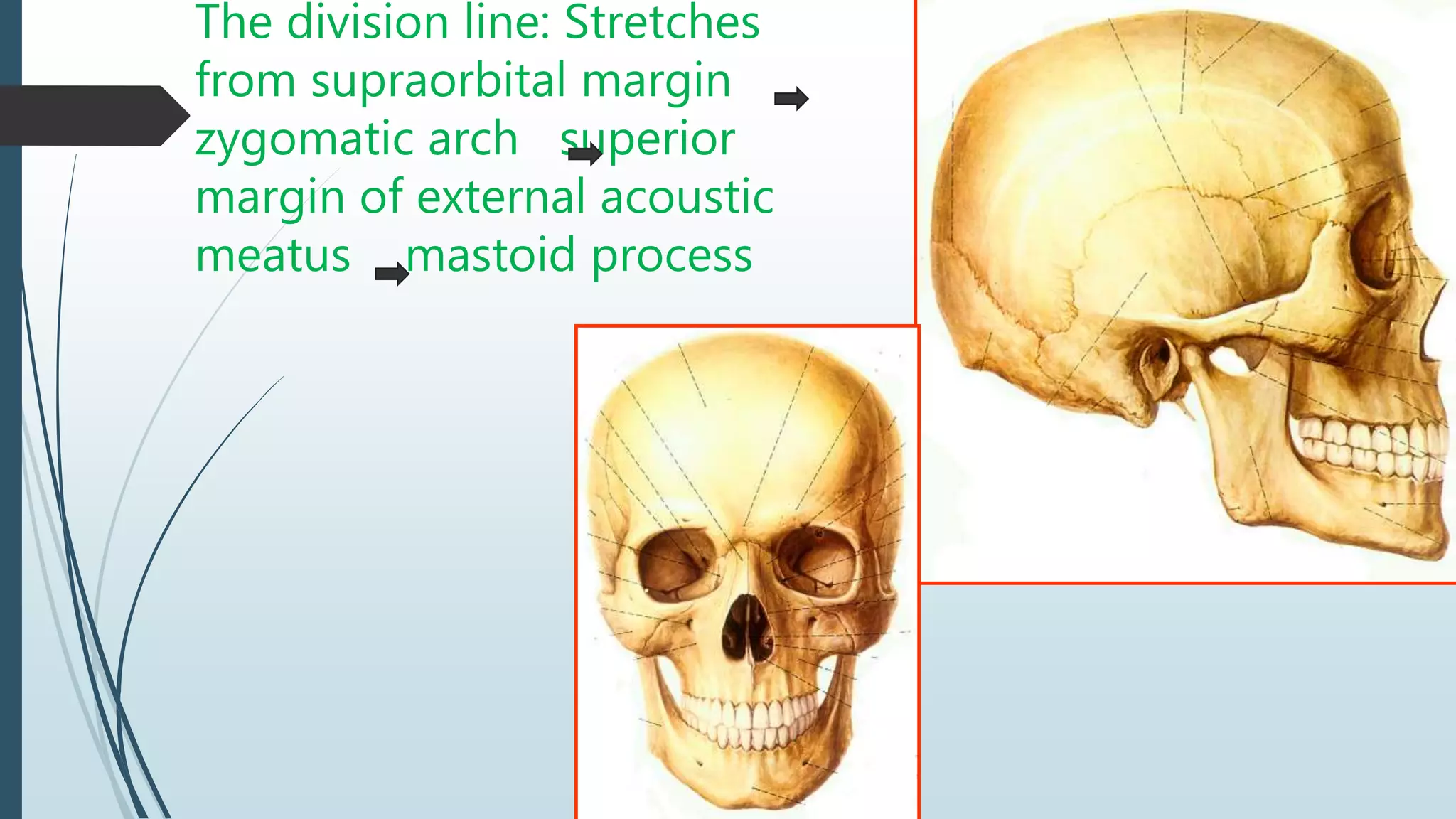
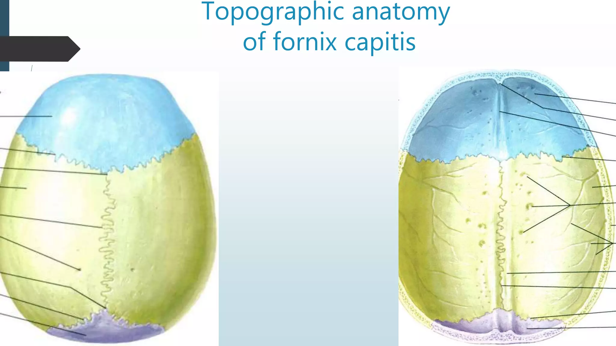
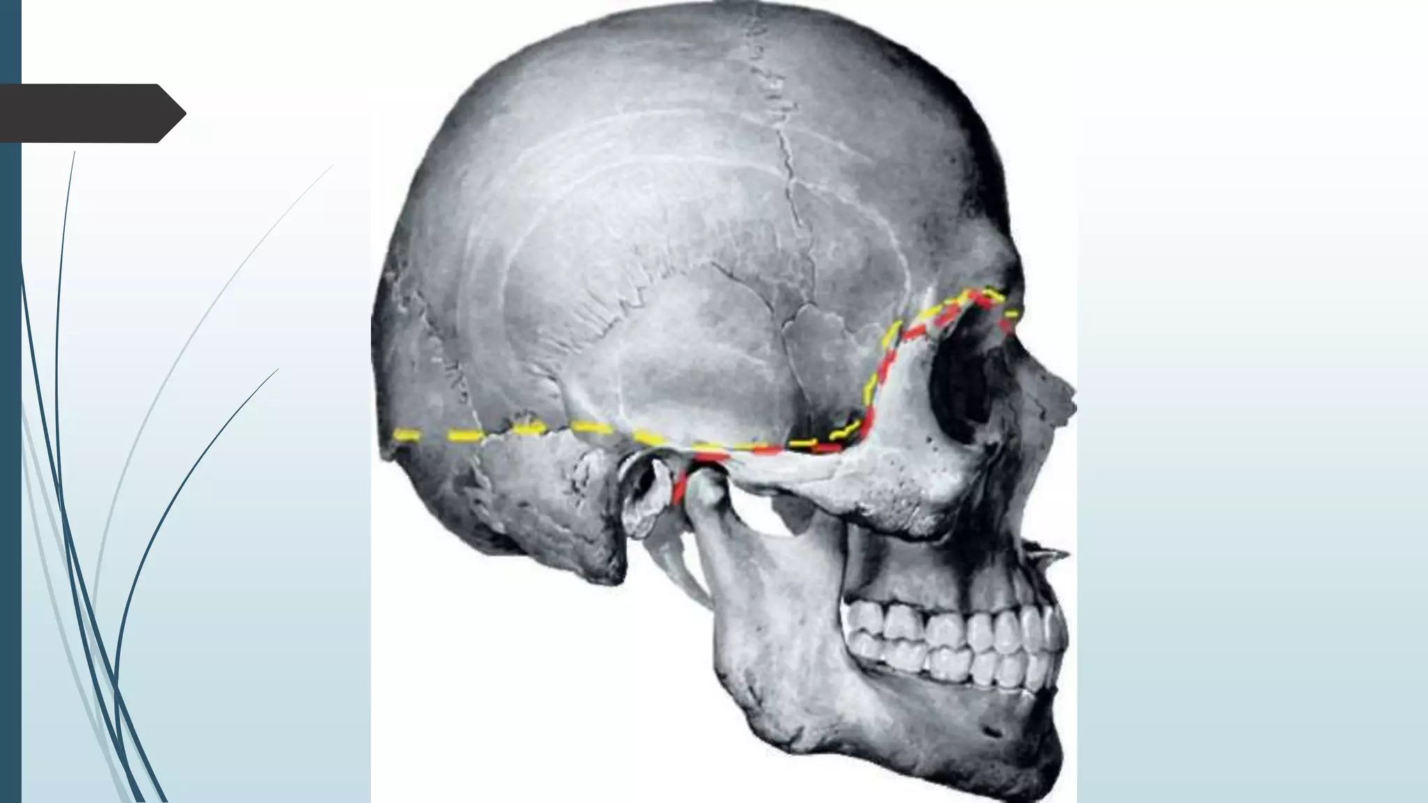
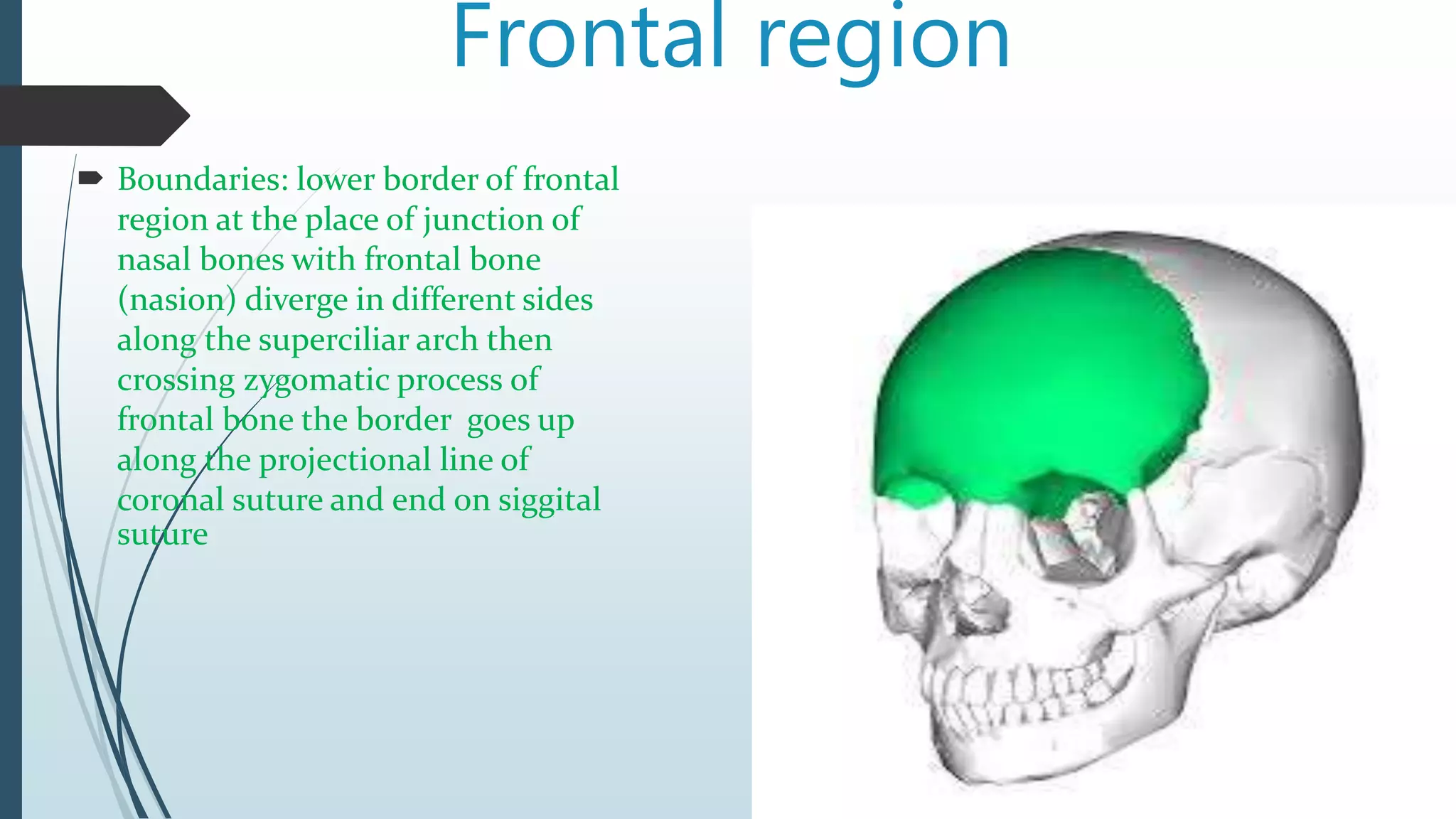
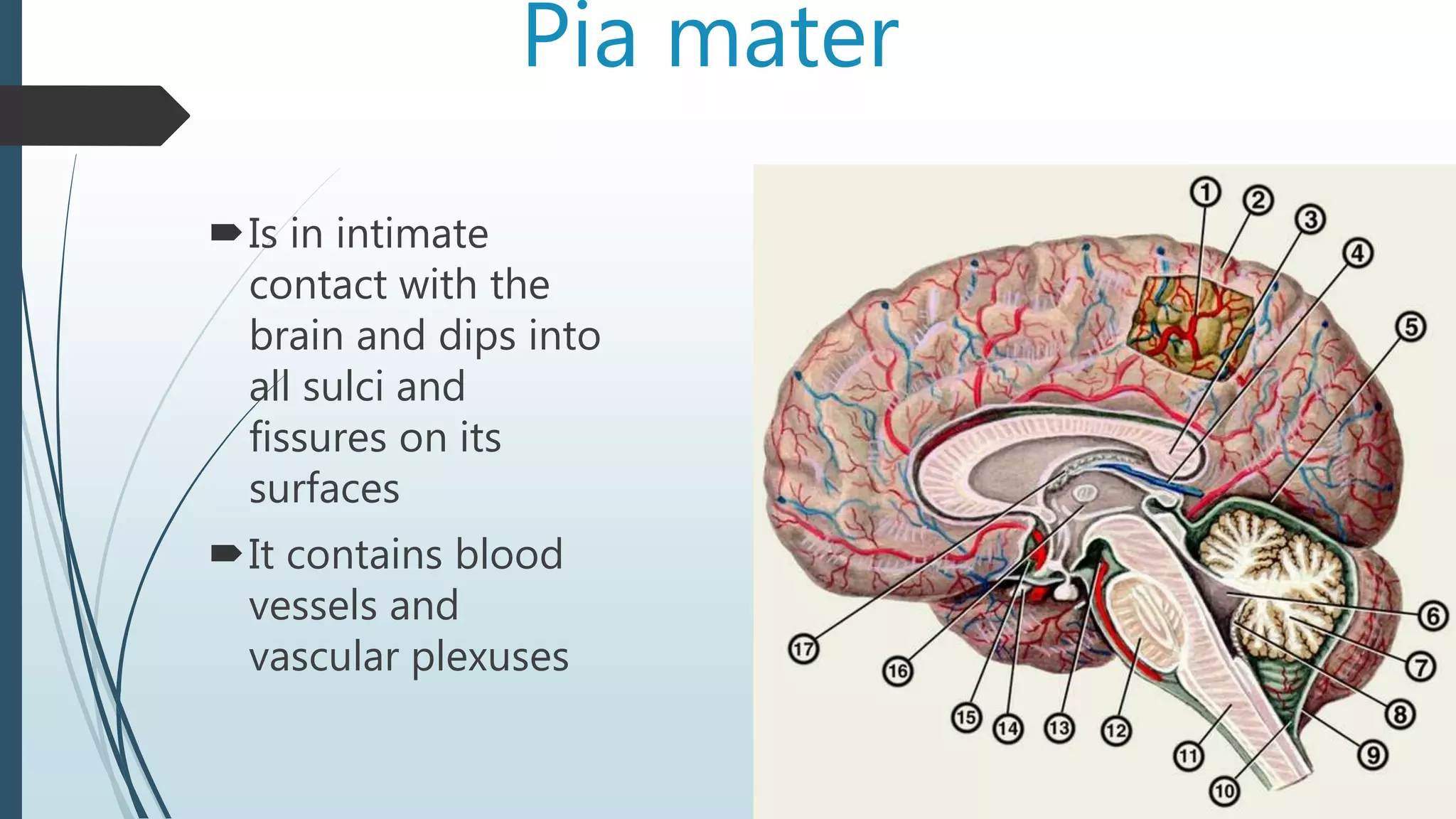
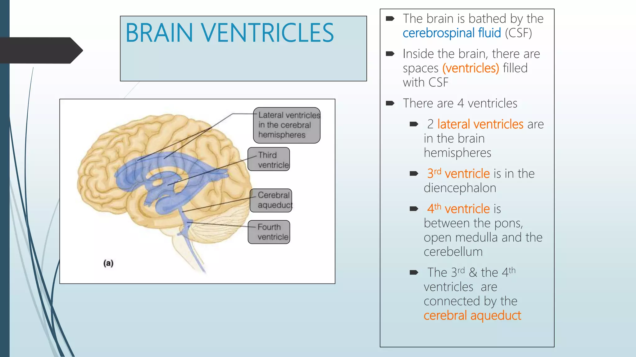
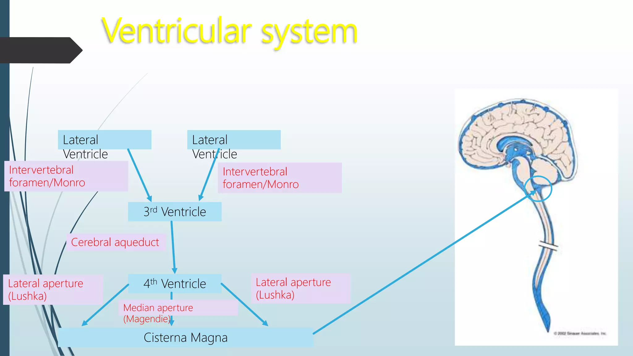
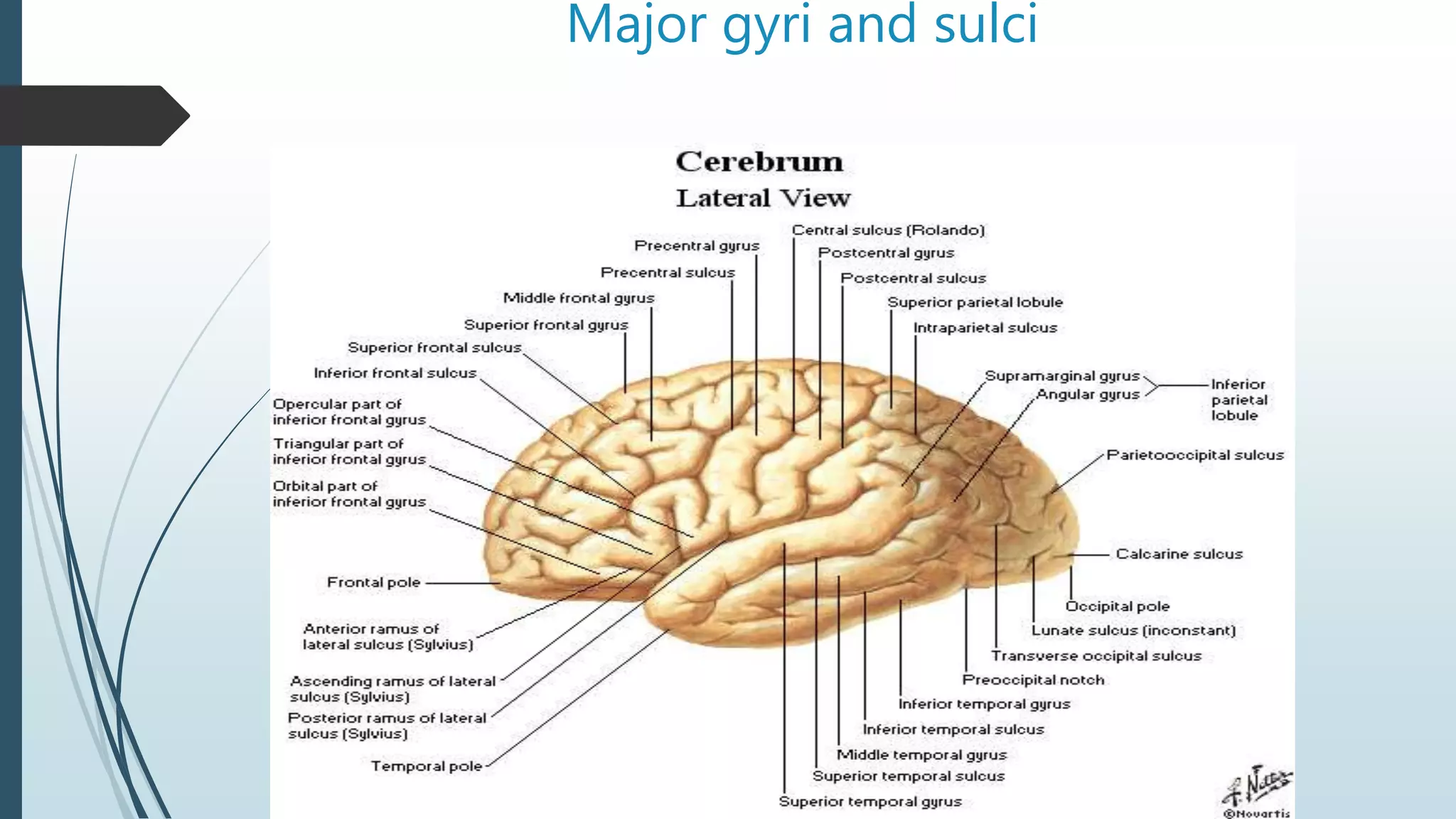
SlideShare a Scribd company logo
Topographic anatomy of the
brain and brain part of the skull
Rustam Sultonov
Borders of the head:
From the chin
along the lower
border of mandible
to its angle and
then from the
mastoid process
along the superior
nuchal line to
external occipital
protuberance
Divisions
The facial part
Includes:
• Orbits
• Initial parts of
respiratory system
• Initial parts of GI
tract
Brain part
Fornix capitis
Consists of several
regions: Frontal,
Parietal, Occipital,
Temporal, and
Mastoidal
Basis
cranii Has
got internal
and external
surfaces
The division line: Stretches
from supraorbital margin
zygomatic arch superior
margin of external acoustic
meatus mastoid process
Topographic anatomy
of fornix capitis
Frontal region
 Boundaries: lower border of frontal
region at the place of junction of
nasal bones with frontal bone
(nasion) diverge in different sides
along the superciliar arch then
crossing zygomatic process of
frontal bone the border goes up
along the projectional line of
coronal suture and end on siggital
suture
 Blood supply: a. supratrochlearis, a. supraorbitalis (from a.
ophtalmica)
 Venous drainage: v. supratrochlearis, v. supraorbitalis
 Lymph drainage: lymph vessels drain into nodi lymphatici
parotidei superficiales
 Innervation: Sensitive nerves are n. supratrochlearis, n.
supraorbitalis (from n. ophtalmicus. Motor nerves: rami
temporalis n. facialis (branches go to m. frontalis)
Parietal region
 Boundaries: good visible only in
the skull. Borders are along the
projectional lines of coronal,
saggital, lambdoidal sutures and
temporal line
Blood supply: branches of a. temporalis
superficialis
Venous drainage: branches of v. temporalis
superficialis
Lymph drainage: lymph vessels drain into nodi
lymphatici mastoidei
Innervation: branches from nn. Supraorbitalis et
frontalis, n. auriculotemporalis, n. occipialis major
Occipital region
 Boundaries: borders of the region from
above and on the sides correspond to
the location of lambdoidal suture;
below borders correspond to the line
that stretches from the bases of one
mastoid process to the basis of another
one through the external occipital
protuberance
Blood supply: a. occipitalis, a. auricularis
posterior
Venous drainage: v. occipitalis, v.
vertebralis
Lymph drainage: lymph vessels drain into
nodi lymphatici occipitalis
Innervation: Sensory nerve is n.
suboccipitalis; Motor nerves are n.
occipitalis major, n. occipitalis minor
Layers and spaces of frontal, parietal and
occipital regions
1. Skin (cutis) – in these areas is thick and covered by hair (except
frontal region), tightly connected with subcutaneous fat and m.
epicranius by CT septa
2. Subcutaneous fat (panniculus adiposus) contains CT septa, fat
drops and vascular net. Vascular and lymphatic vessels are
tightly fixed in septa and that’s why due to injuries they do not
dissipate and severe bleeding might occur
3. M. epicranius – consists of 2 bellies (venter frontalis and venter
occipitalis) and galea aponeuotica between them. Innervation:
n. facialis
4. Stratum fasciale – the layer of the loose tissue localized between
m. epicranius and periostium
5. Periosteum – tightly connected to bones only in the are of
sutures, in other places it can easily be detached
6. Skull bones (ossa cranii) - thickness varies in different zones.
These bones do not possess ability to regenerate ( that’s why
defect after trepanation is closed only by fibrous tissue). Skull
bones have 3 layers: lamina externa, diploe(spongy layer), lamina
interna
7. Epidural space (cavitas epiduralis) – between internal laminae
and dura mater
8. Dura mater encephali – consists of dense fibrous CT
9. Spatium subdurale – a space between dura mater and
arachnoidea encephali containing fluid
10. Arachnoidea encephali – passes from one gyri to another, not
dropping in sulci.
11. Cavitas arachnoidalis – contains cerebrospinal fluid
12. Pia mater encephali – contains many blood vessels. Covering
all the brain enters into sulci
13. Gray matter of brain
Topographic
anatomy of
brain
Brain meninges
Pia mater
encephali
Dura mater encephali
 A thick whitish CT membrane
is outermost in position.
 External surface is in direct
contact with skull bones for
which it serves like periosteum
 The inner surface facing the
brain is lined with endothelium
and is therefore smooth and
shiny
 Between it and cerebral
arachnoid mater is narrow
subdural space filled with a
small amount of fluid
Inner surface gives off several processes, which penetrate between the parts
of the brain and separate one part from another:
The Falx cerebri – lies sagittal between both cerebral hemispheres. Anterior
narrow end grows into the crista galli, while the wide posterior end blend
with the superior surface of tentorium cerebelli
The Tentorium cerebelli – separates the cerebral occipital lobes from the
cerebellum
The Falx cerebelli – lies in saggital plane and separates cerebellar
hemispheres
 Blood supply: a. meningea anterior, a. meningea media, a.
meningea posterior, ramus meningeus a. occipitalis
 Innervation: r. tentorii n. ophtalmici, r. meningeus n.
maxillaris, r. meningeus n. mandibularis, r. meningeus n.
vagi
Arachnoidea encephali
Forms Middle layer of brain
meninges
Separated from dura mater by a
capillary subdural slit-like space
In contrast to pia mater it does not
penetrate into the sulci and
depressions of the brain but
bridges them as a result of which
subarachnoidal spaces filled with
fluid forms
In some places sunarachnoidal
spaces are particularly well
developed and form wide and
deep reserviours filled with
cerebrospinal fluid – cisternae
1. Cisterna cerebromedullaris
2. Cisterna interpeduncularis
3. Cisterna chiasmatis
4. Cisterna fossae lateralis
cerebri
Pia mater
Is in intimate
contact with the
brain and dips into
all sulci and
fissures on its
surfaces
It contains blood
vessels and
vascular plexuses
BRAIN VENTRICLES
 The brain is bathed by the
cerebrospinal fluid (CSF)
 Inside the brain, there are
spaces (ventricles) filled
with CSF
 There are 4 ventricles
 2 lateral ventricles are
in the brain
hemispheres
 3rd ventricle is in the
diencephalon
 4th ventricle is
between the pons,
open medulla and the
cerebellum
 The 3rd & the 4th
ventricles are
connected by the
cerebral aqueduct
Ventricular system
Lateral
Ventricle
Lateral
Ventricle
3rd Ventricle
4th Ventricle
Cisterna Magna
Intervertebral
foramen/Monro
Cerebral aqueduct
Lateral aperture
(Lushka)
Median aperture
(Magendie)
Lateral aperture
(Lushka)
Intervertebral
foramen/Monro
Major gyri and sulci
Lobes
 Frontal lobe
 in front of central sulcus and above lateral sulcus, line from central sulcus down
to corpus collosum
 Parietal lobes
 behind central sulcus and above lateral sulcus. A line between parieto-occipital
sulcus and preoccipital notch and middle of the line above to the lateral sulcus
(See Picture 13-1 on P261)
 Temporal lobes
 lateral sulcus and line described above, a line from anterior end of calcarine
sulcus and preoccipital notch (see Pic 13-2 on P 262)
 Occipital lobe
 medial surface of the hemisphere, separated from temporal lobe.
 Insula
 bottom of lateral sulcus.
THANK YOU
FOR
your attention! Any
question? No ?
Great! Bye!)))

More Related Content

What's hot

Pons Anatomy
Pons AnatomyPons Anatomy
Pons Anatomy
Mehul Tandel
 
Posterior triangle of neck - Powerpoint lecture notes by Dr.N.Mugunthan.
Posterior triangle of neck - Powerpoint lecture notes by Dr.N.Mugunthan.Posterior triangle of neck - Powerpoint lecture notes by Dr.N.Mugunthan.
Posterior triangle of neck - Powerpoint lecture notes by Dr.N.Mugunthan.
mgmcri1234
 
Anatomy of the cranial nerves
Anatomy of the cranial nervesAnatomy of the cranial nerves
Anatomy of the cranial nerves
Mukhtar Khan
 
Functional anatomy of brainstem and clinical correlates
Functional anatomy of brainstem and clinical correlatesFunctional anatomy of brainstem and clinical correlates
Functional anatomy of brainstem and clinical correlates
Dr. Arun Mathai Mani
 
Trachea
TracheaTrachea
Anatomy of thalamus
Anatomy of thalamusAnatomy of thalamus
Anatomy of thalamusMBBS IMS MSU
 
Medulla oplongata 22 04-16
Medulla oplongata 22 04-16Medulla oplongata 22 04-16
Medulla oplongata 22 04-16
mgmcri1234
 
Anatomy of pharynx
Anatomy of pharynxAnatomy of pharynx
Anatomy of pharynx
Dr Gangaprasad Waghmare
 
Esophagus stomach-dr.gosai
Esophagus stomach-dr.gosaiEsophagus stomach-dr.gosai
Esophagus stomach-dr.gosaiDr.B.B. Gosai
 
Anatomy of nose and paranasal sinus
Anatomy of nose and paranasal sinusAnatomy of nose and paranasal sinus
Anatomy of nose and paranasal sinusMBBS IMS MSU
 
Mediastinum & pleurae
Mediastinum & pleurae Mediastinum & pleurae
2. paranasal sinuses
2. paranasal sinuses2. paranasal sinuses
2. paranasal sinuses
Dr. Mohammad Mahmoud
 
Pleura
PleuraPleura
The pleurae
The pleuraeThe pleurae
The pleurae
Idris Siddiqui
 
Hepatic Portal vein and portocaval anatomosis
Hepatic Portal vein and portocaval anatomosisHepatic Portal vein and portocaval anatomosis
Hepatic Portal vein and portocaval anatomosis
Nepalese army institute of health sciences
 
Introduction to the topographical anatomy and operative sugery
Introduction to the topographical anatomy and operative sugeryIntroduction to the topographical anatomy and operative sugery
Introduction to the topographical anatomy and operative sugery
kavanvyas1
 
ANATOMY OF PANCREAS
ANATOMY OF PANCREASANATOMY OF PANCREAS
ANATOMY OF PANCREAS
Aamir Hela
 

What's hot (20)

Pons Anatomy
Pons AnatomyPons Anatomy
Pons Anatomy
 
Posterior triangle of neck - Powerpoint lecture notes by Dr.N.Mugunthan.
Posterior triangle of neck - Powerpoint lecture notes by Dr.N.Mugunthan.Posterior triangle of neck - Powerpoint lecture notes by Dr.N.Mugunthan.
Posterior triangle of neck - Powerpoint lecture notes by Dr.N.Mugunthan.
 
Cerebellum
CerebellumCerebellum
Cerebellum
 
Anatomy of the cranial nerves
Anatomy of the cranial nervesAnatomy of the cranial nerves
Anatomy of the cranial nerves
 
Functional anatomy of brainstem and clinical correlates
Functional anatomy of brainstem and clinical correlatesFunctional anatomy of brainstem and clinical correlates
Functional anatomy of brainstem and clinical correlates
 
Trachea
TracheaTrachea
Trachea
 
Mediastinum
MediastinumMediastinum
Mediastinum
 
Anatomy of thalamus
Anatomy of thalamusAnatomy of thalamus
Anatomy of thalamus
 
Medulla oplongata 22 04-16
Medulla oplongata 22 04-16Medulla oplongata 22 04-16
Medulla oplongata 22 04-16
 
mediastinum
mediastinummediastinum
mediastinum
 
Anatomy of pharynx
Anatomy of pharynxAnatomy of pharynx
Anatomy of pharynx
 
Esophagus stomach-dr.gosai
Esophagus stomach-dr.gosaiEsophagus stomach-dr.gosai
Esophagus stomach-dr.gosai
 
Anatomy of nose and paranasal sinus
Anatomy of nose and paranasal sinusAnatomy of nose and paranasal sinus
Anatomy of nose and paranasal sinus
 
Mediastinum & pleurae
Mediastinum & pleurae Mediastinum & pleurae
Mediastinum & pleurae
 
2. paranasal sinuses
2. paranasal sinuses2. paranasal sinuses
2. paranasal sinuses
 
Pleura
PleuraPleura
Pleura
 
The pleurae
The pleuraeThe pleurae
The pleurae
 
Hepatic Portal vein and portocaval anatomosis
Hepatic Portal vein and portocaval anatomosisHepatic Portal vein and portocaval anatomosis
Hepatic Portal vein and portocaval anatomosis
 
Introduction to the topographical anatomy and operative sugery
Introduction to the topographical anatomy and operative sugeryIntroduction to the topographical anatomy and operative sugery
Introduction to the topographical anatomy and operative sugery
 
ANATOMY OF PANCREAS
ANATOMY OF PANCREASANATOMY OF PANCREAS
ANATOMY OF PANCREAS
 

Viewers also liked

MRI SECTIONAL ANATOMY OF BRAIN
MRI SECTIONAL ANATOMY OF BRAIN MRI SECTIONAL ANATOMY OF BRAIN
MRI SECTIONAL ANATOMY OF BRAIN Vipin Kumar
 
Lower limb anatomylect 2
Lower limb anatomylect 2Lower limb anatomylect 2
Lower limb anatomylect 2
Yasir Jameel
 
Surgical anatomy of microsurgical sulcal key points
Surgical anatomy of microsurgical sulcal key pointsSurgical anatomy of microsurgical sulcal key points
Surgical anatomy of microsurgical sulcal key pointsINUB
 
Anatomy of the Brain
Anatomy of the BrainAnatomy of the Brain
Anatomy of the Brain
Myriam Dimanche
 
The Cerebral Cortex by Dr. NIda Kanwal, Neurosrugery LNH, Karachi
The Cerebral Cortex by Dr. NIda Kanwal, Neurosrugery LNH, KarachiThe Cerebral Cortex by Dr. NIda Kanwal, Neurosrugery LNH, Karachi
The Cerebral Cortex by Dr. NIda Kanwal, Neurosrugery LNH, Karachi
noorulainiqbal
 
topographic anatomy Abdomen
topographic anatomy  Abdomentopographic anatomy  Abdomen
topographic anatomy Abdomen
Medical Students
 
Anatomy of brain
Anatomy of brainAnatomy of brain
Anatomy of brain
Col Mukteshwar Prasad
 
Anatomy of the brain
Anatomy of the brainAnatomy of the brain
Anatomy of the braingarretcj
 
Sulci gyri
Sulci gyriSulci gyri
Sulci gyri
Mradul Srivastava
 
002 Surgical anatomy of the brain
002 Surgical anatomy of the brain002 Surgical anatomy of the brain
002 Surgical anatomy of the brain
Neurosurgery Vajira
 
1 osteology of cranial bones rt(67) Dr. RAHUL TIWARI
1 osteology of cranial bones rt(67) Dr. RAHUL TIWARI1 osteology of cranial bones rt(67) Dr. RAHUL TIWARI
1 osteology of cranial bones rt(67) Dr. RAHUL TIWARI
CLOVE Dental OMNI Hospitals Andhra Hospital
 
Skull the normas
Skull   the normasSkull   the normas
Skull the normas
Akram Jaffar
 
Operative surgery ppt mbbs students
Operative surgery ppt mbbs students Operative surgery ppt mbbs students
Operative surgery ppt mbbs students
TONY SCARIA
 
Sulcal anatomy supratentorial brain, excluding the temporal lobe.
Sulcal anatomy supratentorial brain, excluding the temporal lobe.Sulcal anatomy supratentorial brain, excluding the temporal lobe.
Sulcal anatomy supratentorial brain, excluding the temporal lobe.
Sarbesh Tiwari
 
Anatomical landmarks of the crown
Anatomical landmarks of the crownAnatomical landmarks of the crown
Anatomical landmarks of the crown
Hesham Dameer
 
Osteology of facial skeleton
Osteology of facial skeletonOsteology of facial skeleton
Osteology of facial skeleton
K BHATTACHARJEE
 
Sulci,Gyri & Functional areas of cerebrum Dr.N.Mugunthan.M.S
Sulci,Gyri & Functional areas of cerebrum Dr.N.Mugunthan.M.SSulci,Gyri & Functional areas of cerebrum Dr.N.Mugunthan.M.S
Sulci,Gyri & Functional areas of cerebrum Dr.N.Mugunthan.M.S
mgmcri1234
 
Presentation1.pptx, radiological anatomy of the brain.
Presentation1.pptx, radiological anatomy of the brain.Presentation1.pptx, radiological anatomy of the brain.
Presentation1.pptx, radiological anatomy of the brain.
Abdellah Nazeer
 
Head & neck rs x-rays
Head & neck rs   x-raysHead & neck rs   x-rays
Head & neck rs x-raysAHS_anatomy2
 

Viewers also liked (20)

MRI SECTIONAL ANATOMY OF BRAIN
MRI SECTIONAL ANATOMY OF BRAIN MRI SECTIONAL ANATOMY OF BRAIN
MRI SECTIONAL ANATOMY OF BRAIN
 
Lower limb anatomylect 2
Lower limb anatomylect 2Lower limb anatomylect 2
Lower limb anatomylect 2
 
Surgical anatomy of microsurgical sulcal key points
Surgical anatomy of microsurgical sulcal key pointsSurgical anatomy of microsurgical sulcal key points
Surgical anatomy of microsurgical sulcal key points
 
Anatomy of the Brain
Anatomy of the BrainAnatomy of the Brain
Anatomy of the Brain
 
The Cerebral Cortex by Dr. NIda Kanwal, Neurosrugery LNH, Karachi
The Cerebral Cortex by Dr. NIda Kanwal, Neurosrugery LNH, KarachiThe Cerebral Cortex by Dr. NIda Kanwal, Neurosrugery LNH, Karachi
The Cerebral Cortex by Dr. NIda Kanwal, Neurosrugery LNH, Karachi
 
topographic anatomy Abdomen
topographic anatomy  Abdomentopographic anatomy  Abdomen
topographic anatomy Abdomen
 
Anatomy of brain
Anatomy of brainAnatomy of brain
Anatomy of brain
 
Anatomy of the brain
Anatomy of the brainAnatomy of the brain
Anatomy of the brain
 
Sulci gyri
Sulci gyriSulci gyri
Sulci gyri
 
002 Surgical anatomy of the brain
002 Surgical anatomy of the brain002 Surgical anatomy of the brain
002 Surgical anatomy of the brain
 
1 osteology of cranial bones rt(67) Dr. RAHUL TIWARI
1 osteology of cranial bones rt(67) Dr. RAHUL TIWARI1 osteology of cranial bones rt(67) Dr. RAHUL TIWARI
1 osteology of cranial bones rt(67) Dr. RAHUL TIWARI
 
Benign focal lesions in liver
Benign focal lesions in liverBenign focal lesions in liver
Benign focal lesions in liver
 
Skull the normas
Skull   the normasSkull   the normas
Skull the normas
 
Operative surgery ppt mbbs students
Operative surgery ppt mbbs students Operative surgery ppt mbbs students
Operative surgery ppt mbbs students
 
Sulcal anatomy supratentorial brain, excluding the temporal lobe.
Sulcal anatomy supratentorial brain, excluding the temporal lobe.Sulcal anatomy supratentorial brain, excluding the temporal lobe.
Sulcal anatomy supratentorial brain, excluding the temporal lobe.
 
Anatomical landmarks of the crown
Anatomical landmarks of the crownAnatomical landmarks of the crown
Anatomical landmarks of the crown
 
Osteology of facial skeleton
Osteology of facial skeletonOsteology of facial skeleton
Osteology of facial skeleton
 
Sulci,Gyri & Functional areas of cerebrum Dr.N.Mugunthan.M.S
Sulci,Gyri & Functional areas of cerebrum Dr.N.Mugunthan.M.SSulci,Gyri & Functional areas of cerebrum Dr.N.Mugunthan.M.S
Sulci,Gyri & Functional areas of cerebrum Dr.N.Mugunthan.M.S
 
Presentation1.pptx, radiological anatomy of the brain.
Presentation1.pptx, radiological anatomy of the brain.Presentation1.pptx, radiological anatomy of the brain.
Presentation1.pptx, radiological anatomy of the brain.
 
Head & neck rs x-rays
Head & neck rs   x-raysHead & neck rs   x-rays
Head & neck rs x-rays
 

Similar to Topographic anatomy of the brain and brain part of the skull

2. brain
2. brain2. brain
2. brain
Abitigenet
 
Venous drainageof head and neck ih
Venous drainageof head and neck   ihVenous drainageof head and neck   ih
Venous drainageof head and neck ih
itrat hussain
 
Cranial cavity ( Department of Anatomy)
Cranial cavity ( Department of Anatomy)Cranial cavity ( Department of Anatomy)
Cranial cavity ( Department of Anatomy)
Tarun kumar ubhu
 
Dural venous sinuses.pptx
Dural venous sinuses.pptxDural venous sinuses.pptx
Dural venous sinuses.pptx
Sundip Charmode
 
Meninges.ppt
Meninges.pptMeninges.ppt
Meninges.ppt
RupaSingh83
 
anatomyofmeningesventriclescerebrospinalfluid-100604195832-phpapp02.pdf
anatomyofmeningesventriclescerebrospinalfluid-100604195832-phpapp02.pdfanatomyofmeningesventriclescerebrospinalfluid-100604195832-phpapp02.pdf
anatomyofmeningesventriclescerebrospinalfluid-100604195832-phpapp02.pdf
DipaliTalaviya1
 
Anatomy of the Scalp
Anatomy of the ScalpAnatomy of the Scalp
Anatomy of the Scalp
Rafid Rashid
 
Brain CT Anatomy and Basic Interpretation Part II
Brain CT Anatomy and Basic Interpretation Part IIBrain CT Anatomy and Basic Interpretation Part II
Brain CT Anatomy and Basic Interpretation Part II
Sakher Alkhaderi
 
Ventricular system of brain final
Ventricular system of brain finalVentricular system of brain final
Ventricular system of brain finalfarhan_aq91
 
Meninges covering of the CNS.pdf
Meninges covering of the CNS.pdfMeninges covering of the CNS.pdf
Meninges covering of the CNS.pdf
AdebayoAbayomi3
 
Ventricular system of brain & the CSF
Ventricular system of brain & the CSFVentricular system of brain & the CSF
Ventricular system of brain & the CSF
ABDUL QADEER MEMON
 
Blood supplyand lymphatic drainage to oral cavity
Blood supplyand lymphatic drainage to oral cavityBlood supplyand lymphatic drainage to oral cavity
Blood supplyand lymphatic drainage to oral cavity
Aishwarya Hajare
 
venous supply of head & neck
venous supply of head & neckvenous supply of head & neck
venous supply of head & neckAshish Soni
 
VENTRICLES AND CISTERNS OF HUMAN BRAIN.pptx
VENTRICLES AND CISTERNS OF HUMAN BRAIN.pptxVENTRICLES AND CISTERNS OF HUMAN BRAIN.pptx
VENTRICLES AND CISTERNS OF HUMAN BRAIN.pptx
hulkie8606
 
Meninges
MeningesMeninges
Meninges
Idris Siddiqui
 
arterial supply of head and neck
arterial supply of head and neckarterial supply of head and neck
arterial supply of head and neck
shalini sampreethi
 
Scalp
ScalpScalp
ventricle
ventricleventricle
ventricle
RathiNivedhana
 
Ventricles
VentriclesVentricles
Ventricles
Med Study
 

Similar to Topographic anatomy of the brain and brain part of the skull (20)

2. brain
2. brain2. brain
2. brain
 
Venous drainageof head and neck ih
Venous drainageof head and neck   ihVenous drainageof head and neck   ih
Venous drainageof head and neck ih
 
Cranial cavity ( Department of Anatomy)
Cranial cavity ( Department of Anatomy)Cranial cavity ( Department of Anatomy)
Cranial cavity ( Department of Anatomy)
 
Dural venous sinuses.pptx
Dural venous sinuses.pptxDural venous sinuses.pptx
Dural venous sinuses.pptx
 
Meninges.ppt
Meninges.pptMeninges.ppt
Meninges.ppt
 
Meninges.pptx
Meninges.pptxMeninges.pptx
Meninges.pptx
 
anatomyofmeningesventriclescerebrospinalfluid-100604195832-phpapp02.pdf
anatomyofmeningesventriclescerebrospinalfluid-100604195832-phpapp02.pdfanatomyofmeningesventriclescerebrospinalfluid-100604195832-phpapp02.pdf
anatomyofmeningesventriclescerebrospinalfluid-100604195832-phpapp02.pdf
 
Anatomy of the Scalp
Anatomy of the ScalpAnatomy of the Scalp
Anatomy of the Scalp
 
Brain CT Anatomy and Basic Interpretation Part II
Brain CT Anatomy and Basic Interpretation Part IIBrain CT Anatomy and Basic Interpretation Part II
Brain CT Anatomy and Basic Interpretation Part II
 
Ventricular system of brain final
Ventricular system of brain finalVentricular system of brain final
Ventricular system of brain final
 
Meninges covering of the CNS.pdf
Meninges covering of the CNS.pdfMeninges covering of the CNS.pdf
Meninges covering of the CNS.pdf
 
Ventricular system of brain & the CSF
Ventricular system of brain & the CSFVentricular system of brain & the CSF
Ventricular system of brain & the CSF
 
Blood supplyand lymphatic drainage to oral cavity
Blood supplyand lymphatic drainage to oral cavityBlood supplyand lymphatic drainage to oral cavity
Blood supplyand lymphatic drainage to oral cavity
 
venous supply of head & neck
venous supply of head & neckvenous supply of head & neck
venous supply of head & neck
 
VENTRICLES AND CISTERNS OF HUMAN BRAIN.pptx
VENTRICLES AND CISTERNS OF HUMAN BRAIN.pptxVENTRICLES AND CISTERNS OF HUMAN BRAIN.pptx
VENTRICLES AND CISTERNS OF HUMAN BRAIN.pptx
 
Meninges
MeningesMeninges
Meninges
 
arterial supply of head and neck
arterial supply of head and neckarterial supply of head and neck
arterial supply of head and neck
 
Scalp
ScalpScalp
Scalp
 
ventricle
ventricleventricle
ventricle
 
Ventricles
VentriclesVentricles
Ventricles
 

Recently uploaded

Alcohol_Dr. Jeenal Mistry MD Pharmacology.pdf
Alcohol_Dr. Jeenal Mistry MD Pharmacology.pdfAlcohol_Dr. Jeenal Mistry MD Pharmacology.pdf
Alcohol_Dr. Jeenal Mistry MD Pharmacology.pdf
Dr Jeenal Mistry
 
Charaka Samhita Sutra sthana Chapter 15 Upakalpaniyaadhyaya
Charaka Samhita Sutra sthana Chapter 15 UpakalpaniyaadhyayaCharaka Samhita Sutra sthana Chapter 15 Upakalpaniyaadhyaya
Charaka Samhita Sutra sthana Chapter 15 Upakalpaniyaadhyaya
Dr KHALID B.M
 
ARTIFICIAL INTELLIGENCE IN HEALTHCARE.pdf
ARTIFICIAL INTELLIGENCE IN  HEALTHCARE.pdfARTIFICIAL INTELLIGENCE IN  HEALTHCARE.pdf
ARTIFICIAL INTELLIGENCE IN HEALTHCARE.pdf
Anujkumaranit
 
Are There Any Natural Remedies To Treat Syphilis.pdf
Are There Any Natural Remedies To Treat Syphilis.pdfAre There Any Natural Remedies To Treat Syphilis.pdf
Are There Any Natural Remedies To Treat Syphilis.pdf
Little Cross Family Clinic
 
TEST BANK for Operations Management, 14th Edition by William J. Stevenson, Ve...
TEST BANK for Operations Management, 14th Edition by William J. Stevenson, Ve...TEST BANK for Operations Management, 14th Edition by William J. Stevenson, Ve...
TEST BANK for Operations Management, 14th Edition by William J. Stevenson, Ve...
kevinkariuki227
 
For Better Surat #ℂall #Girl Service ❤85270-49040❤ Surat #ℂall #Girls
For Better Surat #ℂall #Girl Service ❤85270-49040❤ Surat #ℂall #GirlsFor Better Surat #ℂall #Girl Service ❤85270-49040❤ Surat #ℂall #Girls
For Better Surat #ℂall #Girl Service ❤85270-49040❤ Surat #ℂall #Girls
Savita Shen $i11
 
basicmodesofventilation2022-220313203758.pdf
basicmodesofventilation2022-220313203758.pdfbasicmodesofventilation2022-220313203758.pdf
basicmodesofventilation2022-220313203758.pdf
aljamhori teaching hospital
 
Lung Cancer: Artificial Intelligence, Synergetics, Complex System Analysis, S...
Lung Cancer: Artificial Intelligence, Synergetics, Complex System Analysis, S...Lung Cancer: Artificial Intelligence, Synergetics, Complex System Analysis, S...
Lung Cancer: Artificial Intelligence, Synergetics, Complex System Analysis, S...
Oleg Kshivets
 
ANATOMY AND PHYSIOLOGY OF URINARY SYSTEM.pptx
ANATOMY AND PHYSIOLOGY OF URINARY SYSTEM.pptxANATOMY AND PHYSIOLOGY OF URINARY SYSTEM.pptx
ANATOMY AND PHYSIOLOGY OF URINARY SYSTEM.pptx
Swetaba Besh
 
Ocular injury ppt Upendra pal optometrist upums saifai etawah
Ocular injury  ppt  Upendra pal  optometrist upums saifai etawahOcular injury  ppt  Upendra pal  optometrist upums saifai etawah
Ocular injury ppt Upendra pal optometrist upums saifai etawah
pal078100
 
micro teaching on communication m.sc nursing.pdf
micro teaching on communication m.sc nursing.pdfmicro teaching on communication m.sc nursing.pdf
micro teaching on communication m.sc nursing.pdf
Anurag Sharma
 
Report Back from SGO 2024: What’s the Latest in Cervical Cancer?
Report Back from SGO 2024: What’s the Latest in Cervical Cancer?Report Back from SGO 2024: What’s the Latest in Cervical Cancer?
Report Back from SGO 2024: What’s the Latest in Cervical Cancer?
bkling
 
HOT NEW PRODUCT! BIG SALES FAST SHIPPING NOW FROM CHINA!! EU KU DB BK substit...
HOT NEW PRODUCT! BIG SALES FAST SHIPPING NOW FROM CHINA!! EU KU DB BK substit...HOT NEW PRODUCT! BIG SALES FAST SHIPPING NOW FROM CHINA!! EU KU DB BK substit...
HOT NEW PRODUCT! BIG SALES FAST SHIPPING NOW FROM CHINA!! EU KU DB BK substit...
GL Anaacs
 
Couples presenting to the infertility clinic- Do they really have infertility...
Couples presenting to the infertility clinic- Do they really have infertility...Couples presenting to the infertility clinic- Do they really have infertility...
Couples presenting to the infertility clinic- Do they really have infertility...
Sujoy Dasgupta
 
BRACHYTHERAPY OVERVIEW AND APPLICATORS
BRACHYTHERAPY OVERVIEW  AND  APPLICATORSBRACHYTHERAPY OVERVIEW  AND  APPLICATORS
BRACHYTHERAPY OVERVIEW AND APPLICATORS
Krishan Murari
 
Ophthalmology Clinical Tests for OSCE exam
Ophthalmology Clinical Tests for OSCE examOphthalmology Clinical Tests for OSCE exam
Ophthalmology Clinical Tests for OSCE exam
KafrELShiekh University
 
New Directions in Targeted Therapeutic Approaches for Older Adults With Mantl...
New Directions in Targeted Therapeutic Approaches for Older Adults With Mantl...New Directions in Targeted Therapeutic Approaches for Older Adults With Mantl...
New Directions in Targeted Therapeutic Approaches for Older Adults With Mantl...
i3 Health
 
Cervical & Brachial Plexus By Dr. RIG.pptx
Cervical & Brachial Plexus By Dr. RIG.pptxCervical & Brachial Plexus By Dr. RIG.pptx
Cervical & Brachial Plexus By Dr. RIG.pptx
Dr. Rabia Inam Gandapore
 
Physiology of Special Chemical Sensation of Taste
Physiology of Special Chemical Sensation of TastePhysiology of Special Chemical Sensation of Taste
Physiology of Special Chemical Sensation of Taste
MedicoseAcademics
 
Hemodialysis: Chapter 3, Dialysis Water Unit - Dr.Gawad
Hemodialysis: Chapter 3, Dialysis Water Unit - Dr.GawadHemodialysis: Chapter 3, Dialysis Water Unit - Dr.Gawad
Hemodialysis: Chapter 3, Dialysis Water Unit - Dr.Gawad
NephroTube - Dr.Gawad
 

Recently uploaded (20)

Alcohol_Dr. Jeenal Mistry MD Pharmacology.pdf
Alcohol_Dr. Jeenal Mistry MD Pharmacology.pdfAlcohol_Dr. Jeenal Mistry MD Pharmacology.pdf
Alcohol_Dr. Jeenal Mistry MD Pharmacology.pdf
 
Charaka Samhita Sutra sthana Chapter 15 Upakalpaniyaadhyaya
Charaka Samhita Sutra sthana Chapter 15 UpakalpaniyaadhyayaCharaka Samhita Sutra sthana Chapter 15 Upakalpaniyaadhyaya
Charaka Samhita Sutra sthana Chapter 15 Upakalpaniyaadhyaya
 
ARTIFICIAL INTELLIGENCE IN HEALTHCARE.pdf
ARTIFICIAL INTELLIGENCE IN  HEALTHCARE.pdfARTIFICIAL INTELLIGENCE IN  HEALTHCARE.pdf
ARTIFICIAL INTELLIGENCE IN HEALTHCARE.pdf
 
Are There Any Natural Remedies To Treat Syphilis.pdf
Are There Any Natural Remedies To Treat Syphilis.pdfAre There Any Natural Remedies To Treat Syphilis.pdf
Are There Any Natural Remedies To Treat Syphilis.pdf
 
TEST BANK for Operations Management, 14th Edition by William J. Stevenson, Ve...
TEST BANK for Operations Management, 14th Edition by William J. Stevenson, Ve...TEST BANK for Operations Management, 14th Edition by William J. Stevenson, Ve...
TEST BANK for Operations Management, 14th Edition by William J. Stevenson, Ve...
 
For Better Surat #ℂall #Girl Service ❤85270-49040❤ Surat #ℂall #Girls
For Better Surat #ℂall #Girl Service ❤85270-49040❤ Surat #ℂall #GirlsFor Better Surat #ℂall #Girl Service ❤85270-49040❤ Surat #ℂall #Girls
For Better Surat #ℂall #Girl Service ❤85270-49040❤ Surat #ℂall #Girls
 
basicmodesofventilation2022-220313203758.pdf
basicmodesofventilation2022-220313203758.pdfbasicmodesofventilation2022-220313203758.pdf
basicmodesofventilation2022-220313203758.pdf
 
Lung Cancer: Artificial Intelligence, Synergetics, Complex System Analysis, S...
Lung Cancer: Artificial Intelligence, Synergetics, Complex System Analysis, S...Lung Cancer: Artificial Intelligence, Synergetics, Complex System Analysis, S...
Lung Cancer: Artificial Intelligence, Synergetics, Complex System Analysis, S...
 
ANATOMY AND PHYSIOLOGY OF URINARY SYSTEM.pptx
ANATOMY AND PHYSIOLOGY OF URINARY SYSTEM.pptxANATOMY AND PHYSIOLOGY OF URINARY SYSTEM.pptx
ANATOMY AND PHYSIOLOGY OF URINARY SYSTEM.pptx
 
Ocular injury ppt Upendra pal optometrist upums saifai etawah
Ocular injury  ppt  Upendra pal  optometrist upums saifai etawahOcular injury  ppt  Upendra pal  optometrist upums saifai etawah
Ocular injury ppt Upendra pal optometrist upums saifai etawah
 
micro teaching on communication m.sc nursing.pdf
micro teaching on communication m.sc nursing.pdfmicro teaching on communication m.sc nursing.pdf
micro teaching on communication m.sc nursing.pdf
 
Report Back from SGO 2024: What’s the Latest in Cervical Cancer?
Report Back from SGO 2024: What’s the Latest in Cervical Cancer?Report Back from SGO 2024: What’s the Latest in Cervical Cancer?
Report Back from SGO 2024: What’s the Latest in Cervical Cancer?
 
HOT NEW PRODUCT! BIG SALES FAST SHIPPING NOW FROM CHINA!! EU KU DB BK substit...
HOT NEW PRODUCT! BIG SALES FAST SHIPPING NOW FROM CHINA!! EU KU DB BK substit...HOT NEW PRODUCT! BIG SALES FAST SHIPPING NOW FROM CHINA!! EU KU DB BK substit...
HOT NEW PRODUCT! BIG SALES FAST SHIPPING NOW FROM CHINA!! EU KU DB BK substit...
 
Couples presenting to the infertility clinic- Do they really have infertility...
Couples presenting to the infertility clinic- Do they really have infertility...Couples presenting to the infertility clinic- Do they really have infertility...
Couples presenting to the infertility clinic- Do they really have infertility...
 
BRACHYTHERAPY OVERVIEW AND APPLICATORS
BRACHYTHERAPY OVERVIEW  AND  APPLICATORSBRACHYTHERAPY OVERVIEW  AND  APPLICATORS
BRACHYTHERAPY OVERVIEW AND APPLICATORS
 
Ophthalmology Clinical Tests for OSCE exam
Ophthalmology Clinical Tests for OSCE examOphthalmology Clinical Tests for OSCE exam
Ophthalmology Clinical Tests for OSCE exam
 
New Directions in Targeted Therapeutic Approaches for Older Adults With Mantl...
New Directions in Targeted Therapeutic Approaches for Older Adults With Mantl...New Directions in Targeted Therapeutic Approaches for Older Adults With Mantl...
New Directions in Targeted Therapeutic Approaches for Older Adults With Mantl...
 
Cervical & Brachial Plexus By Dr. RIG.pptx
Cervical & Brachial Plexus By Dr. RIG.pptxCervical & Brachial Plexus By Dr. RIG.pptx
Cervical & Brachial Plexus By Dr. RIG.pptx
 
Physiology of Special Chemical Sensation of Taste
Physiology of Special Chemical Sensation of TastePhysiology of Special Chemical Sensation of Taste
Physiology of Special Chemical Sensation of Taste
 
Hemodialysis: Chapter 3, Dialysis Water Unit - Dr.Gawad
Hemodialysis: Chapter 3, Dialysis Water Unit - Dr.GawadHemodialysis: Chapter 3, Dialysis Water Unit - Dr.Gawad
Hemodialysis: Chapter 3, Dialysis Water Unit - Dr.Gawad
 

Topographic anatomy of the brain and brain part of the skull

  • 1. Topographic anatomy of the brain and brain part of the skull Rustam Sultonov
  • 2. Borders of the head: From the chin along the lower border of mandible to its angle and then from the mastoid process along the superior nuchal line to external occipital protuberance
  • 3. Divisions The facial part Includes: • Orbits • Initial parts of respiratory system • Initial parts of GI tract Brain part Fornix capitis Consists of several regions: Frontal, Parietal, Occipital, Temporal, and Mastoidal Basis cranii Has got internal and external surfaces
  • 4. The division line: Stretches from supraorbital margin zygomatic arch superior margin of external acoustic meatus mastoid process
  • 6.
  • 7. Frontal region  Boundaries: lower border of frontal region at the place of junction of nasal bones with frontal bone (nasion) diverge in different sides along the superciliar arch then crossing zygomatic process of frontal bone the border goes up along the projectional line of coronal suture and end on siggital suture
  • 8.  Blood supply: a. supratrochlearis, a. supraorbitalis (from a. ophtalmica)  Venous drainage: v. supratrochlearis, v. supraorbitalis  Lymph drainage: lymph vessels drain into nodi lymphatici parotidei superficiales  Innervation: Sensitive nerves are n. supratrochlearis, n. supraorbitalis (from n. ophtalmicus. Motor nerves: rami temporalis n. facialis (branches go to m. frontalis)
  • 9. Parietal region  Boundaries: good visible only in the skull. Borders are along the projectional lines of coronal, saggital, lambdoidal sutures and temporal line Blood supply: branches of a. temporalis superficialis Venous drainage: branches of v. temporalis superficialis Lymph drainage: lymph vessels drain into nodi lymphatici mastoidei Innervation: branches from nn. Supraorbitalis et frontalis, n. auriculotemporalis, n. occipialis major
  • 10. Occipital region  Boundaries: borders of the region from above and on the sides correspond to the location of lambdoidal suture; below borders correspond to the line that stretches from the bases of one mastoid process to the basis of another one through the external occipital protuberance Blood supply: a. occipitalis, a. auricularis posterior Venous drainage: v. occipitalis, v. vertebralis Lymph drainage: lymph vessels drain into nodi lymphatici occipitalis Innervation: Sensory nerve is n. suboccipitalis; Motor nerves are n. occipitalis major, n. occipitalis minor
  • 11. Layers and spaces of frontal, parietal and occipital regions 1. Skin (cutis) – in these areas is thick and covered by hair (except frontal region), tightly connected with subcutaneous fat and m. epicranius by CT septa 2. Subcutaneous fat (panniculus adiposus) contains CT septa, fat drops and vascular net. Vascular and lymphatic vessels are tightly fixed in septa and that’s why due to injuries they do not dissipate and severe bleeding might occur 3. M. epicranius – consists of 2 bellies (venter frontalis and venter occipitalis) and galea aponeuotica between them. Innervation: n. facialis 4. Stratum fasciale – the layer of the loose tissue localized between m. epicranius and periostium 5. Periosteum – tightly connected to bones only in the are of sutures, in other places it can easily be detached
  • 12. 6. Skull bones (ossa cranii) - thickness varies in different zones. These bones do not possess ability to regenerate ( that’s why defect after trepanation is closed only by fibrous tissue). Skull bones have 3 layers: lamina externa, diploe(spongy layer), lamina interna 7. Epidural space (cavitas epiduralis) – between internal laminae and dura mater 8. Dura mater encephali – consists of dense fibrous CT 9. Spatium subdurale – a space between dura mater and arachnoidea encephali containing fluid 10. Arachnoidea encephali – passes from one gyri to another, not dropping in sulci. 11. Cavitas arachnoidalis – contains cerebrospinal fluid 12. Pia mater encephali – contains many blood vessels. Covering all the brain enters into sulci 13. Gray matter of brain
  • 15. Dura mater encephali  A thick whitish CT membrane is outermost in position.  External surface is in direct contact with skull bones for which it serves like periosteum  The inner surface facing the brain is lined with endothelium and is therefore smooth and shiny  Between it and cerebral arachnoid mater is narrow subdural space filled with a small amount of fluid
  • 16. Inner surface gives off several processes, which penetrate between the parts of the brain and separate one part from another: The Falx cerebri – lies sagittal between both cerebral hemispheres. Anterior narrow end grows into the crista galli, while the wide posterior end blend with the superior surface of tentorium cerebelli The Tentorium cerebelli – separates the cerebral occipital lobes from the cerebellum The Falx cerebelli – lies in saggital plane and separates cerebellar hemispheres  Blood supply: a. meningea anterior, a. meningea media, a. meningea posterior, ramus meningeus a. occipitalis  Innervation: r. tentorii n. ophtalmici, r. meningeus n. maxillaris, r. meningeus n. mandibularis, r. meningeus n. vagi
  • 17. Arachnoidea encephali Forms Middle layer of brain meninges Separated from dura mater by a capillary subdural slit-like space In contrast to pia mater it does not penetrate into the sulci and depressions of the brain but bridges them as a result of which subarachnoidal spaces filled with fluid forms In some places sunarachnoidal spaces are particularly well developed and form wide and deep reserviours filled with cerebrospinal fluid – cisternae 1. Cisterna cerebromedullaris 2. Cisterna interpeduncularis 3. Cisterna chiasmatis 4. Cisterna fossae lateralis cerebri
  • 18. Pia mater Is in intimate contact with the brain and dips into all sulci and fissures on its surfaces It contains blood vessels and vascular plexuses
  • 19. BRAIN VENTRICLES  The brain is bathed by the cerebrospinal fluid (CSF)  Inside the brain, there are spaces (ventricles) filled with CSF  There are 4 ventricles  2 lateral ventricles are in the brain hemispheres  3rd ventricle is in the diencephalon  4th ventricle is between the pons, open medulla and the cerebellum  The 3rd & the 4th ventricles are connected by the cerebral aqueduct
  • 20. Ventricular system Lateral Ventricle Lateral Ventricle 3rd Ventricle 4th Ventricle Cisterna Magna Intervertebral foramen/Monro Cerebral aqueduct Lateral aperture (Lushka) Median aperture (Magendie) Lateral aperture (Lushka) Intervertebral foramen/Monro
  • 21. Major gyri and sulci
  • 22. Lobes  Frontal lobe  in front of central sulcus and above lateral sulcus, line from central sulcus down to corpus collosum  Parietal lobes  behind central sulcus and above lateral sulcus. A line between parieto-occipital sulcus and preoccipital notch and middle of the line above to the lateral sulcus (See Picture 13-1 on P261)  Temporal lobes  lateral sulcus and line described above, a line from anterior end of calcarine sulcus and preoccipital notch (see Pic 13-2 on P 262)  Occipital lobe  medial surface of the hemisphere, separated from temporal lobe.  Insula  bottom of lateral sulcus.
  • 23.
  • 24. THANK YOU FOR your attention! Any question? No ? Great! Bye!)))