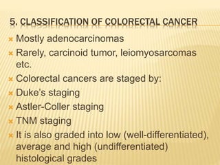
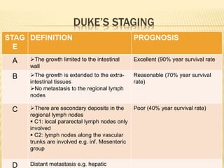
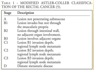
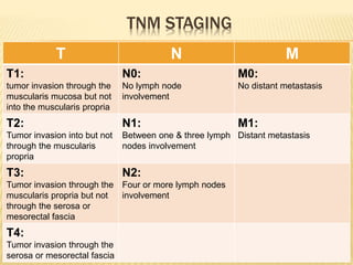
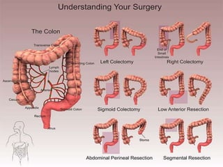
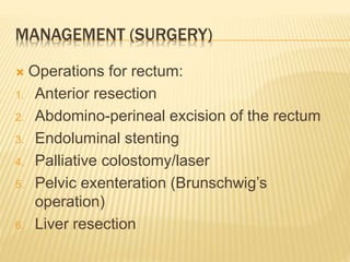
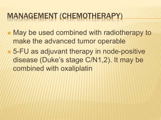
COLORECTAL
NEOPLASMS
By
Dr. Abdul Qadeer
MBBS; FCPS; FICS
Assistant Professor in Surgery
King Faisal University College of Medicine
OBJECTIVES
1. Clinical anatomy of colon & rectum
2. Physiology of colon
3. Colorectal polyps
4. Adenoma-carcinoma sequence
5. Classification of colorectal cancer
6. Signs & symptoms
7. Causes
8. Pathogenesis
OBJECTIVES (CONTD….)
9. Diagnosis
10. Prevention: Lifestyle, medication, screening
11. Management: surgery, chemotherapy,
radiation, palliative care
12. Prognosis
13. Follow-up
14. Epidemiology
1. CLINICAL ANATOMY OF COLON & RECTUM
ļƒ’ Large intestine (Colon & rectum) begins at ileo-
caecal valve and extends up to the anus
ļƒ’ Consists of:
1. Caecum (with appendix attached),
2. Ascending colon,
3. Hepatic flexure,
4. Transverse colon with attached greater
omentum,
5. Splenic flexure,
6. Descending colon,
7. Sigmoid colon and
8. Rectum (begins at sacral promontry)
ļƒ’ Transverse and sigmoid colon are mobile
ļƒ’ Ascending, descending colon and rectum are
fixed
ļƒ’ Apendices epiploicae
ļƒ’ Taenia coli
ļƒ’ Haustrations
ANATOMICAL RELATIONS
ļƒ’ Posterior relations of cecum & ascending
colon are:
1. Right ureter
2. Right gonadal vessels
3. Duodenum
ļƒ’ Posterior relations of left colon are:
1. Left ureter
2. Left gonadal vessels
3. Tail of pancreas
ļƒ’ Rectum is 12-18 cm long in adults, encircled
by the puborectalis muscle, creating an
anorectal ring (120o)
ļƒ’ It has three curves, 2 on right side and 1 on
left side
ļƒ’ Theses curves make semicircular folds
(Houston’s valves) from inside
ANATOMICAL RELATIONS OF THE RECTUM
ļƒ’ Its upper 1/3rd = peritoneum anteriorly +
laterally
ļƒ’ Its middle 1/3rd = peritoneum anteriorly
ļƒ’ Its lower 1/3rd = no peritoneum covering
ļƒ’ Anteriorly = prostate/vagina separated by the
Denonvilliers’ fascia
ļƒ’ Posteriorly = sacrum & coccyx separated by
Waldeyer’s fascia
BLOOD SUPPLY OF LARGE INTESTINE
ļƒ’ Branches of:
a) Superior and inferior mesenteric arteries
b) Marginal artery of Drummond
ļƒ’ Watershed area at the splenic flexure
renders it vulnerable to ischemic colitis
ļƒ’ Venous and lymphatic drainage of colon
follow the arterial supply
ļƒ’ Venous blood is drained into the portal
system
BLOOD SUPPLY OF RECTUM
1. Superior rectal artery: a direct continuation
of inferior mesenteric artery
2. Middle rectal artery: on each side from the
internal iliac artery
3. Inferior rectal artery: on each side from the
internal iliac artery
4. Upper ½: superior hemorrhoidal veins
superior rectal vein inferior
mesenteric vein
5. Middle rectal veins (unimportant)
occasionally exist
LYMPHATIC DRAINAGE OF RECTUM
ļƒ’ Follow the upward route of superior rectal
vessels (the inferior mesenteric vessels),
hence important for surgical clearance in
malignancy
ļƒ’ If it is blocked, then reverse flow may occur
to the nodes along with middle rectal vessels
or the inguinal region via inferior rectal
vessels
NERVE SUPPLY OF THE LARGE INTESTINE
ļƒ’ Sympathetic nerves via splanchnic nerves
surrounding the superior and inferior
mesenteric arteries
ļƒ’ Parasympathetic nerves via vagus and pelvic
splanchnic (S2-4) nerves
2. PYHSIOLOGY OF LARGE INTESTINE
ļƒ’ Absorption of water is the principal function
of the colon
ļƒ’ Out of 1000 ml of ileal contents, 150-250 is
excreted as feces.
ļƒ’ Sodium is actively absorbed
ļƒ’ Chloride and water are passively absorbed
by the sodium pump
ļƒ’ A tiny amount of glucose, fatty acids, amino
acids and vitamins are also absorbed
ļƒ’ Dietary fiber is fermented by normal colonic
microflora (anaerobes e.g. bacteroides and
bifidobacteria) producing short chain fatty
acids (SCFAs) e.g. butyrate which provides
fuel for colonic mucosa
ļƒ’ Diversion of the fecal stream e.g. loop
ileostomy may cause diversion colitis
COLONIC MOTILITY
ļƒ’ It is variable
ļƒ’ Fecal residue reaches the caecum 4 hours,
and rectum 24 hours after meals
ļƒ’ Some residue may still be passed after 4
days through the rectum
3. CLASSIFICATION OF INTESTINAL POLYPS
NO
.
TYPE OF
POLYP
EXAMPLE
1. Inflammatory Pseudopolyps in ulcerative colitis
2. Metaplastic Metaplastic or hyperplastic polyps
3. Hamartomatous Peutz-Jeghers polyp, Juvenile polyp
4. Neoplastic A. Adenoma i. Tubular
ii. Tubulovillous
iii. Villous
B. Adenocarcinoma
C. Carcinoid tumor
4. ADENOMA-CARCINOMA SEQUENCE
ļƒ’ Colorectal cancer arises in a stepwise
progression from adenomas in which
increasing dysplasia in the adenoma is due
to an accumulation of genetic abnormalities.
ļƒ’ This is known as adenoma-carcinoma
sequence
5. CLASSIFICATION OF COLORECTAL CANCER
ļƒ’ Mostly adenocarcinomas
ļƒ’ Rarely, carcinoid tumor, leiomyosarcomas
etc.
ļƒ’ Colorectal cancers are staged by:
ļƒ’ Duke’s staging
ļƒ’ Astler-Coller staging
ļƒ’ TNM staging
ļƒ’ It is also graded into low (well-differentiated),
average and high (undifferentiated)
histological grades
DUKE’S STAGING
STAG
E
DEFINITION PROGNOSIS
A The growth limited to the intestinal
wall
Excellent (90% year survival rate
B The growth is extended to the extra-
intestinal tissues
No metastasis to the regional lymph
nodes
Reasonable (70% year survival
rate)
C There are secondary deposits in the
regional lymph nodes
ļ‚§ C1: local pararectal lymph nodes only
involved
ļ‚§ C2: lymph nodes along the vascular
trunks are involved e.g. inf. Mesenteric
group
Poor (40% year survival rate)
D Distant metastasis e.g. hepatic
TNM STAGING
T N M
T1:
tumor invasion through the
muscularis mucosa but not
into the muscularis propria
N0:
No lymph node
involvement
M0:
No distant metastasis
T2:
Tumor invasion into but not
through the muscularis
propria
N1:
Between one & three lymph
nodes involvement
M1:
Distant metastasis
T3:
Tumor invasion through the
muscularis propria but not
through the serosa or
mesorectal fascia
N2:
Four or more lymph nodes
involvement
T4:
Tumor invasion through the
serosa or mesorectal fascia
6. SIGNS & SYMPTOMS
ļƒ’ Age: usually above 55
ļƒ’ bleeding per rectum
ļƒ’ Sense of incomplete defecation:
tenesmus, spurious diarrhea, bloody slime
ļƒ’ Alteration in bowel habit: early morning
bloody diarrhea
ļƒ’ Pain: a late symptom
ļƒ’ Weight loss: suggests liver metastasis
7. ETIOLOGY
ļƒ’ Adenoma-carcinoma sequence
ļƒ’ Mutation of APC gene, K-ras gene, p53 gene
ļƒ’ Diet: red meat, animal fat. Fiber is protective
ļƒ’ Smoking
ļƒ’ Alcohol
ļƒ’ Long-standing ulcerative colitis & Crohn’s
disease
ļƒ’ After cholecystectomy and
ureterosigmoidostomy?
8. PATHOGENESIS
ļƒ’ Macroscopically: 4 forms
1. Annular
2. Tubular
3. Ulcerative
4. Cauliflower
ļƒ’ Microscopically:
1. Adenocarcinoma commonly
2. Lymphoma, GIST (leiomyosarcoma) rarely
SPREAD OF COLORECTAL CARCINOMA
1. Direct spread: longitudinal, transverse or
radial
2. Lymphatic spread
3. Hematogenous spread: commonly to the
liver, lungs, brian, bone, ovary, kidney are
rare
4. Transcoelomic spread: other structures
within the peritoneum e.g. omentum
9. DIAGNOSIS / INVESTIGATIONS
ļƒ’ Screening: testing for fecal occult blood
followed by colonoscopy in 60-69 aged
people. A guaiac-based test is used which
detects the peroxidase like activity of fecal
hematin.
ļƒ’ Endoscopy: fiberoptic, flexible
sigmoidoscopy / colonoscopy ± biopsy
ļƒ’ Radiology: double-contrast barium enema
may show a filling defect. CT scan of
abdomen / chest
CONTRAST RADIOGRAPHY (APPLE-CORE
APPEARANCE)
10. PREVENTION
ļƒ’ Change the dietary habits
ļƒ’ Quit smoking and alcohol
ļƒ’ Early diagnosis of intestinal polyps
11. MANAGEMENT (SURGERY)
ļƒ’ Pre-operative preparation±
ļƒ’ Prevention of thromboembolism by anti-
embolic stockings or s/c LMW heparin
ļƒ’ Operations for colon:
1. Right hemicolectomy
2. Extended right hemicolectomy
3. Left hemicolectomy
4. Emergency surgery e.g. Hartmann’s
procedure
MANAGEMENT (SURGERY)
ļƒ’ Operations for rectum:
1. Anterior resection
2. Abdomino-perineal excision of the rectum
3. Endoluminal stenting
4. Palliative colostomy/laser
5. Pelvic exenteration (Brunschwig’s
operation)
6. Liver resection
MANAGEMENT (CHEMOTHERAPY)
ļƒ’ May be used combined with radiotherapy to
make the advanced tumor operable
ļƒ’ 5-FU as adjuvant therapy in node-positive
disease (Duke’s stage C/N1,2). It may be
combined with oxaliplatin
MANAGEMENT (RADIOTHERAPY)
ļƒ’ Palliative irradiation for inoperable, painful
tumors
ļƒ’ Brachyradiotherapy
12. PROGNOSIS (5-YEAR SURVIVAL RATE)
ļƒ’ Overall 5-year survival for colorectal cancer
is 50%
ļƒ’ Duke’s stage A: by surgical resection alone,
90%
ļƒ’ Tumor spread beyond the bowel wall: 60-
70%
ļƒ’ Tumor with lymph node metastasis: 30%
ļƒ’ Metastatic disease: 10%
13. FOLLOW-UP
ļƒ’ Surveillance colonoscopy
ļƒ’ Regular imaging of liver (US, CTS) for
metastasis
ļƒ’ Measurement of CEA
14. EPIDEMIOLOGY
ļƒ’ In UK, colorectal cancer is second most
cause of cancer death
ļƒ’ About 35000 patients are diagnosed with
colorectal cancer every year in UK; 1/3rd of
rectum and 2/3rd of colon
ļƒ’ M:F = same
ļƒ’ Less frequent in developing countries than in
industrialized countries
Colorectal neoplasms

Colorectal neoplasms

  • 1.
    COLORECTAL NEOPLASMS By Dr. Abdul Qadeer MBBS;FCPS; FICS Assistant Professor in Surgery King Faisal University College of Medicine
  • 2.
    OBJECTIVES 1. Clinical anatomyof colon & rectum 2. Physiology of colon 3. Colorectal polyps 4. Adenoma-carcinoma sequence 5. Classification of colorectal cancer 6. Signs & symptoms 7. Causes 8. Pathogenesis
  • 3.
    OBJECTIVES (CONTD….) 9. Diagnosis 10.Prevention: Lifestyle, medication, screening 11. Management: surgery, chemotherapy, radiation, palliative care 12. Prognosis 13. Follow-up 14. Epidemiology
  • 4.
    1. CLINICAL ANATOMYOF COLON & RECTUM ļƒ’ Large intestine (Colon & rectum) begins at ileo- caecal valve and extends up to the anus ļƒ’ Consists of: 1. Caecum (with appendix attached), 2. Ascending colon, 3. Hepatic flexure, 4. Transverse colon with attached greater omentum, 5. Splenic flexure, 6. Descending colon, 7. Sigmoid colon and 8. Rectum (begins at sacral promontry)
  • 6.
    ļƒ’ Transverse andsigmoid colon are mobile ļƒ’ Ascending, descending colon and rectum are fixed ļƒ’ Apendices epiploicae ļƒ’ Taenia coli ļƒ’ Haustrations
  • 8.
    ANATOMICAL RELATIONS ļƒ’ Posteriorrelations of cecum & ascending colon are: 1. Right ureter 2. Right gonadal vessels 3. Duodenum ļƒ’ Posterior relations of left colon are: 1. Left ureter 2. Left gonadal vessels 3. Tail of pancreas
  • 10.
    ļƒ’ Rectum is12-18 cm long in adults, encircled by the puborectalis muscle, creating an anorectal ring (120o) ļƒ’ It has three curves, 2 on right side and 1 on left side ļƒ’ Theses curves make semicircular folds (Houston’s valves) from inside
  • 12.
    ANATOMICAL RELATIONS OFTHE RECTUM ļƒ’ Its upper 1/3rd = peritoneum anteriorly + laterally ļƒ’ Its middle 1/3rd = peritoneum anteriorly ļƒ’ Its lower 1/3rd = no peritoneum covering ļƒ’ Anteriorly = prostate/vagina separated by the Denonvilliers’ fascia ļƒ’ Posteriorly = sacrum & coccyx separated by Waldeyer’s fascia
  • 13.
    BLOOD SUPPLY OFLARGE INTESTINE ļƒ’ Branches of: a) Superior and inferior mesenteric arteries b) Marginal artery of Drummond ļƒ’ Watershed area at the splenic flexure renders it vulnerable to ischemic colitis ļƒ’ Venous and lymphatic drainage of colon follow the arterial supply ļƒ’ Venous blood is drained into the portal system
  • 15.
    BLOOD SUPPLY OFRECTUM 1. Superior rectal artery: a direct continuation of inferior mesenteric artery 2. Middle rectal artery: on each side from the internal iliac artery 3. Inferior rectal artery: on each side from the internal iliac artery 4. Upper ½: superior hemorrhoidal veins superior rectal vein inferior mesenteric vein 5. Middle rectal veins (unimportant) occasionally exist
  • 17.
    LYMPHATIC DRAINAGE OFRECTUM ļƒ’ Follow the upward route of superior rectal vessels (the inferior mesenteric vessels), hence important for surgical clearance in malignancy ļƒ’ If it is blocked, then reverse flow may occur to the nodes along with middle rectal vessels or the inguinal region via inferior rectal vessels
  • 18.
    NERVE SUPPLY OFTHE LARGE INTESTINE ļƒ’ Sympathetic nerves via splanchnic nerves surrounding the superior and inferior mesenteric arteries ļƒ’ Parasympathetic nerves via vagus and pelvic splanchnic (S2-4) nerves
  • 19.
    2. PYHSIOLOGY OFLARGE INTESTINE ļƒ’ Absorption of water is the principal function of the colon ļƒ’ Out of 1000 ml of ileal contents, 150-250 is excreted as feces. ļƒ’ Sodium is actively absorbed ļƒ’ Chloride and water are passively absorbed by the sodium pump ļƒ’ A tiny amount of glucose, fatty acids, amino acids and vitamins are also absorbed
  • 20.
    ļƒ’ Dietary fiberis fermented by normal colonic microflora (anaerobes e.g. bacteroides and bifidobacteria) producing short chain fatty acids (SCFAs) e.g. butyrate which provides fuel for colonic mucosa ļƒ’ Diversion of the fecal stream e.g. loop ileostomy may cause diversion colitis
  • 21.
    COLONIC MOTILITY ļƒ’ Itis variable ļƒ’ Fecal residue reaches the caecum 4 hours, and rectum 24 hours after meals ļƒ’ Some residue may still be passed after 4 days through the rectum
  • 22.
    3. CLASSIFICATION OFINTESTINAL POLYPS NO . TYPE OF POLYP EXAMPLE 1. Inflammatory Pseudopolyps in ulcerative colitis 2. Metaplastic Metaplastic or hyperplastic polyps 3. Hamartomatous Peutz-Jeghers polyp, Juvenile polyp 4. Neoplastic A. Adenoma i. Tubular ii. Tubulovillous iii. Villous B. Adenocarcinoma C. Carcinoid tumor
  • 23.
    4. ADENOMA-CARCINOMA SEQUENCE ļƒ’Colorectal cancer arises in a stepwise progression from adenomas in which increasing dysplasia in the adenoma is due to an accumulation of genetic abnormalities. ļƒ’ This is known as adenoma-carcinoma sequence
  • 25.
    5. CLASSIFICATION OFCOLORECTAL CANCER ļƒ’ Mostly adenocarcinomas ļƒ’ Rarely, carcinoid tumor, leiomyosarcomas etc. ļƒ’ Colorectal cancers are staged by: ļƒ’ Duke’s staging ļƒ’ Astler-Coller staging ļƒ’ TNM staging ļƒ’ It is also graded into low (well-differentiated), average and high (undifferentiated) histological grades
  • 26.
    DUKE’S STAGING STAG E DEFINITION PROGNOSIS AThe growth limited to the intestinal wall Excellent (90% year survival rate B The growth is extended to the extra- intestinal tissues No metastasis to the regional lymph nodes Reasonable (70% year survival rate) C There are secondary deposits in the regional lymph nodes ļ‚§ C1: local pararectal lymph nodes only involved ļ‚§ C2: lymph nodes along the vascular trunks are involved e.g. inf. Mesenteric group Poor (40% year survival rate) D Distant metastasis e.g. hepatic
  • 28.
    TNM STAGING T NM T1: tumor invasion through the muscularis mucosa but not into the muscularis propria N0: No lymph node involvement M0: No distant metastasis T2: Tumor invasion into but not through the muscularis propria N1: Between one & three lymph nodes involvement M1: Distant metastasis T3: Tumor invasion through the muscularis propria but not through the serosa or mesorectal fascia N2: Four or more lymph nodes involvement T4: Tumor invasion through the serosa or mesorectal fascia
  • 29.
    6. SIGNS &SYMPTOMS ļƒ’ Age: usually above 55 ļƒ’ bleeding per rectum ļƒ’ Sense of incomplete defecation: tenesmus, spurious diarrhea, bloody slime ļƒ’ Alteration in bowel habit: early morning bloody diarrhea ļƒ’ Pain: a late symptom ļƒ’ Weight loss: suggests liver metastasis
  • 30.
    7. ETIOLOGY ļƒ’ Adenoma-carcinomasequence ļƒ’ Mutation of APC gene, K-ras gene, p53 gene ļƒ’ Diet: red meat, animal fat. Fiber is protective ļƒ’ Smoking ļƒ’ Alcohol ļƒ’ Long-standing ulcerative colitis & Crohn’s disease ļƒ’ After cholecystectomy and ureterosigmoidostomy?
  • 31.
    8. PATHOGENESIS ļƒ’ Macroscopically:4 forms 1. Annular 2. Tubular 3. Ulcerative 4. Cauliflower ļƒ’ Microscopically: 1. Adenocarcinoma commonly 2. Lymphoma, GIST (leiomyosarcoma) rarely
  • 32.
    SPREAD OF COLORECTALCARCINOMA 1. Direct spread: longitudinal, transverse or radial 2. Lymphatic spread 3. Hematogenous spread: commonly to the liver, lungs, brian, bone, ovary, kidney are rare 4. Transcoelomic spread: other structures within the peritoneum e.g. omentum
  • 33.
    9. DIAGNOSIS /INVESTIGATIONS ļƒ’ Screening: testing for fecal occult blood followed by colonoscopy in 60-69 aged people. A guaiac-based test is used which detects the peroxidase like activity of fecal hematin. ļƒ’ Endoscopy: fiberoptic, flexible sigmoidoscopy / colonoscopy ± biopsy ļƒ’ Radiology: double-contrast barium enema may show a filling defect. CT scan of abdomen / chest
  • 34.
  • 35.
    10. PREVENTION ļƒ’ Changethe dietary habits ļƒ’ Quit smoking and alcohol ļƒ’ Early diagnosis of intestinal polyps
  • 36.
    11. MANAGEMENT (SURGERY) ļƒ’Pre-operative preparation± ļƒ’ Prevention of thromboembolism by anti- embolic stockings or s/c LMW heparin ļƒ’ Operations for colon: 1. Right hemicolectomy 2. Extended right hemicolectomy 3. Left hemicolectomy 4. Emergency surgery e.g. Hartmann’s procedure
  • 38.
    MANAGEMENT (SURGERY) ļƒ’ Operationsfor rectum: 1. Anterior resection 2. Abdomino-perineal excision of the rectum 3. Endoluminal stenting 4. Palliative colostomy/laser 5. Pelvic exenteration (Brunschwig’s operation) 6. Liver resection
  • 40.
    MANAGEMENT (CHEMOTHERAPY) ļƒ’ Maybe used combined with radiotherapy to make the advanced tumor operable ļƒ’ 5-FU as adjuvant therapy in node-positive disease (Duke’s stage C/N1,2). It may be combined with oxaliplatin
  • 41.
    MANAGEMENT (RADIOTHERAPY) ļƒ’ Palliativeirradiation for inoperable, painful tumors ļƒ’ Brachyradiotherapy
  • 42.
    12. PROGNOSIS (5-YEARSURVIVAL RATE) ļƒ’ Overall 5-year survival for colorectal cancer is 50% ļƒ’ Duke’s stage A: by surgical resection alone, 90% ļƒ’ Tumor spread beyond the bowel wall: 60- 70% ļƒ’ Tumor with lymph node metastasis: 30% ļƒ’ Metastatic disease: 10%
  • 43.
    13. FOLLOW-UP ļƒ’ Surveillancecolonoscopy ļƒ’ Regular imaging of liver (US, CTS) for metastasis ļƒ’ Measurement of CEA
  • 44.
    14. EPIDEMIOLOGY ļƒ’ InUK, colorectal cancer is second most cause of cancer death ļƒ’ About 35000 patients are diagnosed with colorectal cancer every year in UK; 1/3rd of rectum and 2/3rd of colon ļƒ’ M:F = same ļƒ’ Less frequent in developing countries than in industrialized countries