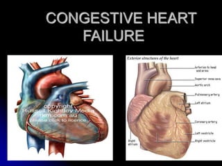
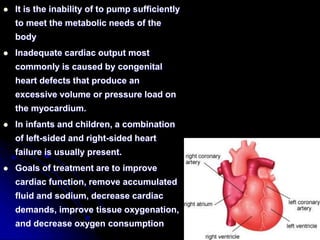
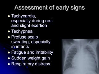
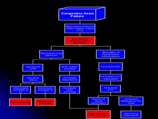
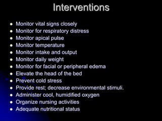
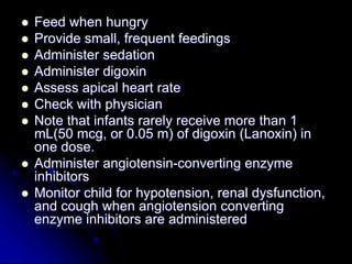
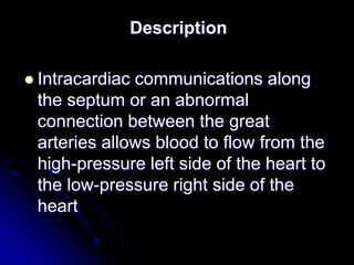
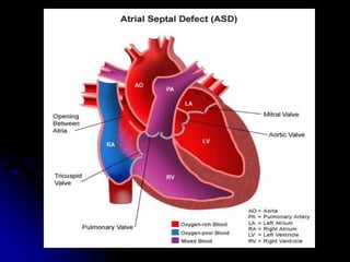
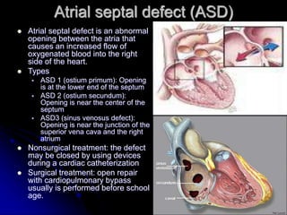
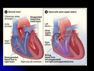
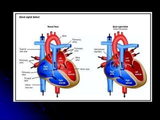
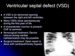
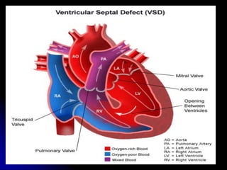
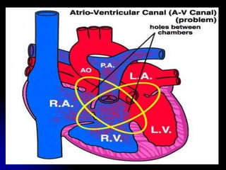
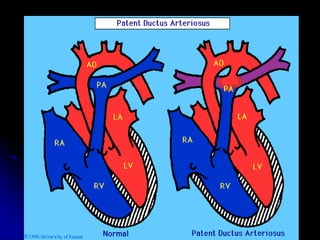
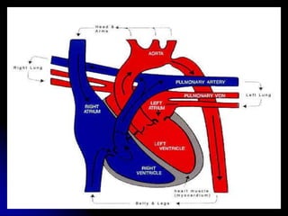
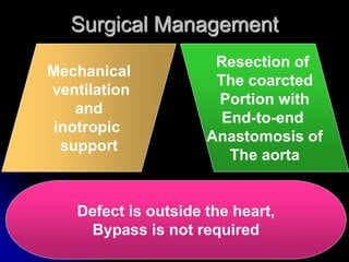
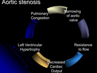
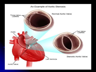
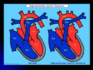
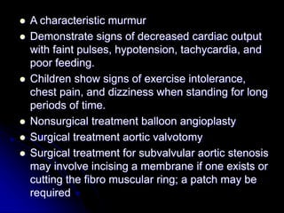
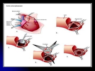
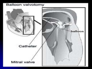
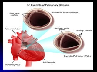
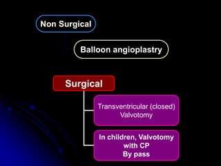
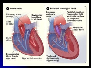
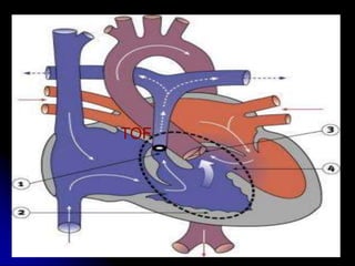
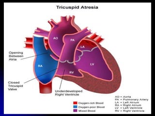
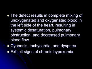
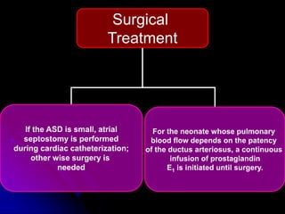
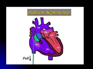
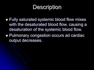
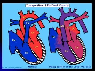
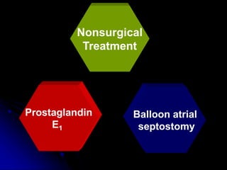
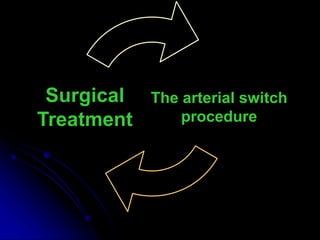
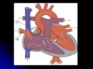
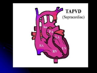
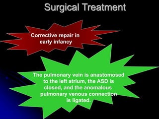
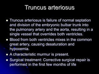
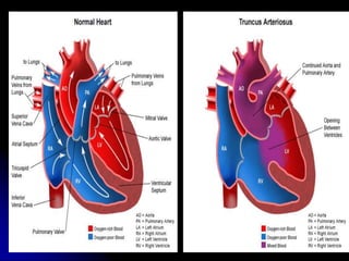
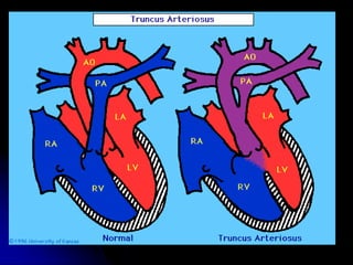
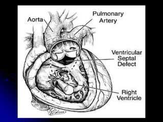
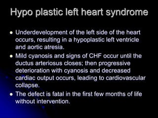
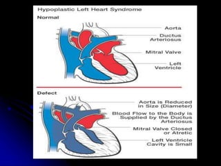
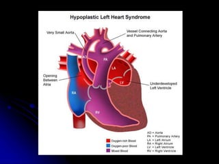
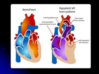
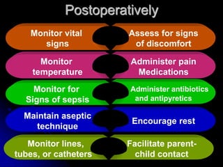
CARDIO-VASCULAR
DISORDERS
CONGESTIVE HEART
FAILURE
 It is the inability of to pump sufficiently
to meet the metabolic needs of the
body
 Inadequate cardiac output most
commonly is caused by congenital
heart defects that produce an
excessive volume or pressure load on
the myocardium.
 In infants and children, a combination
of left-sided and right-sided heart
failure is usually present.
 Goals of treatment are to improve
cardiac function, remove accumulated
fluid and sodium, decrease cardiac
demands, improve tissue oxygenation,
and decrease oxygen consumption
Assessment of early signs
 Tachycardia,
especially during rest
and slight exertion
 Tachypnea
 Profuse scalp
sweating, especially
in infants
 Fatigue and irritability
 Sudden weight gain
 Respiratory distress
Interventions
 Monitor vital signs closely
 Monitor for respiratory distress
 Monitor apical pulse
 Monitor temperature
 Monitor intake and output
 Monitor daily weight
 Monitor for facial or peripheral edema
 Elevate the head of the bed
 Prevent cold stress
 Provide rest; decrease environmental stimuli.
 Administer cool, humidified oxygen
 Organize nursing activities
 Adequate nutritional status
 Feed when hungry
 Provide small, frequent feedings
 Administer sedation
 Administer digoxin
 Assess apical heart rate
 Check with physician
 Note that infants rarely receive more than 1
mL(50 mcg, or 0.05 m) of digoxin (Lanoxin) in
one dose.
 Administer angiotensin-converting enzyme
inhibitors
 Monitor child for hypotension, renal dysfunction,
and cough when angiotension converting
enzyme inhibitors are administered
 Administer diuretics as prescribed
 Administer potassium supplements
 Monitor serum electrolytes
 Restrict fluid as prescribed in the acute
stage
 Check with the physician regarding
sodium restriction.
 Instruct the parents regarding the
description of the diagnosis and
administration of medications
 Cardiopulmonary resuscitation.
Defects with
increased
pulmonary
Blood Flow
Description
 Intracardiac communications along
the septum or an abnormal
connection between the great
arteries allows blood to flow from the
high-pressure left side of the heart to
the low-pressure right side of the
heart
Atrial septal defect (ASD)
 Atrial septal defect is an abnormal
opening between the atria that
causes an increased flow of
oxygenated blood into the right
side of the heart.
 Types
 ASD 1 (ostium primum): Opening
is at the lower end of the septum
 ASD 2 (ostium secundum):
Opening is near the center of the
septum
 ASD3 (sinus venosus defect):
Opening is near the junction of the
superior vena cava and the right
atrium
 Nonsurgical treatment: the defect
may be closed by using devices
during a cardiac catheterization
 Surgical treatment: open repair
with cardiopulmonary bypass
usually is performed before school
age.
Ventricular septal defect (VSD)
 A VSD is an abnormal opening
between the right and left ventricles.
 Many VSDs close spontaneously
during the first year of life
 A characteristic murmur is present;
CHF is common.
 Nonsurgical treatment: Device
closure during cardiac
catheterization may be possible.
 Surgical treatment: Open repair is
done with cardiopulmonary bypass.
Atrioventricular canal defect
 The defect results from incomplete fusion of the
endocardial cushions.
 The defect is the most common cardiac defect in
Down syndrome.
 A characteristic murmur is present.
 The infant usually has mild to moderate CHF;
cyanosis increases with crying.
 Surgical treatment can include pulmonary artery
banding for infants with severe symptoms
(palliative) or complete repair via
cardiopulmonary bypass.
Patent ductus arteriosus
 Patent ductus arteriosus is failure of the fetal
ductus arteriosus (artery connecting the aorta
and the pulmonary artery) to close within the first
weeks of life.
 A characteristic machinery-like murmur
 A widened pulse pressure and bounding pulses
are present.
 Medical management: Indomethacin (Indocin), a
prostaglandin inhibitor, may be administered to
close a patent ductus arteriosus in premature
infants and some newborns.
 Management: Coils may be used to occlude the
patent ductus arteriosus via a cardiac
catheterization procedure or the defect may
require surgical management.
OBSTRUCTIVE
DEFECTS
Description
 Blood exiting the heart meets an area of
anatomic narrowing (stenosis), causing
obstruction to blood flow.
 The location of narrowing is usually near
the valve
Coarctation of the aorta
 Coarctation of the aorta is localized narrowing
near the insertion of the ductus arteriousus.
 Collateral circulation develops during fetal life to
maintain flow from the ascending to the
descending aorta.
 Signs of CHF occur in infants
 High blood pressure and bounding pulses in the
arms, weak or absent femoral pulses, and cool
lower extremities may be present.
 Children may experience headaches,
dizziness, fainting, and epistaxis resulting
from hypertension.
 Nonsurgical treatment is balloon angioplasty
in children; restenosis can occur.
Coarctation of the aorta is a constriction (narrowing) of a part of
the aorta, the main artery carrying "red blood" to the body. This
generally occurs close to the region where arteries to the head
and neck arise. The constriction obstructs blood flow to the
lower parts of the body. It causes blood pressure to increase
above the coarctation, resulting in higher blood pressure in the
upper part of the body compared with the lower part of the
body.
The left side of the heart works hard as it tries to pump blood
past the coarctation. This may cause the left ventricle to
enlarge. If the coarctation is very severe, an infant's heart may
fail, resulting in rapid heart rate, rapid breathing and poor
feeding. In less severe narrowing, the child may have no
noticeable side effects except increased blood pressure. If the
high blood pressure continues, damage results to other blood
vessels in the body.
Surgical Management
Mechanical
ventilation
and
inotropic
support
Resection of
The coarcted
Portion with
End-to-end
Anastomosis of
The aorta
Defect is outside the heart,
Bypass is not required
Aortic stenosis
Narrowing
of aortic
valve
Left Ventricular
Hypertrophy
Pulmonary
Congestion
Decreased
Cardiac
Output
Resistance
to flow
Supravalvular stenosis
Subvalvular stenosis
Types
Valvular stenosis
Valvular stenosis
ECHOCARDIOGRAPHY
 A characteristic murmur
 Demonstrate signs of decreased cardiac output
with faint pulses, hypotension, tachycardia, and
poor feeding.
 Children show signs of exercise intolerance,
chest pain, and dizziness when standing for long
periods of time.
 Nonsurgical treatment balloon angioplasty
 Surgical treatment aortic valvotomy
 Surgical treatment for subvalvular aortic stenosis
may involve incising a membrane if one exists or
cutting the fibro muscular ring; a patch may be
required
Pulmonary stenosis
 Pulmonary stenosis is narrowing at the entrance
to the pulmonary artery.
 Resistance to blood flow causes right ventricular
hypertrophy and decreased pulmonary blood
flow;
 Pulmonary atresia is the extreme form of
pulmonary stenosis
 A characteristic murmur
 The infant or child may be asymptomatic
 Severe narrowing will be cyanotic.
Non Surgical
Balloon angioplastry
Surgical
Transventricular (closed)
Valvotomy
In children, Valvotomy
with CP
By pass
DEFECTS WITH
DECREASED
PULMONARY BLOOD
FLOW
Description
 Obstructed pulmonary blood flow and an
anatomic defect (ASD or VD) between the
right and left sides of the heart are
present.
 Pressure on the right side of the heart
increases exceeding pressure on the left
side, which allows desaturated blood to
shunt right to left, causing desaturation in
the left side of the heart and in the
systemic circulation.
 Hypoxemia and Cyanosis
Tetralogy of Fallot
 The tetralogy of Fillet includes four
defects: VSD, pulmonary stenosis,
overriding aorta, and right ventricular
hypertrophy.
 If pulmonary vascular resistance is higher
than systemic resistance, the shut is from
right to left; if systemic resistance is higher
than pulmonary resistance, the shunt is
left to right.
TOF
 Infants
 The infant may be acutely cyanotic at birth or may
have mild cyanosis that progresses over the first year
of life as the pulmonic stenosis worsens.
 Characteristic murmur
 Called blue spells or tet spells
 X-ray shows boot shaped heart
 Children: With increasing cyanosis, squatting,
clubbing of the fingers, and poor growth
 Surgery: Ballock Taussing procedure-
anastomosis of subclavian to pulmonary artery
 Pott’s procedure – Anastomosis of lt.pul. Artery
with descending aorta
 Watterson procedure – Ascending aorta with rt
pulmonary artery
Surgical Treatment
Palliative shunt Complete repair
•The shunt increases
pulmonary blood flow
and increases oxygen
saturation in infants who
cannot undergo primary
repair
• First year of life.
• Complete repair
involves closure of the
VSD and resection of
the stenosis, with a
pericardial patch to
enlarge the right
ventricular outflow tract.
Tricuspid Artesia
 Tricuspid Artesia is failure of the tricuspid
valve to develop.
 No communication exists from the right
atrium to the right ventricle
 Blood flows through an ASD or a patent
foramen ovale to the left side of the heart
and through a VSD to the right ventricle
and out to the lungs.
 Associated with pulmonic stenosis and
transposition of the great arteries.
 The defect results in complete mixing of
unoxygenated and oxygenated blood in
the left side of the heart, resulting in
systemic desaturation, pulmonary
obstruction, and decreased pulmonary
blood flow.
 Cyanosis, tachycardia, and dyspnea
 Exhibit signs of chronic hypoxemia
Surgical
Treatment
If the ASD is small, atrial
septostomy is performed
during cardiac catheterization;
other wise surgery is
needed
For the neonate whose pulmonary
blood flow depends on the patency
of the ductus arteriosus, a continuous
infusion of prostaglandin
E1 is initiated until surgery.
atrial
septostomy
MIXED
DEFECTS
Description
 Fully saturated systemic blood flow mixes
with the desaturated blood flow, causing a
desaturation of the systemic blood flow.
 Pulmonary congestion occurs ad cardiac
output decreases.
 The pulmonary artery leaves the left ventricle,
and the aorta exits from the right ventricle.
 No communication exists between the systemic
and pulmonary circulation
 Infants with minimal communication are severely
cyanotic and depressed at birth.
 Infants with large septal defects or a patent
ductus arteriosus may be less severely cyanotic
but may have symptoms of CHF.
 Cardiomegaly is evident a few weeks after birth
Transposition of the great vessels.
Nonsurgical
Treatment
Prostaglandin
E1
Balloon atrial
septostomy
The arterial switch
procedure
Surgical
Treatment
Total anomalous pulmonary venous
connection
 The defect is a failure of the pulmonary
veins to join the left atrium
 The defect results in mixed blood being
returned to the right atrium and shunted
from the right to the left through an ASD
 May remain small.
Surgical Treatment
Corrective repair in
early infancy
The pulmonary vein is anastomosed
to the left atrium, the ASD is
closed, and the anomalous
pulmonary venous connection
is ligated.
Truncus arteriosus
 Truncus arteriosus is failure of normal septation
and division of the embryonic bulbar trunk into
the pulmonary artery and the aorta, resulting in a
single vessel that overrides both ventricles.
 Blood from both ventricles mixes in the common
great artery, causing desaturation and
hypoxemia.
 A characteristic murmur is present.
 Surgical treatment: Corrective surgical repair is
performed in the first few months of life
Hypo plastic left heart syndrome
 Underdevelopment of the left side of the heart
occurs, resulting in a hypoplastic left ventricle
and aortic atresia.
 Mild cyanosis and signs of CHF occur until the
ductus arteriosus closes; then progressive
deterioration with cyanosis and decreased
cardiac output occurs, leading to cardiovascular
collapse.
 The defect is fatal in the first few months of life
without intervention.
Surgical treatment
In the preoperative period, the neonate
requires mechanical ventilation and a
continuous infusion of prostaglandin E1 to
maintain ductal patency, ensuring
adequate systemic blood flow.
Postoperatively
Monitor vital
signs
Assess for signs
of discomfort
Monitor
temperature
Administer pain
Medications
Monitor for
Signs of sepsis
Administer antibiotics
and antipyretics
Maintain aseptic
technique
Encourage rest
Monitor lines,
tubes, or catheters
Facilitate parent-
child contact

C.V.S.ppt

  • 1.
  • 3.
  • 4.
     It isthe inability of to pump sufficiently to meet the metabolic needs of the body  Inadequate cardiac output most commonly is caused by congenital heart defects that produce an excessive volume or pressure load on the myocardium.  In infants and children, a combination of left-sided and right-sided heart failure is usually present.  Goals of treatment are to improve cardiac function, remove accumulated fluid and sodium, decrease cardiac demands, improve tissue oxygenation, and decrease oxygen consumption
  • 5.
    Assessment of earlysigns  Tachycardia, especially during rest and slight exertion  Tachypnea  Profuse scalp sweating, especially in infants  Fatigue and irritability  Sudden weight gain  Respiratory distress
  • 7.
    Interventions  Monitor vitalsigns closely  Monitor for respiratory distress  Monitor apical pulse  Monitor temperature  Monitor intake and output  Monitor daily weight  Monitor for facial or peripheral edema  Elevate the head of the bed  Prevent cold stress  Provide rest; decrease environmental stimuli.  Administer cool, humidified oxygen  Organize nursing activities  Adequate nutritional status
  • 8.
     Feed whenhungry  Provide small, frequent feedings  Administer sedation  Administer digoxin  Assess apical heart rate  Check with physician  Note that infants rarely receive more than 1 mL(50 mcg, or 0.05 m) of digoxin (Lanoxin) in one dose.  Administer angiotensin-converting enzyme inhibitors  Monitor child for hypotension, renal dysfunction, and cough when angiotension converting enzyme inhibitors are administered
  • 9.
     Administer diureticsas prescribed  Administer potassium supplements  Monitor serum electrolytes  Restrict fluid as prescribed in the acute stage  Check with the physician regarding sodium restriction.  Instruct the parents regarding the description of the diagnosis and administration of medications  Cardiopulmonary resuscitation.
  • 10.
  • 11.
    Description  Intracardiac communicationsalong the septum or an abnormal connection between the great arteries allows blood to flow from the high-pressure left side of the heart to the low-pressure right side of the heart
  • 13.
    Atrial septal defect(ASD)  Atrial septal defect is an abnormal opening between the atria that causes an increased flow of oxygenated blood into the right side of the heart.  Types  ASD 1 (ostium primum): Opening is at the lower end of the septum  ASD 2 (ostium secundum): Opening is near the center of the septum  ASD3 (sinus venosus defect): Opening is near the junction of the superior vena cava and the right atrium  Nonsurgical treatment: the defect may be closed by using devices during a cardiac catheterization  Surgical treatment: open repair with cardiopulmonary bypass usually is performed before school age.
  • 16.
    Ventricular septal defect(VSD)  A VSD is an abnormal opening between the right and left ventricles.  Many VSDs close spontaneously during the first year of life  A characteristic murmur is present; CHF is common.  Nonsurgical treatment: Device closure during cardiac catheterization may be possible.  Surgical treatment: Open repair is done with cardiopulmonary bypass.
  • 18.
    Atrioventricular canal defect The defect results from incomplete fusion of the endocardial cushions.  The defect is the most common cardiac defect in Down syndrome.  A characteristic murmur is present.  The infant usually has mild to moderate CHF; cyanosis increases with crying.  Surgical treatment can include pulmonary artery banding for infants with severe symptoms (palliative) or complete repair via cardiopulmonary bypass.
  • 20.
    Patent ductus arteriosus Patent ductus arteriosus is failure of the fetal ductus arteriosus (artery connecting the aorta and the pulmonary artery) to close within the first weeks of life.  A characteristic machinery-like murmur  A widened pulse pressure and bounding pulses are present.  Medical management: Indomethacin (Indocin), a prostaglandin inhibitor, may be administered to close a patent ductus arteriosus in premature infants and some newborns.  Management: Coils may be used to occlude the patent ductus arteriosus via a cardiac catheterization procedure or the defect may require surgical management.
  • 22.
  • 23.
    Description  Blood exitingthe heart meets an area of anatomic narrowing (stenosis), causing obstruction to blood flow.  The location of narrowing is usually near the valve
  • 24.
    Coarctation of theaorta  Coarctation of the aorta is localized narrowing near the insertion of the ductus arteriousus.  Collateral circulation develops during fetal life to maintain flow from the ascending to the descending aorta.  Signs of CHF occur in infants  High blood pressure and bounding pulses in the arms, weak or absent femoral pulses, and cool lower extremities may be present.  Children may experience headaches, dizziness, fainting, and epistaxis resulting from hypertension.  Nonsurgical treatment is balloon angioplasty in children; restenosis can occur.
  • 25.
    Coarctation of theaorta is a constriction (narrowing) of a part of the aorta, the main artery carrying "red blood" to the body. This generally occurs close to the region where arteries to the head and neck arise. The constriction obstructs blood flow to the lower parts of the body. It causes blood pressure to increase above the coarctation, resulting in higher blood pressure in the upper part of the body compared with the lower part of the body. The left side of the heart works hard as it tries to pump blood past the coarctation. This may cause the left ventricle to enlarge. If the coarctation is very severe, an infant's heart may fail, resulting in rapid heart rate, rapid breathing and poor feeding. In less severe narrowing, the child may have no noticeable side effects except increased blood pressure. If the high blood pressure continues, damage results to other blood vessels in the body.
  • 29.
    Surgical Management Mechanical ventilation and inotropic support Resection of Thecoarcted Portion with End-to-end Anastomosis of The aorta Defect is outside the heart, Bypass is not required
  • 30.
    Aortic stenosis Narrowing of aortic valve LeftVentricular Hypertrophy Pulmonary Congestion Decreased Cardiac Output Resistance to flow
  • 33.
  • 34.
  • 37.
     A characteristicmurmur  Demonstrate signs of decreased cardiac output with faint pulses, hypotension, tachycardia, and poor feeding.  Children show signs of exercise intolerance, chest pain, and dizziness when standing for long periods of time.  Nonsurgical treatment balloon angioplasty  Surgical treatment aortic valvotomy  Surgical treatment for subvalvular aortic stenosis may involve incising a membrane if one exists or cutting the fibro muscular ring; a patch may be required
  • 40.
    Pulmonary stenosis  Pulmonarystenosis is narrowing at the entrance to the pulmonary artery.  Resistance to blood flow causes right ventricular hypertrophy and decreased pulmonary blood flow;  Pulmonary atresia is the extreme form of pulmonary stenosis  A characteristic murmur  The infant or child may be asymptomatic  Severe narrowing will be cyanotic.
  • 42.
    Non Surgical Balloon angioplastry Surgical Transventricular(closed) Valvotomy In children, Valvotomy with CP By pass
  • 46.
  • 47.
    Description  Obstructed pulmonaryblood flow and an anatomic defect (ASD or VD) between the right and left sides of the heart are present.  Pressure on the right side of the heart increases exceeding pressure on the left side, which allows desaturated blood to shunt right to left, causing desaturation in the left side of the heart and in the systemic circulation.  Hypoxemia and Cyanosis
  • 48.
    Tetralogy of Fallot The tetralogy of Fillet includes four defects: VSD, pulmonary stenosis, overriding aorta, and right ventricular hypertrophy.  If pulmonary vascular resistance is higher than systemic resistance, the shut is from right to left; if systemic resistance is higher than pulmonary resistance, the shunt is left to right.
  • 50.
  • 51.
     Infants  Theinfant may be acutely cyanotic at birth or may have mild cyanosis that progresses over the first year of life as the pulmonic stenosis worsens.  Characteristic murmur  Called blue spells or tet spells  X-ray shows boot shaped heart  Children: With increasing cyanosis, squatting, clubbing of the fingers, and poor growth  Surgery: Ballock Taussing procedure- anastomosis of subclavian to pulmonary artery  Pott’s procedure – Anastomosis of lt.pul. Artery with descending aorta  Watterson procedure – Ascending aorta with rt pulmonary artery
  • 52.
    Surgical Treatment Palliative shuntComplete repair •The shunt increases pulmonary blood flow and increases oxygen saturation in infants who cannot undergo primary repair • First year of life. • Complete repair involves closure of the VSD and resection of the stenosis, with a pericardial patch to enlarge the right ventricular outflow tract.
  • 53.
    Tricuspid Artesia  TricuspidArtesia is failure of the tricuspid valve to develop.  No communication exists from the right atrium to the right ventricle  Blood flows through an ASD or a patent foramen ovale to the left side of the heart and through a VSD to the right ventricle and out to the lungs.  Associated with pulmonic stenosis and transposition of the great arteries.
  • 55.
     The defectresults in complete mixing of unoxygenated and oxygenated blood in the left side of the heart, resulting in systemic desaturation, pulmonary obstruction, and decreased pulmonary blood flow.  Cyanosis, tachycardia, and dyspnea  Exhibit signs of chronic hypoxemia
  • 56.
    Surgical Treatment If the ASDis small, atrial septostomy is performed during cardiac catheterization; other wise surgery is needed For the neonate whose pulmonary blood flow depends on the patency of the ductus arteriosus, a continuous infusion of prostaglandin E1 is initiated until surgery.
  • 58.
  • 59.
  • 60.
    Description  Fully saturatedsystemic blood flow mixes with the desaturated blood flow, causing a desaturation of the systemic blood flow.  Pulmonary congestion occurs ad cardiac output decreases.
  • 61.
     The pulmonaryartery leaves the left ventricle, and the aorta exits from the right ventricle.  No communication exists between the systemic and pulmonary circulation  Infants with minimal communication are severely cyanotic and depressed at birth.  Infants with large septal defects or a patent ductus arteriosus may be less severely cyanotic but may have symptoms of CHF.  Cardiomegaly is evident a few weeks after birth Transposition of the great vessels.
  • 64.
  • 65.
  • 66.
    Total anomalous pulmonaryvenous connection  The defect is a failure of the pulmonary veins to join the left atrium  The defect results in mixed blood being returned to the right atrium and shunted from the right to the left through an ASD  May remain small.
  • 69.
    Surgical Treatment Corrective repairin early infancy The pulmonary vein is anastomosed to the left atrium, the ASD is closed, and the anomalous pulmonary venous connection is ligated.
  • 70.
    Truncus arteriosus  Truncusarteriosus is failure of normal septation and division of the embryonic bulbar trunk into the pulmonary artery and the aorta, resulting in a single vessel that overrides both ventricles.  Blood from both ventricles mixes in the common great artery, causing desaturation and hypoxemia.  A characteristic murmur is present.  Surgical treatment: Corrective surgical repair is performed in the first few months of life
  • 74.
    Hypo plastic leftheart syndrome  Underdevelopment of the left side of the heart occurs, resulting in a hypoplastic left ventricle and aortic atresia.  Mild cyanosis and signs of CHF occur until the ductus arteriosus closes; then progressive deterioration with cyanosis and decreased cardiac output occurs, leading to cardiovascular collapse.  The defect is fatal in the first few months of life without intervention.
  • 78.
    Surgical treatment In thepreoperative period, the neonate requires mechanical ventilation and a continuous infusion of prostaglandin E1 to maintain ductal patency, ensuring adequate systemic blood flow.
  • 79.
    Postoperatively Monitor vital signs Assess forsigns of discomfort Monitor temperature Administer pain Medications Monitor for Signs of sepsis Administer antibiotics and antipyretics Maintain aseptic technique Encourage rest Monitor lines, tubes, or catheters Facilitate parent- child contact