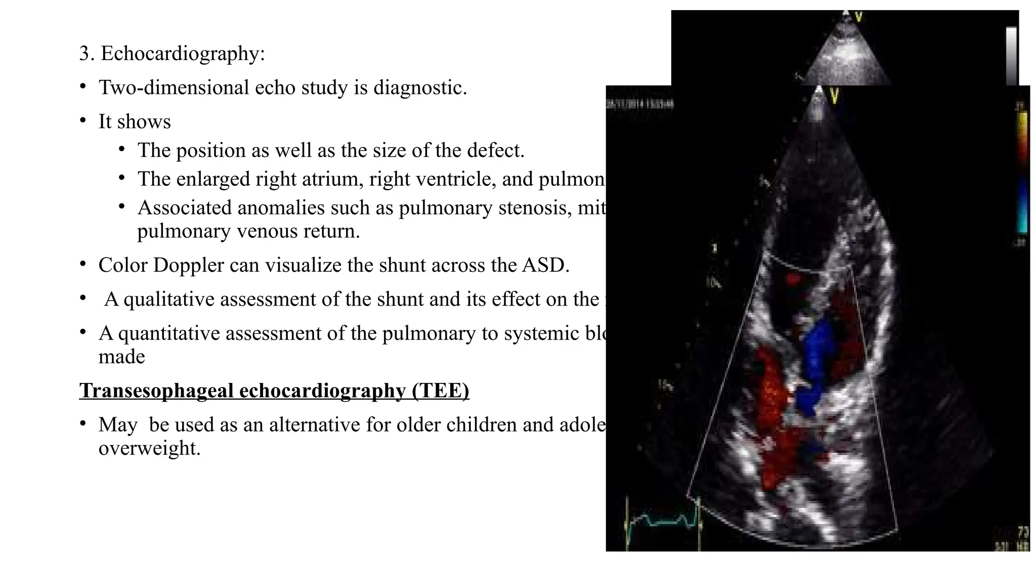
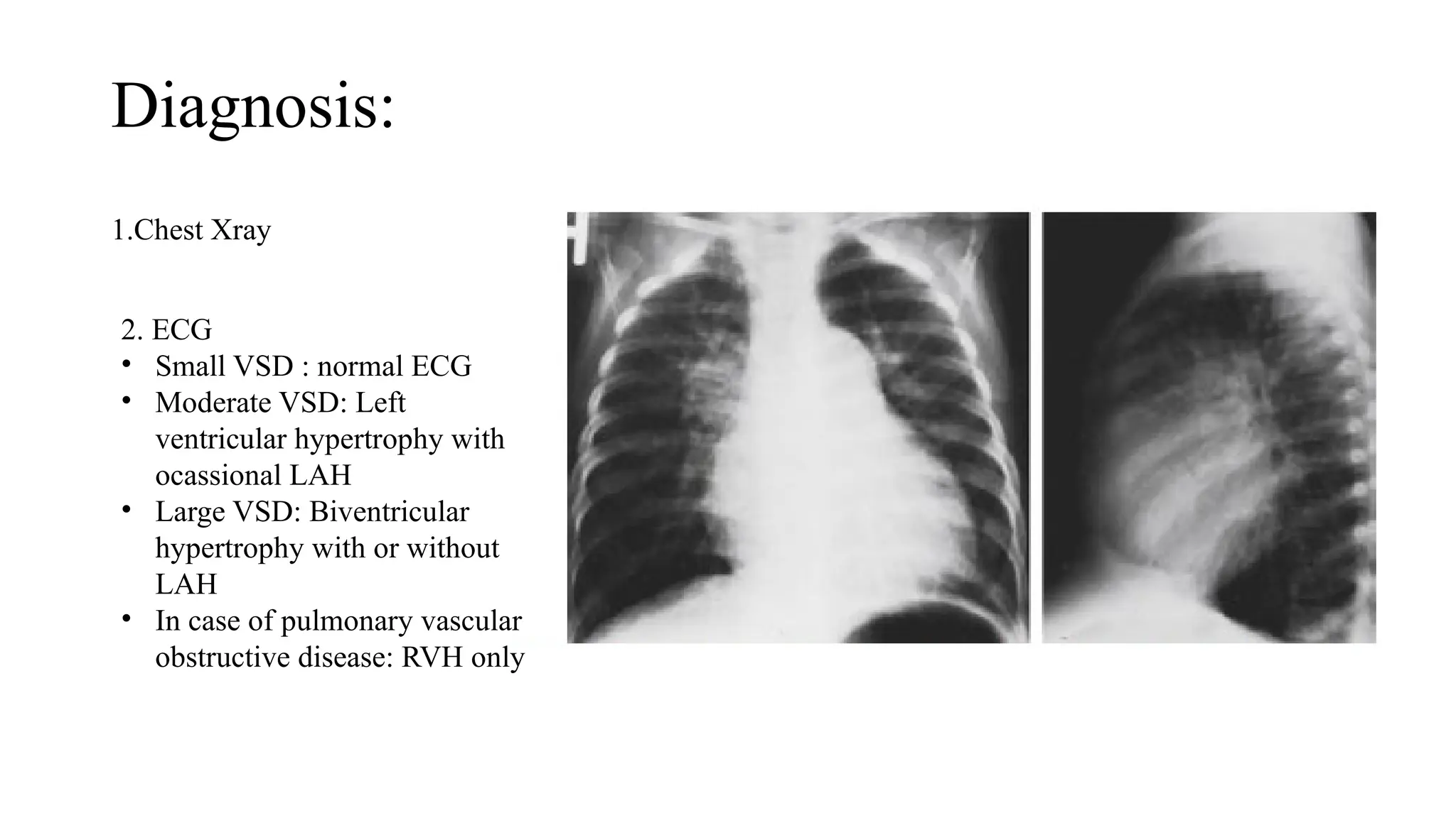
ACYANOTIC CONGENITAL HEART
DISEASE-ATRIAL SEPTAL DEFECT
AND VENTRICULAR SEPTAL
DEFECT
Presenter- Dr Ira KC
Moderator – Dr Sameera Thapa
Atrial septal defect
• ASDs constitute 8% to 10% of congenital heart defects in children.
• The recent incidence of ASDs has been estimated to be 100 per 100,000 live births.
• The female:male ratio for secundum ASDs is 2:1, but for the sinus venosus ASDs it is
1:1 .
• About 30% to 50% of children with congenital heart defects have an ASD as part of the
cardiac defect.
• The majority of cases of ASD are sporadic.
• Autosomal dominant inheritance does occur as part of the
• Holt-Oram syndrome
• Families with secundum ASD and heart block.
Types of ASD
1. Secundum ASD (50- 70%)
2. Ostium primum ASD (15-20%)
3. Sinus venosus ASD (10%)
• SVC type
• IVC type
4. Coronary sinus ASD (<1%)
MVP in 20% of ostium primum
And sinus venosus ASD
Pathophysiology
The degree of left to right shunting is dependent on:
• The size of the defect
• The relative compliance of the right ventricle and left ventricle and
• The relative vascular resistance in the pulmonary and systemic circulations.
Clinical presentation
HISTORY:
• Asymptomatic
• Infants
• Features of pulmonary overcirculation
• Recurrent respiratory infections
• Failure to thrive
• Older children
• Mild fatigue
• Dyspnea that may worsen with age
PHYSICAL EXAMINATION
• Slender body built (less than 10th
centile)
• Examination of precordium
• Inspection:
Left precordial bulge.
• Palpation:
Prominent right ventricular impulse felt along
the lower left sternal border and the subcostal
area
Auscultation:
• Wide, fixed splitting of the second heart sound (S2).
• A systolic ejection murmur best heard at the left upper sternal border.
• A short, rumbling mid-diastolic murmur is often audible at the lower left sternal border.
Diagnosis
1. Chest X-RAY:
• A small shunt across the ASD
normal-appearing CXR
• Patients with significant shunts
• Cardiomegaly, due to right atrial
and right ventricular enlargement
• Increased pulmonary vascular markings
• A prominent pulmonary artery (PA) segment.
2. ECG:
• A small left-to-right shunt and no right atrial or ventricular dilation, the ECG is normal.
• Secundum ASD : A significant left-to-right shunt
• Right axis deviation
• Right ventricular hypertrophy
• rsR′ pattern in V1 (RBBB)
3. Echocardiography:
• Two-dimensional echo study is diagnostic.
• It shows
• The position as well as the size of the defect.
• The enlarged right atrium, right ventricle, and pulmonary arteries.
• Associated anomalies such as pulmonary stenosis, mitral valve prolapse, and anomalous
pulmonary venous return.
• Color Doppler can visualize the shunt across the ASD.
• A qualitative assessment of the shunt and its effect on the right sided cardiac chambers.
• A quantitative assessment of the pulmonary to systemic blood flow ratio (Qp: Qs) also can be
made
Transesophageal echocardiography (TEE)
• May be used as an alternative for older children and adolescents, especially in those who are
overweight.
4. Cardiac catheterization
Indication
• If pulmonary vascular disease is suspected.
• Diagnosing associated lesions such as
• Partial anomalous pulmonary venous return or
• Mitral stenosis.
Advantages
• Direct measurement of intracardiac and pulmonary artery pressure can be performed.
• Pulmonary vascular resistance can be calculated.
Natural History
• Overall rate of spontaneous closure 87%
• < 3mm 100% closure bye age of 1.5 years
• 3-8mm 80% closure bye age of 1.5 years
• Size >8mm is less likely to close
• Spontaneous closure is less likely after 5 years of age
• If a large defect is untreated, CHF and pulmonary hypertension begin to develop in adults
• Atrial arrhythmias (flutter or fibrillation) may occur in adults
• Infective endocarditis does not occur in isolated ASDs.
• Cerebrovascular accident, resulting from paradoxical embolization through an ASD, is a rare
complication.
Management
• General:
1.Exercise restriction is unnecessary.
2. Asymptomatic infant and children are followed up at yearly interval until surgery is planned.
3. Symptomatic infant: anti-congestive therapy with diuretics maybe indicated until closure is accomplished.
• Nonsurgical closure:
• Closure of an ASD is indicated if there is a large shunt.
• Indicators of a large shunt include
• Qp:Qs ≥ 1.5.
• A diastolic flow rumble in the tricuspid area
• ECG evidence of right ventricular hypertrophy
• Chest x-ray evidence of cardiomegaly or increased pulmonary vascular markings
• Echocardiographic evidence of right ventricular enlargement and/or paradoxical septal motion.
• Secundum ASD, measuring 5 mm or more in diameter (but less than 32 mm).
• A significant left-to-right shunt with clinical evidence of right ventricular volume overload (i.e., Qp/Qs
ratio of 1.5:1 or greater or RV enlargement).
• There must be enough rim (4 mm) of septal tissue around the defect for appropriate placement of the
device.
Devices available for clinical use:
A. Amplatzer septal occlude
B. Gore Helix septal occlude
C. CardioSEAL device
D. BioSTAR device
Advantages
• Complete avoidance of cardiopulmonary bypass.
• Avoidance of pain and residual thoracotomy scars.
• A less than 24-hour hospital stay, and rapid recovery.
Post-device closure follow-up:
• Aspirin 5 mg/kg/day or 81mg for 6 months.
• Post procedure echo studies check for
• Residual atrial shunt
• Unobstructed flow of pulmonary veins, coronary sinus, and venae cava
• Proper function of the mitral and tricuspid valves.
Complications
• Early ECG abnormalities are common within the first 24 hours after
implant.
• Most feared complication with the Amplatzer device is early or late
erosion of the device into the aortic root, with subsequent pericardial
tamponade and rarely death.
• Thrombus formation in the right and left atrium occurs (2%–3%).
• Release of nickel from the device (with peak at 1 month after implant)
is rare complication.
Surgical closure
• Indications and Timing
• A left-to-right shunt with a pulmonary-to-systemic blood flow ratio ( Qp/ Qs) of ≥1.5:1 only after
2-4 years and if device closure is not considered appropriate.
• If CHF does not respond to medical management, surgery is performed during infancy
• If oxygen and other medical therapy are needed for infants with associated bronchopulmonary
dysplasia, surgery is performed during infancy.
• ASD with other associations
• Pulmonary vascular resistance >10units/m2
is contraindication for surgery.
VSD -Introduction
• The most common form of CHD, accounting for up to 20% of patients diagnosed with CHD.
• 3-3.8 per 1000 live births
• An echocardiographic study revealed a high incidence of 5-50 VSDs per 1000 newborns.
• VSDs are the common lesion chromosomal syndromes like trisomy 13, trisomy 18, trisomy 21.
• VSDs are slightly more common in female patients than in male patients (56% vs 44%)
Classification
• Morphological
• Hemodynamic
• Compared to aortic annulus
Morphological
• Perimembranous(membranous/ infracristal )-70-80%
• Inlet
• Outlet
• trabecular
• Muscular- 5-20%
Central- mid muscular
Apical
Marginal- along RV septal junction
Swiss cheese septum – multiple defects
• Inlet/ AV canal type-5-8%
• Supracrital/ subaortic/ Outlet- 5-7%
Hemodynamic classification
• Restrictive- Resistance that limits the shunt at the site of VSD
LVSP > RVSP
pulmonary /aortic systolic pressure ratio < 0.3
Qp / Qs<1.4/1
• Moderately restrictive - RVSP high, but less than LVSP
Qp/Qs 1.4/2.2
• Non restrictive - Shunt not limited at the site of defect
RVSP , LVSP, PA , Aortic systolic pressures equal
Qp/Qs >2.2
Flow determined by PVR
PATHOPHYSIOLOGY
• Defect compared to aortic annulus
• Large: > 50% of annulus size
• Significant pressure gradient exists between the LV and RV (high velocity),
• Medium: 25-50% of annulus size
• Small: <25% of annulus size
SMALL VSD
• <1/3rd
or 25% size of aortic root
• Shunt limited by size of the defect
• Shunt entirely during ventricular systole
• L R shunt <50% LV output
• Pulmonary:systemic flow ratio < 2:1
MODERATE VSD
• VSD size about half or 25-50% – equal to the
size of the aortic orifice
• When PA & RVSP are > 50% of systemic
arterial pressure
• Moderate-large L R shunt develops
Large VSD
• Size equal to the aortic root
• Equalization of pressures in RV& LV
• Increased LA pressure opening of foramen ovale
• LV volume overload earlier in life with progressive pulmonary hypertension and ultimately Eisenmenger’s
syndrome
Mechanism of Closure
• Growth & hypertrophy of septum around the defect
• By development of subacute bacterial endocarditis
• Ventricular septal aneurysm
• Prolapse of aortic cusp
• Intrusion of a sinus of valsalva aneurysm
Natural History and Complication
• Spontaneous closure of perimembranous and muscular VSDs can
occur, more frequently with small defects and during the first 6
months of life
• 60% of small to moderate muscular VSDs close spontaneously but not
after 8 years of age.
• 35% of small perimembranous VSDs close spontaneously but not after
5 years of age.
• 90% of spontaneous closure occurs by 3years of age
• Risk factors for decreased survival
1. Shortness of breath, fatigue,
2. Cardiomegaly
3. PASP >60mm Hg or >1/2 of systemic pressure
• Good prognosticators
1. Lack of symptoms
2. Normal LV size & function
3. Small LR shunt
4. Normal pulmonary pressures / resistance
History
• Small VSD:
• Asymptomatic with normal growth and development
• Moderate to large VSD:
• Delayed growth and development
• Repeated pulmonary infection
• CHF around 6-10 weeks
• With long standing Pulmonary hypertension:
• Cyanosis
• Respiratory distress
• Decreased activity
Physical examination
• Small VSD
Large VSD
Diagnosis:
1.Chest Xray
2. ECG
• Small VSD : normal ECG
• Moderate VSD: Left
ventricular hypertrophy with
ocassional LAH
• Large VSD: Biventricular
hypertrophy with or without
LAH
• In case of pulmonary vascular
obstructive disease: RVH only
3. ECHOCARDIOGRAPHY
Management
• General:
• No exercise restriction is required in the absence of pulmonary hypertension
• Small VSD
• Frequent feedings of high-calorie formulas, by nasogastric tube or oral feeding
• Anemia, if present, should be corrected by oral iron therapy.
• Medical management:
• To treat Congestive heart failure:
Diuretics to decrease preload
Spironolactone minimize potassium loss
With or without digoxin
Captopril reduce afterload
Drugs:
• Digoxin 10-20mcg/kg per day
• Furosemide 1–3 mg/kg per day
• Captopril 0.5–2 mg/kg per day
• Enalapril 0.1mg/kg per day
Indications of surgical intervention
• Growth failure not be improved by medical therapy
• VSD should be operated on within the first 6 months of life, preferably by 3 to 4 months of
age.
• PA pressure is more than 50% of systemic pressure, surgical closure should be done by the end of
the first year.
• After 1 year of age, a significant left-to-right shunt with Qp/Qs ratio of at least 2:1
• Older infants with large VSDs and evidence of elevated PVR should be operated on as soon as
possible.
Timing:
• <3months - if symptomatic
• 3-6 months - symptomatic, growth failure, increasing PAH
• >6 months – primarily based on PAH
• We can wait till 1 year, if no features of PAH are seen.
Surgery
1. PA banding
• palliative procedure , when additional lesions make repair difficult
• Done in multiple VSDs
• 30-50% of original diameter is narrowed
2. Direct closure of the defect
• carried out under hypothermic cardiopulmonary bypass, preferably without right ventriculotomy.
• Perimembranous and inlet VSD
• Repaired by a transatrial approach.
• Outlet(conal) defects
• Repaired through an incision in the main pulmonary artery.
• Apical VSD
• Require apical right ventriculotomy.
Complications
• Right bundle branch block(RBBB)
• RBBB and left anterior hemiblock 10%
• Complete heart block 1-2%
• Requiring pacemaker
• Residual shunt 5%
• Neurological complication

Congenital heart disease - ASD and VSD .

  • 1.
    ACYANOTIC CONGENITAL HEART DISEASE-ATRIALSEPTAL DEFECT AND VENTRICULAR SEPTAL DEFECT Presenter- Dr Ira KC Moderator – Dr Sameera Thapa
  • 2.
    Atrial septal defect •ASDs constitute 8% to 10% of congenital heart defects in children. • The recent incidence of ASDs has been estimated to be 100 per 100,000 live births. • The female:male ratio for secundum ASDs is 2:1, but for the sinus venosus ASDs it is 1:1 . • About 30% to 50% of children with congenital heart defects have an ASD as part of the cardiac defect. • The majority of cases of ASD are sporadic. • Autosomal dominant inheritance does occur as part of the • Holt-Oram syndrome • Families with secundum ASD and heart block.
  • 3.
    Types of ASD 1.Secundum ASD (50- 70%) 2. Ostium primum ASD (15-20%) 3. Sinus venosus ASD (10%) • SVC type • IVC type 4. Coronary sinus ASD (<1%) MVP in 20% of ostium primum And sinus venosus ASD
  • 4.
    Pathophysiology The degree ofleft to right shunting is dependent on: • The size of the defect • The relative compliance of the right ventricle and left ventricle and • The relative vascular resistance in the pulmonary and systemic circulations.
  • 5.
    Clinical presentation HISTORY: • Asymptomatic •Infants • Features of pulmonary overcirculation • Recurrent respiratory infections • Failure to thrive • Older children • Mild fatigue • Dyspnea that may worsen with age PHYSICAL EXAMINATION • Slender body built (less than 10th centile) • Examination of precordium • Inspection: Left precordial bulge. • Palpation: Prominent right ventricular impulse felt along the lower left sternal border and the subcostal area
  • 6.
    Auscultation: • Wide, fixedsplitting of the second heart sound (S2). • A systolic ejection murmur best heard at the left upper sternal border. • A short, rumbling mid-diastolic murmur is often audible at the lower left sternal border.
  • 7.
    Diagnosis 1. Chest X-RAY: •A small shunt across the ASD normal-appearing CXR • Patients with significant shunts • Cardiomegaly, due to right atrial and right ventricular enlargement • Increased pulmonary vascular markings • A prominent pulmonary artery (PA) segment. 2. ECG: • A small left-to-right shunt and no right atrial or ventricular dilation, the ECG is normal. • Secundum ASD : A significant left-to-right shunt • Right axis deviation • Right ventricular hypertrophy • rsR′ pattern in V1 (RBBB)
  • 8.
    3. Echocardiography: • Two-dimensionalecho study is diagnostic. • It shows • The position as well as the size of the defect. • The enlarged right atrium, right ventricle, and pulmonary arteries. • Associated anomalies such as pulmonary stenosis, mitral valve prolapse, and anomalous pulmonary venous return. • Color Doppler can visualize the shunt across the ASD. • A qualitative assessment of the shunt and its effect on the right sided cardiac chambers. • A quantitative assessment of the pulmonary to systemic blood flow ratio (Qp: Qs) also can be made Transesophageal echocardiography (TEE) • May be used as an alternative for older children and adolescents, especially in those who are overweight.
  • 9.
    4. Cardiac catheterization Indication •If pulmonary vascular disease is suspected. • Diagnosing associated lesions such as • Partial anomalous pulmonary venous return or • Mitral stenosis. Advantages • Direct measurement of intracardiac and pulmonary artery pressure can be performed. • Pulmonary vascular resistance can be calculated.
  • 10.
    Natural History • Overallrate of spontaneous closure 87% • < 3mm 100% closure bye age of 1.5 years • 3-8mm 80% closure bye age of 1.5 years • Size >8mm is less likely to close • Spontaneous closure is less likely after 5 years of age • If a large defect is untreated, CHF and pulmonary hypertension begin to develop in adults • Atrial arrhythmias (flutter or fibrillation) may occur in adults • Infective endocarditis does not occur in isolated ASDs. • Cerebrovascular accident, resulting from paradoxical embolization through an ASD, is a rare complication.
  • 11.
    Management • General: 1.Exercise restrictionis unnecessary. 2. Asymptomatic infant and children are followed up at yearly interval until surgery is planned. 3. Symptomatic infant: anti-congestive therapy with diuretics maybe indicated until closure is accomplished. • Nonsurgical closure: • Closure of an ASD is indicated if there is a large shunt. • Indicators of a large shunt include • Qp:Qs ≥ 1.5. • A diastolic flow rumble in the tricuspid area • ECG evidence of right ventricular hypertrophy • Chest x-ray evidence of cardiomegaly or increased pulmonary vascular markings • Echocardiographic evidence of right ventricular enlargement and/or paradoxical septal motion. • Secundum ASD, measuring 5 mm or more in diameter (but less than 32 mm). • A significant left-to-right shunt with clinical evidence of right ventricular volume overload (i.e., Qp/Qs ratio of 1.5:1 or greater or RV enlargement). • There must be enough rim (4 mm) of septal tissue around the defect for appropriate placement of the device.
  • 12.
    Devices available forclinical use: A. Amplatzer septal occlude B. Gore Helix septal occlude C. CardioSEAL device D. BioSTAR device
  • 13.
    Advantages • Complete avoidanceof cardiopulmonary bypass. • Avoidance of pain and residual thoracotomy scars. • A less than 24-hour hospital stay, and rapid recovery. Post-device closure follow-up: • Aspirin 5 mg/kg/day or 81mg for 6 months. • Post procedure echo studies check for • Residual atrial shunt • Unobstructed flow of pulmonary veins, coronary sinus, and venae cava • Proper function of the mitral and tricuspid valves.
  • 14.
    Complications • Early ECGabnormalities are common within the first 24 hours after implant. • Most feared complication with the Amplatzer device is early or late erosion of the device into the aortic root, with subsequent pericardial tamponade and rarely death. • Thrombus formation in the right and left atrium occurs (2%–3%). • Release of nickel from the device (with peak at 1 month after implant) is rare complication.
  • 15.
    Surgical closure • Indicationsand Timing • A left-to-right shunt with a pulmonary-to-systemic blood flow ratio ( Qp/ Qs) of ≥1.5:1 only after 2-4 years and if device closure is not considered appropriate. • If CHF does not respond to medical management, surgery is performed during infancy • If oxygen and other medical therapy are needed for infants with associated bronchopulmonary dysplasia, surgery is performed during infancy. • ASD with other associations • Pulmonary vascular resistance >10units/m2 is contraindication for surgery.
  • 16.
    VSD -Introduction • Themost common form of CHD, accounting for up to 20% of patients diagnosed with CHD. • 3-3.8 per 1000 live births • An echocardiographic study revealed a high incidence of 5-50 VSDs per 1000 newborns. • VSDs are the common lesion chromosomal syndromes like trisomy 13, trisomy 18, trisomy 21. • VSDs are slightly more common in female patients than in male patients (56% vs 44%) Classification • Morphological • Hemodynamic • Compared to aortic annulus
  • 17.
    Morphological • Perimembranous(membranous/ infracristal)-70-80% • Inlet • Outlet • trabecular • Muscular- 5-20% Central- mid muscular Apical Marginal- along RV septal junction Swiss cheese septum – multiple defects • Inlet/ AV canal type-5-8% • Supracrital/ subaortic/ Outlet- 5-7%
  • 18.
    Hemodynamic classification • Restrictive-Resistance that limits the shunt at the site of VSD LVSP > RVSP pulmonary /aortic systolic pressure ratio < 0.3 Qp / Qs<1.4/1 • Moderately restrictive - RVSP high, but less than LVSP Qp/Qs 1.4/2.2 • Non restrictive - Shunt not limited at the site of defect RVSP , LVSP, PA , Aortic systolic pressures equal Qp/Qs >2.2 Flow determined by PVR
  • 19.
    PATHOPHYSIOLOGY • Defect comparedto aortic annulus • Large: > 50% of annulus size • Significant pressure gradient exists between the LV and RV (high velocity), • Medium: 25-50% of annulus size • Small: <25% of annulus size
  • 20.
    SMALL VSD • <1/3rd or25% size of aortic root • Shunt limited by size of the defect • Shunt entirely during ventricular systole • L R shunt <50% LV output • Pulmonary:systemic flow ratio < 2:1 MODERATE VSD • VSD size about half or 25-50% – equal to the size of the aortic orifice • When PA & RVSP are > 50% of systemic arterial pressure • Moderate-large L R shunt develops Large VSD • Size equal to the aortic root • Equalization of pressures in RV& LV • Increased LA pressure opening of foramen ovale • LV volume overload earlier in life with progressive pulmonary hypertension and ultimately Eisenmenger’s syndrome
  • 21.
    Mechanism of Closure •Growth & hypertrophy of septum around the defect • By development of subacute bacterial endocarditis • Ventricular septal aneurysm • Prolapse of aortic cusp • Intrusion of a sinus of valsalva aneurysm
  • 22.
    Natural History andComplication • Spontaneous closure of perimembranous and muscular VSDs can occur, more frequently with small defects and during the first 6 months of life • 60% of small to moderate muscular VSDs close spontaneously but not after 8 years of age. • 35% of small perimembranous VSDs close spontaneously but not after 5 years of age. • 90% of spontaneous closure occurs by 3years of age
  • 23.
    • Risk factorsfor decreased survival 1. Shortness of breath, fatigue, 2. Cardiomegaly 3. PASP >60mm Hg or >1/2 of systemic pressure • Good prognosticators 1. Lack of symptoms 2. Normal LV size & function 3. Small LR shunt 4. Normal pulmonary pressures / resistance
  • 24.
    History • Small VSD: •Asymptomatic with normal growth and development • Moderate to large VSD: • Delayed growth and development • Repeated pulmonary infection • CHF around 6-10 weeks • With long standing Pulmonary hypertension: • Cyanosis • Respiratory distress • Decreased activity
  • 25.
  • 26.
  • 27.
    Diagnosis: 1.Chest Xray 2. ECG •Small VSD : normal ECG • Moderate VSD: Left ventricular hypertrophy with ocassional LAH • Large VSD: Biventricular hypertrophy with or without LAH • In case of pulmonary vascular obstructive disease: RVH only
  • 28.
  • 29.
    Management • General: • Noexercise restriction is required in the absence of pulmonary hypertension • Small VSD • Frequent feedings of high-calorie formulas, by nasogastric tube or oral feeding • Anemia, if present, should be corrected by oral iron therapy. • Medical management: • To treat Congestive heart failure: Diuretics to decrease preload Spironolactone minimize potassium loss With or without digoxin Captopril reduce afterload Drugs: • Digoxin 10-20mcg/kg per day • Furosemide 1–3 mg/kg per day • Captopril 0.5–2 mg/kg per day • Enalapril 0.1mg/kg per day
  • 30.
    Indications of surgicalintervention • Growth failure not be improved by medical therapy • VSD should be operated on within the first 6 months of life, preferably by 3 to 4 months of age. • PA pressure is more than 50% of systemic pressure, surgical closure should be done by the end of the first year. • After 1 year of age, a significant left-to-right shunt with Qp/Qs ratio of at least 2:1 • Older infants with large VSDs and evidence of elevated PVR should be operated on as soon as possible. Timing: • <3months - if symptomatic • 3-6 months - symptomatic, growth failure, increasing PAH • >6 months – primarily based on PAH • We can wait till 1 year, if no features of PAH are seen.
  • 31.
    Surgery 1. PA banding •palliative procedure , when additional lesions make repair difficult • Done in multiple VSDs • 30-50% of original diameter is narrowed 2. Direct closure of the defect • carried out under hypothermic cardiopulmonary bypass, preferably without right ventriculotomy. • Perimembranous and inlet VSD • Repaired by a transatrial approach. • Outlet(conal) defects • Repaired through an incision in the main pulmonary artery. • Apical VSD • Require apical right ventriculotomy.
  • 32.
    Complications • Right bundlebranch block(RBBB) • RBBB and left anterior hemiblock 10% • Complete heart block 1-2% • Requiring pacemaker • Residual shunt 5% • Neurological complication

Editor's Notes

  • #3 1This defect is present at the site of fossa ovalis,.Anomalous pulmonary venous return is present in about 10% of cases. 3 The former is commonly associated with anomalous drainage of the right upper pulmonary vein (into the RA), and the latter is often associated with anomalous drainage of the right lung into the IVC (“scimitar syndrome”) 4 there is a defect in the roof of the coronary sinus and the LA blood shunts through the defect and empties into the RA through the coronary sinus ostium, producing clinical pictures similar to other types of ASDs.
  • #6 With a large left-to-right shunt, a mid-diastolic rumble resulting from relative tricuspid stenosis may be audible at the lower left sternal border. 3. Classic auscultatory findings (and electrocardiographic [ECG] and radiography findings) of ASD are not present unless the shunt is reasonabl large (at least Qp/Qs of 1.5 or greater). The typical auscultatory findings may be absent in infants and toddlers, even in those with a large defect because theRV is poorly compliant.
  • #10 1.who are in their 20s and 30s, and it becomes common after 40 years of age. 2 . The incidence of atrial arrhythmias increases to as high as 13% in patients older than 40 years of age.
  • #13 If 1-month and 1-year follow-up echocardiographic findings are normal, yearly or biennial follow-up will suffice.
  • #16 A Developmental defect of heart occurs in which cyanosis doesnot ensueinspite of the fact that a communication exist inbetween the cavities of two ventricle.
  • #17 1.According to the accompanying defect in the adjacent muscular septum, perimembranous VSDs have been called perimembranous inlet (atrioventricular [AV] canal type), perimembranous trabecular, or perimembranous outlet (tetralogy type) defects Inlet: The defect is located posterior and inferior to the perimembranous defect beneath the septal leaflet of the tricuspid valve OUTLET: The defect is located within the outlet (conal) septum, and part of its rim is formed by the aortic and pulmonary annulus. An aortic leaflet can prolapse through the VSD and cause aortic insufficiency
  • #25 2-4/6 systolic mr, medium frequency
  • #26 Respiratory distress at 2-8 wks Cardiomegaly Systolic thrill , along LSB
  • #27 In pulmonary vascular obstructive disease, the main PA and the hilar PAs enlarge noticeably, but the peripheral lung fields are ischemic. The heart size is usually normal
  • #29 for 2 to 4 months to see if growth failure can be improved Angiotensin-converting enzyme (ACE) inhibitors may raise the serum potassium level, and spironolactone or potassium supplementation should be discontinued.
  • #30 1> Surgery should be delayed for infants who respond to medical therapy. 3>Surgery is not indicated for small VSD with Qp/Qs ratio less than 1.5:1. Surgery is contraindicated in patients with a pulmonary to systemic vascular resistance ratio of greater than 0.5 or with pulmonary vascular obstructive disease with a predominant right-to-left shunt.
  • #31 Systolic pressure of 25-30 mmHg beyond the constriction RV/PA pressure gradient > 45 associated with hypoxemia
  • #32 Disrupttion of purkenji fiber or direct injury to right bundle can cause sudden cardiac death 4.Intraoperative TEE reduce the incidence 5. Related to circulatory arrest time