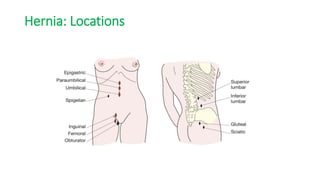
Ventral hernia
Prepared by
Dr. Pranjal Rokaya
Resident
General Surgery
Moderator
Dr. Ellina Dangol
25th September 2022
Outline
• Anatomy of the ventral abdominal wall
• Introduction
• Classification
• History and examination of hernia
• Specific hernia: Management.
Anatomy of anterior abdominal wall
Anatomy: Rectus sheath
Hernia
• Protrusion or bulging of part of the contents of abdominal cavitiy through a
weakness in the abdominal wall.
Biomechanical basis of herniation
Pascal’s law of pressure Laplace law of wall tension
…Pathophysiology
• The Abdominopelvic cavity is a cylinder enveloped by muscles,
tendons, and bony structures.
• If intraabdominal pressure > abdominal wall pressure, the wall
ruptures at the weakest point causing herniation.
• Once hernia forms will continue to enlarge due to increased wall
tension.
Anatomical causes of hernia
• Excessive intraabdominal pressure
• Basic design weakness
• Weakness due to structures entering and leaving the abdomen.
• Developmental failures.
• Genetic weakness of collagen.
• Sharp and blunt trauma.
• Weakness due to aging and pregnancy.
Risk factors
• Constipation
• Prostatic symptoms
• Chronic cough
• Obesity
Types of hernia
Based on Complexity
 Occult
Reducible
Irreducible
Strangulated
 Infarcted
Based on location
 Internal
 External
Ritcher’s hernia: Only a part of bowel enters the hernia
Based on location
Ventral wall hernia
Epigastric hernia
Umbilcal hernia
Spiegelian hernia
Incisional hernia
Parastomal hernia
Flank hernia
Superior lumbar hernia
Inferior lumbar hernia
 Groin hernia
 Inguinal hernia
 Femoral hernia
Pelvic hernia
 Sciatic hernia
 Obturator hernia
 Perineal hernia
Hernia: Locations
European Hernia Society Classification
History
• Self-diagnosis common
• Usually painless but may complain of aching or heavy sensation.
• Severe pain suggests strangulation.
• Symptoms of bowel obstruction.
• History of previous surgery.
Examination
• Examined lying down initially, then standing.
• Cough impulse
• Reducibility
• Tenderness
• Overlying skin color changes
• Multiple defects
• Signs of the previous repair.
• Scrotal content for groin hernia
Investigation
• For most, no investigation required, just clinical examination.
• Chest Xray: diaphragmatic hernia/ hiatus hernia.
• Ultrasound: In irreducible hernia when differential includes mass or fluid
collection.
• CT scan: In complex incisional hernia (number and size of defects,
identifying content, presence of adhesions).
• Laparoscopy: Feasibility of lap repair.
Principles of hernia repair
• Reduction of hernia content into the abdominal cavity with the removal of any
nonviable tissue and bowel repair if necessary.
• Excision and closure of peritoneal sac or replacing it back.
• Reapproximation of walls of neck of hernia.
• Permanent reinforcement of wall defect with sutures or mesh.
Mesh in hernia repair
To bridge a defect
To plug a defect
To augment a repair
• Should have good overlap, at least 2 cm up to 5 cm.
Mesh characteristics
• Woven, knitted, or sheet.
• Synthetic vs biologic.
• Light, medium, or heavyweight.
• Large pore vs small pore
• Intraperitoneal use or not.
• Noabdorbable vs absorbable.
Locations of mesh placement
Ventral hernias
a. Umbilical hernia: Children
• Occurs in upto 10% of infants.
• Within few weeks of birth, often symptomless.
• Increases in size on crying and assumes conical shape.
• Obstruction extremely rare below 3 years.
• 95% will resolve spontaneously.
• If persist beyond 2 years, sugical repair indicated.
Umbilical hernia: Adult
• Conditions that stretch linea alba predisposes to opening of umbilical defect.
• Defect rounded with well defined fibrous margin.
• Small hernia: Peritoneal fat or omentum
• Larger hernia : small or large bowel
Clinical picture: Umbilical hernia
• Commonly overweight with thinned and attenuated midline raphae.
• Bulge typically to one side of umbilical depression creating crescent appearance.
• Women > Men
• Pain due to tissue tension or symptoms of bowel obstruction.
• Overlying skin may become thinned, stretched and develop dermatitis.
Treatment: Umbilical hernia
a. Open umbilical hernia repair
• Defects less than 2 cm
Very small defects (<1 cm)closed with the simple figure of 8 suture or darn
technique.
Mayo’s repair of small umbilical hernia
 Defects larger than 2 cm: Mesh repair recommended
i. Within peritoneal cavity
• Tissue separating mesh placed through defect on the underside of abdominal
wall.
• Ideally 5 cm overlap.
• Expensive mesh.
ii.In retromuscular space
• Linea alba opened vertically, b/l posterior sheath incised exposing rectus.
• Muscle elevated, mesh placed and sutured.
• Mesh should overlap midline by 5 cm laterally and umbilicus vertically.
• Very secure but requires extensive dissection.
iii. In extraperitoneal space
Plane between posterior rectus sheath and peritoneum.
Care taken to avoid button holing peritoneum.
If peritoneum damaged suring repair, alternative sought.
d. In subcutaneous plane (Onlay)
Mesh placed over anterior rectus sheath and sutured.
Prone to infection
b. Lap umbilical hernia repair
• Three ports placed laterally.
• Contents of hernia reduced by traction and external pressure.
• Falciform ligament and median umbilical fold may be taken down.
• Nonadherent mesh introduced, centered and fixed to peritoneum
and posterior rectus sheath.
• May cause severe pain lasting 24-48 hours.
b. Epigastric hernia
• Through linea alba between xiphoid process and the umbilicus.
• Defect where small blood vessels pierce linea alba or due to abnormal
decussation of aponeurotic fibres.
• Usually less than 1 cmm, contains usually extraperitoneal fat .
• Rarely contains bowel.
• More than one hernia may be present.
• Frequency: 3-5% among all hernias
Clinical picture: Epigastric hernia
• Fit healthy men aged 20 to 45 years.
• May be very painful due to partial strangulation of fat.
• Soft midline swelling often felt rather than seen.
• Unlikely to be reduced.
• May resemble lipoma.
• Cough impulse may or may not be felt.
Treatment: Epigastric hernia
• Very small hernia known to diappear due to infarction of fat.
• Small to moderate sized hernia w/o sac not inherently dangerous.
• Surgery for symptomatic hernia: open or laparoscopic.
• At open surgery, vertical or transverse incision made.
• Protuding extraperitoneal fat simply excised or pushed back.
Treatment: Epigastric hernia
• Defect closed with nonabsorbable sutures in adult and absorbable in children.
• In large hernia, approach similar to umbilical hernia.
• Lap repair also similar except defect is hidden behind falciform ligament.
Repair of small
epigastric hernia
c. Spiegelian hernia
• Spiegelian fascia extend between transversus
muscle and lateral edge of rectus sheath.
• Most hernia appear below the level of umbilicus
but can be found anywhere along spiegelian line.
• More common in elderly, male and female
equally affected.
Clinical picture: Spiegelian
• Intermittent pain due to pinching of fat.
• Lump may or may not be palpable.
• Older patients: reducible swelling at edge of rectus
sheath with symptoms of intermittent obstruction.
• Diagnosis usually confirmed with CT scan.
Treatment: Spiegelian hernia
• Surgery recommended as narrow and fibrous neck predisposes to strangulation.
• In open surgery, no abnormality seen until external oblique (EO) opened.
• Sac and contents dealt with, defect repaired with suture or mesh, laid deep to EO
aponeurosis.
• Lap approach if no sac visible.
d. Incisional hernia
• Arise through defect in musculofascial layers of abdominal wall in region of postoperative
scar.
• Reported in 10-50% of laparotomy incisions and 1-5% of laparoscopic port sites.
• Predisposing factors:
Patient factors Wound factors Surgical factors
Obesity SSI Inappropriate suture
material
Malnutrition Incorrect suture placement
Steroid therapy
Chronic cough
Clinical picture: Incisional hernia
• Localized swelling in small scar but may also bulge in the
entire incision length.
• May have several discrete hernias along the length of the
incision.
• Unsuspected defects may be found during surgery.
• Attacks of partial bowel obstruction.
• Most incisional hernia broad necked and thus low risk of
strangulation.
Incisional hernia grading system
Stage
I : Low recurrence, low SSO Les than 10 cm, clean
II: Moderate recurrence, moderate SSO Less than 10 cm, contaminated
10-20 cm, clean
III: High recurrence, high SSO More than 10 cm, contaminated
Any more than 20 cm
Combined from Ventral hernia working group grading scale and EHS classification
Treatment: Incisional hernia
• Asymptomatic hernia may not require treatment.
• Use of abdominal binder prevent increase in size.
• Surgical repair should cover whole length of previous incision.
• Approximation done with minimal tension and mesh placed.
• Mesh contraindicated in a contaminated field.
Treatment: Incisional hernia
Open repair
• Previous incision opened to full length to
reveal unsuspected defects.
• Hernial sac isolated, sac opened, contents
reduced and local adhesions divided.
• Mesh can be placed in several planes,
retrorectus sublay popular.
Incisional hernia: Retrorectus sublay repair
• Vertical incision through the fascia to separate and elevate muscles.
• Medial edges of posterior sheath sutured.
• Mesh must be large enough to ensure a 5 cm overlap.
• Anterior sheaths are sutured together over the mesh.
• Suction drainage to prevent seroma.
Incisional hernia: Laparoscopic repair
• Laparoscopy and division of adhesions.
• Hernial content reduced and margins of defect exposed.
• Falciform ligament and median umbilical fold taken down.
• Fix the mesh under defect with adequate overlap.
• Tissue separating mesh is essential.
• Mesh fixed with staples or transfascial sutures passing through all muscle layers.
Very large incisional hernia
• If volume of sac more than 25% of volume of abdominal cavity.
• Content may not fit back or if they do, result in high tension.
Techniques:
Preoperative abdominal expansion with progressive preoperative
pneumoperitoneum over several weeks.
Resection of omentum and colon.
Use of mesh to span unclosable gap.
Relaxing incisions in EO aponeurosis or posterior sheath
Risk reduction: Incisional hernia
• Optimize patients prior to surgery.
• Closure of fascial layers with nonabsorbable or slowly absorbable suture.
• The optimal length of suture length to wound length is 4:1.
• Classical teaching: suture 1 cm deep and 1 cm wide is out of favor.
• Drains are brought out through the separate incision.
• Placement of prophylactic mesh.
e. Parastomal hernia
• Muscle defect created suring stoma formation tend to increase in size.
• Rate of formation over 50%.
• Ideal solution is to rejoin bowel and remove stoma altogether.
• Stoma may be resited but recurrence likely.
• Mesh best placed in retromuscular space.
• Use of prophylactic mesh at time of stoma creation.
Pros and Cons of Lap repair
PROS CONS
1. Accurately identifies all fascial defects. 1. Adhesiolysis may be difficult with
increase potential for enterotomy.
2. May identify unsuspected
intraperitoneal pathology.
2. Hard to get good fixation for defects at
margin of cavity.
3. Approaches fascia through ‘ clean
field’
References
• Bailey and Love 27th edition.
• Maingot’s abdominal operation.
• Sabiston’s textbook of surgery.
Thank You.

Ventral hernia

  • 1.
    Ventral hernia Prepared by Dr.Pranjal Rokaya Resident General Surgery Moderator Dr. Ellina Dangol 25th September 2022
  • 2.
    Outline • Anatomy ofthe ventral abdominal wall • Introduction • Classification • History and examination of hernia • Specific hernia: Management.
  • 3.
    Anatomy of anteriorabdominal wall
  • 5.
  • 6.
    Hernia • Protrusion orbulging of part of the contents of abdominal cavitiy through a weakness in the abdominal wall.
  • 7.
    Biomechanical basis ofherniation Pascal’s law of pressure Laplace law of wall tension
  • 8.
    …Pathophysiology • The Abdominopelviccavity is a cylinder enveloped by muscles, tendons, and bony structures. • If intraabdominal pressure > abdominal wall pressure, the wall ruptures at the weakest point causing herniation. • Once hernia forms will continue to enlarge due to increased wall tension.
  • 9.
    Anatomical causes ofhernia • Excessive intraabdominal pressure • Basic design weakness • Weakness due to structures entering and leaving the abdomen. • Developmental failures. • Genetic weakness of collagen. • Sharp and blunt trauma. • Weakness due to aging and pregnancy.
  • 10.
    Risk factors • Constipation •Prostatic symptoms • Chronic cough • Obesity
  • 11.
    Types of hernia Basedon Complexity  Occult Reducible Irreducible Strangulated  Infarcted Based on location  Internal  External Ritcher’s hernia: Only a part of bowel enters the hernia
  • 12.
    Based on location Ventralwall hernia Epigastric hernia Umbilcal hernia Spiegelian hernia Incisional hernia Parastomal hernia Flank hernia Superior lumbar hernia Inferior lumbar hernia  Groin hernia  Inguinal hernia  Femoral hernia Pelvic hernia  Sciatic hernia  Obturator hernia  Perineal hernia
  • 13.
  • 14.
  • 16.
    History • Self-diagnosis common •Usually painless but may complain of aching or heavy sensation. • Severe pain suggests strangulation. • Symptoms of bowel obstruction. • History of previous surgery.
  • 17.
    Examination • Examined lyingdown initially, then standing. • Cough impulse • Reducibility • Tenderness • Overlying skin color changes • Multiple defects • Signs of the previous repair. • Scrotal content for groin hernia
  • 18.
    Investigation • For most,no investigation required, just clinical examination. • Chest Xray: diaphragmatic hernia/ hiatus hernia. • Ultrasound: In irreducible hernia when differential includes mass or fluid collection. • CT scan: In complex incisional hernia (number and size of defects, identifying content, presence of adhesions). • Laparoscopy: Feasibility of lap repair.
  • 19.
    Principles of herniarepair • Reduction of hernia content into the abdominal cavity with the removal of any nonviable tissue and bowel repair if necessary. • Excision and closure of peritoneal sac or replacing it back. • Reapproximation of walls of neck of hernia. • Permanent reinforcement of wall defect with sutures or mesh.
  • 20.
    Mesh in herniarepair To bridge a defect To plug a defect To augment a repair • Should have good overlap, at least 2 cm up to 5 cm.
  • 21.
    Mesh characteristics • Woven,knitted, or sheet. • Synthetic vs biologic. • Light, medium, or heavyweight. • Large pore vs small pore • Intraperitoneal use or not. • Noabdorbable vs absorbable.
  • 22.
  • 23.
  • 24.
    a. Umbilical hernia:Children • Occurs in upto 10% of infants. • Within few weeks of birth, often symptomless. • Increases in size on crying and assumes conical shape. • Obstruction extremely rare below 3 years. • 95% will resolve spontaneously. • If persist beyond 2 years, sugical repair indicated.
  • 25.
    Umbilical hernia: Adult •Conditions that stretch linea alba predisposes to opening of umbilical defect. • Defect rounded with well defined fibrous margin. • Small hernia: Peritoneal fat or omentum • Larger hernia : small or large bowel
  • 26.
    Clinical picture: Umbilicalhernia • Commonly overweight with thinned and attenuated midline raphae. • Bulge typically to one side of umbilical depression creating crescent appearance. • Women > Men • Pain due to tissue tension or symptoms of bowel obstruction. • Overlying skin may become thinned, stretched and develop dermatitis.
  • 27.
    Treatment: Umbilical hernia a.Open umbilical hernia repair • Defects less than 2 cm Very small defects (<1 cm)closed with the simple figure of 8 suture or darn technique.
  • 28.
    Mayo’s repair ofsmall umbilical hernia
  • 29.
     Defects largerthan 2 cm: Mesh repair recommended i. Within peritoneal cavity • Tissue separating mesh placed through defect on the underside of abdominal wall. • Ideally 5 cm overlap. • Expensive mesh.
  • 30.
    ii.In retromuscular space •Linea alba opened vertically, b/l posterior sheath incised exposing rectus. • Muscle elevated, mesh placed and sutured. • Mesh should overlap midline by 5 cm laterally and umbilicus vertically. • Very secure but requires extensive dissection.
  • 31.
    iii. In extraperitonealspace Plane between posterior rectus sheath and peritoneum. Care taken to avoid button holing peritoneum. If peritoneum damaged suring repair, alternative sought. d. In subcutaneous plane (Onlay) Mesh placed over anterior rectus sheath and sutured. Prone to infection
  • 32.
    b. Lap umbilicalhernia repair • Three ports placed laterally. • Contents of hernia reduced by traction and external pressure. • Falciform ligament and median umbilical fold may be taken down. • Nonadherent mesh introduced, centered and fixed to peritoneum and posterior rectus sheath. • May cause severe pain lasting 24-48 hours.
  • 33.
    b. Epigastric hernia •Through linea alba between xiphoid process and the umbilicus. • Defect where small blood vessels pierce linea alba or due to abnormal decussation of aponeurotic fibres. • Usually less than 1 cmm, contains usually extraperitoneal fat . • Rarely contains bowel. • More than one hernia may be present. • Frequency: 3-5% among all hernias
  • 34.
    Clinical picture: Epigastrichernia • Fit healthy men aged 20 to 45 years. • May be very painful due to partial strangulation of fat. • Soft midline swelling often felt rather than seen. • Unlikely to be reduced. • May resemble lipoma. • Cough impulse may or may not be felt.
  • 35.
    Treatment: Epigastric hernia •Very small hernia known to diappear due to infarction of fat. • Small to moderate sized hernia w/o sac not inherently dangerous. • Surgery for symptomatic hernia: open or laparoscopic. • At open surgery, vertical or transverse incision made. • Protuding extraperitoneal fat simply excised or pushed back.
  • 36.
    Treatment: Epigastric hernia •Defect closed with nonabsorbable sutures in adult and absorbable in children. • In large hernia, approach similar to umbilical hernia. • Lap repair also similar except defect is hidden behind falciform ligament.
  • 37.
  • 38.
    c. Spiegelian hernia •Spiegelian fascia extend between transversus muscle and lateral edge of rectus sheath. • Most hernia appear below the level of umbilicus but can be found anywhere along spiegelian line. • More common in elderly, male and female equally affected.
  • 39.
    Clinical picture: Spiegelian •Intermittent pain due to pinching of fat. • Lump may or may not be palpable. • Older patients: reducible swelling at edge of rectus sheath with symptoms of intermittent obstruction. • Diagnosis usually confirmed with CT scan.
  • 40.
    Treatment: Spiegelian hernia •Surgery recommended as narrow and fibrous neck predisposes to strangulation. • In open surgery, no abnormality seen until external oblique (EO) opened. • Sac and contents dealt with, defect repaired with suture or mesh, laid deep to EO aponeurosis. • Lap approach if no sac visible.
  • 41.
    d. Incisional hernia •Arise through defect in musculofascial layers of abdominal wall in region of postoperative scar. • Reported in 10-50% of laparotomy incisions and 1-5% of laparoscopic port sites. • Predisposing factors: Patient factors Wound factors Surgical factors Obesity SSI Inappropriate suture material Malnutrition Incorrect suture placement Steroid therapy Chronic cough
  • 42.
    Clinical picture: Incisionalhernia • Localized swelling in small scar but may also bulge in the entire incision length. • May have several discrete hernias along the length of the incision. • Unsuspected defects may be found during surgery. • Attacks of partial bowel obstruction. • Most incisional hernia broad necked and thus low risk of strangulation.
  • 43.
    Incisional hernia gradingsystem Stage I : Low recurrence, low SSO Les than 10 cm, clean II: Moderate recurrence, moderate SSO Less than 10 cm, contaminated 10-20 cm, clean III: High recurrence, high SSO More than 10 cm, contaminated Any more than 20 cm Combined from Ventral hernia working group grading scale and EHS classification
  • 44.
    Treatment: Incisional hernia •Asymptomatic hernia may not require treatment. • Use of abdominal binder prevent increase in size. • Surgical repair should cover whole length of previous incision. • Approximation done with minimal tension and mesh placed. • Mesh contraindicated in a contaminated field.
  • 45.
    Treatment: Incisional hernia Openrepair • Previous incision opened to full length to reveal unsuspected defects. • Hernial sac isolated, sac opened, contents reduced and local adhesions divided. • Mesh can be placed in several planes, retrorectus sublay popular.
  • 47.
    Incisional hernia: Retrorectussublay repair • Vertical incision through the fascia to separate and elevate muscles. • Medial edges of posterior sheath sutured. • Mesh must be large enough to ensure a 5 cm overlap. • Anterior sheaths are sutured together over the mesh. • Suction drainage to prevent seroma.
  • 48.
    Incisional hernia: Laparoscopicrepair • Laparoscopy and division of adhesions. • Hernial content reduced and margins of defect exposed. • Falciform ligament and median umbilical fold taken down. • Fix the mesh under defect with adequate overlap. • Tissue separating mesh is essential. • Mesh fixed with staples or transfascial sutures passing through all muscle layers.
  • 49.
    Very large incisionalhernia • If volume of sac more than 25% of volume of abdominal cavity. • Content may not fit back or if they do, result in high tension. Techniques: Preoperative abdominal expansion with progressive preoperative pneumoperitoneum over several weeks. Resection of omentum and colon. Use of mesh to span unclosable gap. Relaxing incisions in EO aponeurosis or posterior sheath
  • 50.
    Risk reduction: Incisionalhernia • Optimize patients prior to surgery. • Closure of fascial layers with nonabsorbable or slowly absorbable suture. • The optimal length of suture length to wound length is 4:1. • Classical teaching: suture 1 cm deep and 1 cm wide is out of favor. • Drains are brought out through the separate incision. • Placement of prophylactic mesh.
  • 51.
    e. Parastomal hernia •Muscle defect created suring stoma formation tend to increase in size. • Rate of formation over 50%. • Ideal solution is to rejoin bowel and remove stoma altogether. • Stoma may be resited but recurrence likely. • Mesh best placed in retromuscular space. • Use of prophylactic mesh at time of stoma creation.
  • 52.
    Pros and Consof Lap repair PROS CONS 1. Accurately identifies all fascial defects. 1. Adhesiolysis may be difficult with increase potential for enterotomy. 2. May identify unsuspected intraperitoneal pathology. 2. Hard to get good fixation for defects at margin of cavity. 3. Approaches fascia through ‘ clean field’
  • 53.
    References • Bailey andLove 27th edition. • Maingot’s abdominal operation. • Sabiston’s textbook of surgery.
  • 54.