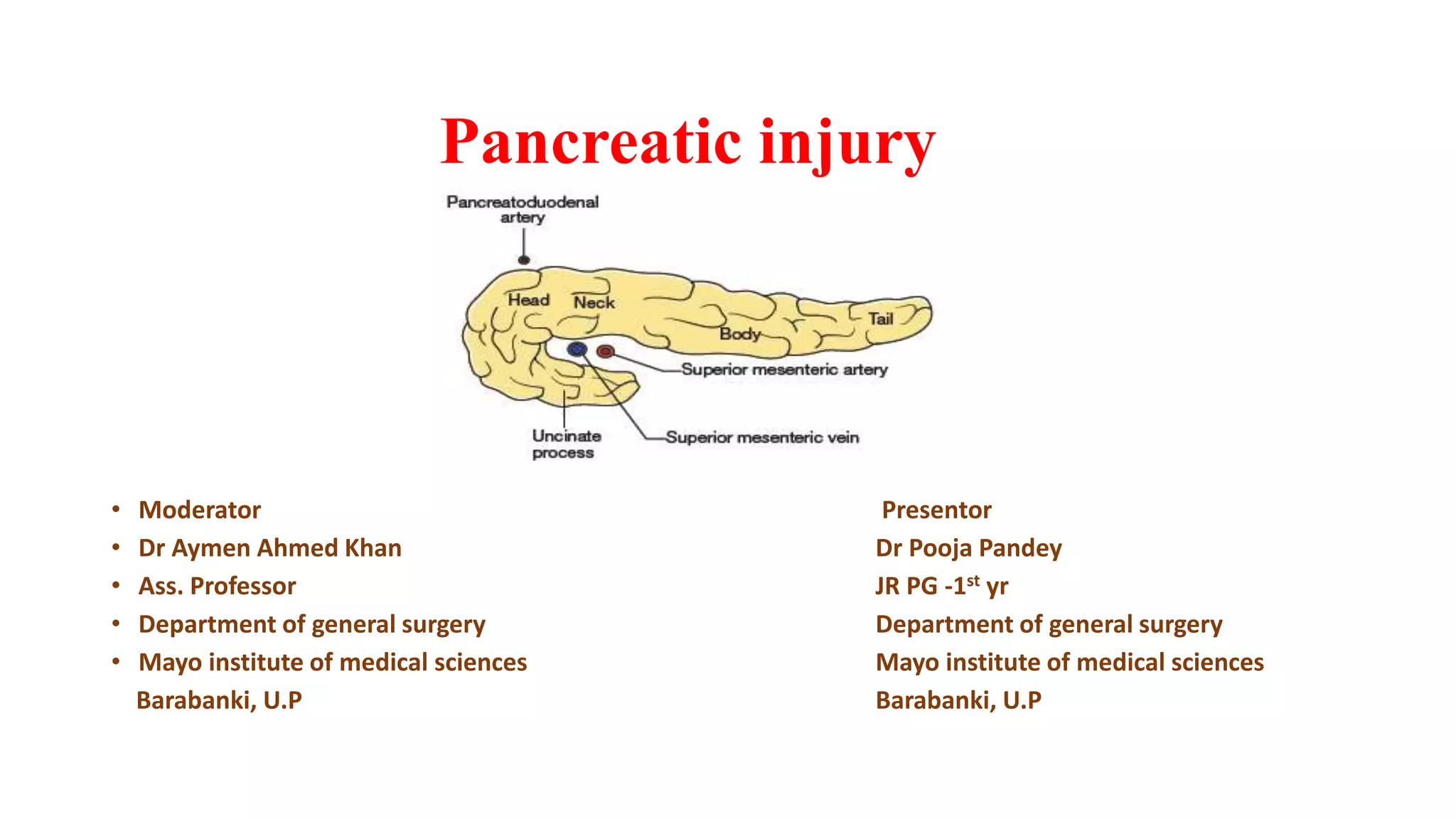
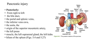
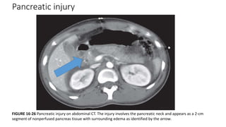
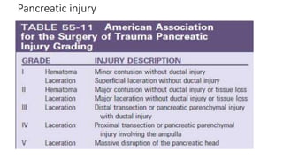
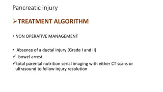
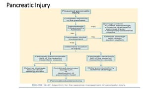
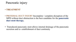
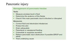
Pancreatic injury
• Moderator Presentor
• Dr Aymen Ahmed Khan Dr Pooja Pandey
• Ass. Professor JR PG -1st yr
• Department of general surgery Department of general surgery
• Mayo institute of medical sciences Mayo institute of medical sciences
Barabanki, U.P Barabanki, U.P
Pancreatic injury
Learning objectives
• Introduction
• Mechanism of injury
• Clinical presentation
• Management
• Follow up
• References
Pancreatic injury
Introduction to injury
• Major public health problem world wide
• Every 5sec one individual dies in the world because of injury
• 10% of the world's deaths, 32% more than the number of fatalities that
result from malaria, tuberculosis, and HIV/AIDS combined.
• For people between the ages of 5 and 44 years, injuries are 1 of the top
3 causes of death
Agarwal, Harshit MS, MCh; Gupta, Amit MS, FRCS; Kumar, Subodh MS, FRCS∗ An overview of pancreatic trauma, Journal of Pancreatology: September 2020 - Volume 3 - Issue 3 - p 139-
146 doi: 10.1097/JP9.0000000000000044
Pancreatic injury
Incidence
• 7 to 10 % of abdominal truma in a polyrauma patient .
• Injuries to the pancreas occur in approximately
• 5% of patients with blunt abdominal trauma,
• 6% of patients with gunshot wounds to the abdomen,
• and 2% of patients with stab wounds to the abdomen.
• The reported incidence ranges from 0.4% to 12%.
Agarwal, Harshit MS, MCh; Gupta, Amit MS, FRCS; Kumar, Subodh MS, FRCS∗ An overview of pancreatic trauma, Journal of Pancreatology: September 2020 - Volume 3 - Issue 3 - p 139-146 doi:
10.1097/JP9.0000000000000044
Pancreatic injury
Incidence conrd..
• In India Pancreatic injuries were present in 1.18 % of all trauma
admissions(Indian scenario)
• Mortality for pancreatic injuries ranges from 9% to 34%; however, only
5% of the pancreatic injuries are directly related to the fatal outcome.
Uma Debi, Ravinder Kaur, Kaushal Kishor Prasad, Saroj Kant Sinha, Anindita Sinha, Kartar Singh World J Gastroenterol. 2013 Dec 21; 19(47): 9003–9011. Published online 2013 Dec
21. doi: 10.3748/wjg.v19.i47.9003
Pancreatic injury
Mortality
• Most of the mortality within 48hrs due to acute haemorrhage of
traumatized vasulature including-
• Splenic vein
• Portal vein
• Inferior vena cava
• Pt surviving the initial haemorrhage ,nearly half will have a
complication of their pancreatic wound such as abscess ,fistula
,pseudocyst , false aneurysm or anastomotic leak .
• Trauma for surgery
Pancreatic injury
• Isolated pancreatic injuries are not common.
• Up to 90% of patients present with associated hepatic, gastric, splenic,
renal, colonic, or vascular lesions.
• Pancreatic injuries commonly occur in association with injury to the
duodenum because of their proximity.
Relations
• Anteriorly: From right to left: the transverse colon and
• the attachment of the transverse mesocolon, the lesser
• sac, and the stomach
Pancreatic injury
Snell’s clinical anatomy pg no 159
Pancreatic injury
• Posteriorly:
• From right to left:
• the bile duct,
• the portal and splenic veins,
• the inferior vena cava,
• the aorta, the
• origin of the superior mesenteric artery,
• the left psoas
• muscle, the left suprarenal gland, the left kidney, and the
• hilum of the spleen (Figs. 5.4 and 5.27)
Pancreatic injury
 Causes of injury
• Road traffic accident
• Steering wheel injury(neck > body)
• Fall from height
• Penetrating injury.(head=body=tail)
Pancreatic injury
.
183
 Mechanism of injury
• Direct compression of the epigastrium against the vertebral column
• varies according to the age of the patient
• Infants and children – child abuse & bicycle handlebar injury
• Adult –Road traffic accident and penetrating injury.
• Most common in children and young adult because of decreased intra-
abdominal fat .
Pancreatic injury
Diagnosis
• The identification of pancreas injuries can be challenging, particularly
because available imaging modalities are not highly effective.
• Based on the technology employed, the ability to detect parenchymal
injury and pancreatic duct disruption remained below 60%.
• Peitzman and colleagues reported better performance by CT in a
prospective study in which the sensitivity of CT was approximately
80%.
Pancreatic injury
CT Scan
• The modality of choice
• Findings peripancreatic hematomas,
free fluid in the lesser sac, and abnormal
thickening of Gerota fascia
Pancreatic injury
FIGURE 16-26 Pancreatic injury on abdominal CT. The injury involves the pancreatic neck and appears as a 2-cm
segment of nonperfused pancreas tissue with surrounding edema as identified by the arrow.
Pancreatic injury
• DIAGNOSIS SERUM AMYLASE LEVEL
Suggest only pancreatic injury Cannot predict or correlate with the
degree of injury
• SERUM LIPASE LEVEL
nonspecific and a poor indication of injury elevated levels may
provide a clue to a severe injury requiring further investigation
Pancreatic injury
• Isolated pancreatic amylase measurement is not recommended because
up to 40% of patients with transected pancreatic duct have normal
serum amylase levels.
• When it is obtained more than 3 hours after injury occurrence, an
elevated serum amylase level may reflect pancreatic trauma.
• Used in this way, serum amylase levels are reasonably sensitive but
are lacking in specificity and therefore are of limited value.
• Serial quantification levels increase the sensitivity of the assay.
Pancreatic injury
MRCP
• High sensitivity and specificity
• Provides excellent visualization of the pancreatic duct, peripancreatic
fluid contiguous to fractured segments of the pancreas, and
hemorrhage after nonpenetrating trauma.
• Main limitations include high cost, availability, and amount of time
required to perform the study.
Pancreatic injury
ENDOSCOPIC RETROGRADE
CHOLANGIOPANCREATOGRAPHY (ERCP)
• sensitivity and specificity of 100%
• demonstrate clearly the site of duct disruption and the grade of duct injury
effective and safe non- operative treatment tool leakages of the pancreatic
duct, trans papillary stent insertion might seal the injury and stabilize it
Pancreatic injury
•ERCP
• The most reliable test to demonstrate pancreatic duct integrity.
• limited by the risk of inducing pancreatitis, availability, and severity of
the trauma
Pancreatic injury
 Lucas classification:
• Type I: Minor contusion (small hematoma) with minimal parenchymal
damage. Main Pancreatic duct intact.
• Type II: Major contusion, laceration or transection, confined to body
and tail of pancreas with or without ductal disruptiion.
• Type III: Major contusion, laceration or transection involving head of
pancreas with or without ductal disruption.
• Type IV: Combined pancreaticoduodenal injury.
• A- Duodenal injury with minor pancreatic injury.
• B-Duodenal injury with severe pancreatic injury with duct disruption
Pancreatic injury
Pancreatic injury
TREATMENT ALGORITHM
• NON OPERATIVE MANAGEMENT
• Absence of a ductal injury (Grade I and II)
 bowel arrest
total parental nutrition serial imaging with either CT scans or
ultrasound to follow injury resolution
Pancreatic Injury
Pancreatic injury
• TREATMENT
• PROXIMAL DUCT INJURY Incomplete / complete disruption of the
MPD without duct obstruction is the best candidate for the pancreatic
duct stent therapy.
• Transductal pancreatic stent allows internal drainage of the pancreatic
secretion and re- establishment of duct continuity
Pancreatic injury
• TREATMENT
• GRADE IV INJURIES WITH PDI In stable patients
pancreaticoduodenectomy is the best definite treatment .
• In unstable patients, exploration and placing of external drainage may be the
best choice for damage control surgery .
Pancreatic injury
• TREATMENT
• DISTAL PANCREATIC INJURY WITH DUCT INVOLVEMENT wounds in the
body or tail of the pancreas with an obvious duct injury or transection
of more than half the width of the pancreas these grade III injuries
are best treated by Distal pancreatectomy .
• Complete transection of the pancreatic body from the head, a distal
Pancreaticojejunostomy and closure of the proximal end of the
pancreas rupture
Pancreatic injury
Complications
• Fistula
• Pancreatic abscess
• Pseudocyst formation and
• Sepsis
Pancreatic injury
•Pancreatic fistula
• 25% of patients following external drainage for pancreatic injuries.
• Drainage between 100–300 ml should be allowed to continue with the
drain left in place and majority of these fistula closes in 6–12 weeks
time.
• The closure of the fistula may be hastened by a short course of
octreotide for 5 days.
• The drain is removed when the fistula discharge becomes less than 30
ml per day.
Pancreatic injury
Pancreatic fistula
• Premature removal of the drain may result in abscess formation.
• Drainage in excess of 300 ml per day is unlikely to close spontaneously.
This is usually
associated with major ductal disruption.
If the fistula persists due to ductal disruption, this should be treated by
internal drainage into a Roux-en-Y loop of jejunum.
Pancreatic injury
References
• Agarwal, Harshit MS, MCh; Gupta, Amit MS, FRCS; Kumar, Subodh MS, FRCS∗ An
overview of pancreatic trauma, Journal of Pancreatology: September 2020 - Volume
3 - Issue 3 - p 139-146 doi: 10.1097/JP9.0000000000000044
• Uma Debi, Ravinder Kaur, Kaushal Kishor Prasad, Saroj Kant Sinha, Anindita Sinha,
Kartar Singh World J Gastroenterol. 2013 Dec 21; 19(47): 9003–9011. Published
online 2013 Dec 21. doi: 10.3748/wjg.v19.i47.9003
• Sabiston text book of surgery vol II 4rth edition
• Bailey and love short practice of surgery 27th edition
Thank you

Pancreatic injury

  • 1.
    Pancreatic injury • ModeratorPresentor • Dr Aymen Ahmed Khan Dr Pooja Pandey • Ass. Professor JR PG -1st yr • Department of general surgery Department of general surgery • Mayo institute of medical sciences Mayo institute of medical sciences Barabanki, U.P Barabanki, U.P
  • 2.
    Pancreatic injury Learning objectives •Introduction • Mechanism of injury • Clinical presentation • Management • Follow up • References
  • 3.
    Pancreatic injury Introduction toinjury • Major public health problem world wide • Every 5sec one individual dies in the world because of injury • 10% of the world's deaths, 32% more than the number of fatalities that result from malaria, tuberculosis, and HIV/AIDS combined. • For people between the ages of 5 and 44 years, injuries are 1 of the top 3 causes of death Agarwal, Harshit MS, MCh; Gupta, Amit MS, FRCS; Kumar, Subodh MS, FRCS∗ An overview of pancreatic trauma, Journal of Pancreatology: September 2020 - Volume 3 - Issue 3 - p 139- 146 doi: 10.1097/JP9.0000000000000044
  • 4.
    Pancreatic injury Incidence • 7to 10 % of abdominal truma in a polyrauma patient . • Injuries to the pancreas occur in approximately • 5% of patients with blunt abdominal trauma, • 6% of patients with gunshot wounds to the abdomen, • and 2% of patients with stab wounds to the abdomen. • The reported incidence ranges from 0.4% to 12%. Agarwal, Harshit MS, MCh; Gupta, Amit MS, FRCS; Kumar, Subodh MS, FRCS∗ An overview of pancreatic trauma, Journal of Pancreatology: September 2020 - Volume 3 - Issue 3 - p 139-146 doi: 10.1097/JP9.0000000000000044
  • 5.
    Pancreatic injury Incidence conrd.. •In India Pancreatic injuries were present in 1.18 % of all trauma admissions(Indian scenario) • Mortality for pancreatic injuries ranges from 9% to 34%; however, only 5% of the pancreatic injuries are directly related to the fatal outcome. Uma Debi, Ravinder Kaur, Kaushal Kishor Prasad, Saroj Kant Sinha, Anindita Sinha, Kartar Singh World J Gastroenterol. 2013 Dec 21; 19(47): 9003–9011. Published online 2013 Dec 21. doi: 10.3748/wjg.v19.i47.9003
  • 6.
    Pancreatic injury Mortality • Mostof the mortality within 48hrs due to acute haemorrhage of traumatized vasulature including- • Splenic vein • Portal vein • Inferior vena cava • Pt surviving the initial haemorrhage ,nearly half will have a complication of their pancreatic wound such as abscess ,fistula ,pseudocyst , false aneurysm or anastomotic leak . • Trauma for surgery
  • 7.
    Pancreatic injury • Isolatedpancreatic injuries are not common. • Up to 90% of patients present with associated hepatic, gastric, splenic, renal, colonic, or vascular lesions. • Pancreatic injuries commonly occur in association with injury to the duodenum because of their proximity. Relations • Anteriorly: From right to left: the transverse colon and • the attachment of the transverse mesocolon, the lesser • sac, and the stomach
  • 8.
  • 9.
    Pancreatic injury • Posteriorly: •From right to left: • the bile duct, • the portal and splenic veins, • the inferior vena cava, • the aorta, the • origin of the superior mesenteric artery, • the left psoas • muscle, the left suprarenal gland, the left kidney, and the • hilum of the spleen (Figs. 5.4 and 5.27)
  • 10.
    Pancreatic injury  Causesof injury • Road traffic accident • Steering wheel injury(neck > body) • Fall from height • Penetrating injury.(head=body=tail)
  • 11.
    Pancreatic injury . 183  Mechanismof injury • Direct compression of the epigastrium against the vertebral column • varies according to the age of the patient • Infants and children – child abuse & bicycle handlebar injury • Adult –Road traffic accident and penetrating injury. • Most common in children and young adult because of decreased intra- abdominal fat .
  • 12.
    Pancreatic injury Diagnosis • Theidentification of pancreas injuries can be challenging, particularly because available imaging modalities are not highly effective. • Based on the technology employed, the ability to detect parenchymal injury and pancreatic duct disruption remained below 60%. • Peitzman and colleagues reported better performance by CT in a prospective study in which the sensitivity of CT was approximately 80%.
  • 13.
    Pancreatic injury CT Scan •The modality of choice • Findings peripancreatic hematomas, free fluid in the lesser sac, and abnormal thickening of Gerota fascia
  • 14.
    Pancreatic injury FIGURE 16-26Pancreatic injury on abdominal CT. The injury involves the pancreatic neck and appears as a 2-cm segment of nonperfused pancreas tissue with surrounding edema as identified by the arrow.
  • 15.
    Pancreatic injury • DIAGNOSISSERUM AMYLASE LEVEL Suggest only pancreatic injury Cannot predict or correlate with the degree of injury • SERUM LIPASE LEVEL nonspecific and a poor indication of injury elevated levels may provide a clue to a severe injury requiring further investigation
  • 16.
    Pancreatic injury • Isolatedpancreatic amylase measurement is not recommended because up to 40% of patients with transected pancreatic duct have normal serum amylase levels. • When it is obtained more than 3 hours after injury occurrence, an elevated serum amylase level may reflect pancreatic trauma. • Used in this way, serum amylase levels are reasonably sensitive but are lacking in specificity and therefore are of limited value. • Serial quantification levels increase the sensitivity of the assay.
  • 17.
    Pancreatic injury MRCP • Highsensitivity and specificity • Provides excellent visualization of the pancreatic duct, peripancreatic fluid contiguous to fractured segments of the pancreas, and hemorrhage after nonpenetrating trauma. • Main limitations include high cost, availability, and amount of time required to perform the study.
  • 18.
    Pancreatic injury ENDOSCOPIC RETROGRADE CHOLANGIOPANCREATOGRAPHY(ERCP) • sensitivity and specificity of 100% • demonstrate clearly the site of duct disruption and the grade of duct injury effective and safe non- operative treatment tool leakages of the pancreatic duct, trans papillary stent insertion might seal the injury and stabilize it
  • 19.
    Pancreatic injury •ERCP • Themost reliable test to demonstrate pancreatic duct integrity. • limited by the risk of inducing pancreatitis, availability, and severity of the trauma
  • 20.
    Pancreatic injury  Lucasclassification: • Type I: Minor contusion (small hematoma) with minimal parenchymal damage. Main Pancreatic duct intact. • Type II: Major contusion, laceration or transection, confined to body and tail of pancreas with or without ductal disruptiion. • Type III: Major contusion, laceration or transection involving head of pancreas with or without ductal disruption. • Type IV: Combined pancreaticoduodenal injury. • A- Duodenal injury with minor pancreatic injury. • B-Duodenal injury with severe pancreatic injury with duct disruption
  • 21.
  • 22.
    Pancreatic injury TREATMENT ALGORITHM •NON OPERATIVE MANAGEMENT • Absence of a ductal injury (Grade I and II)  bowel arrest total parental nutrition serial imaging with either CT scans or ultrasound to follow injury resolution
  • 23.
  • 24.
    Pancreatic injury • TREATMENT •PROXIMAL DUCT INJURY Incomplete / complete disruption of the MPD without duct obstruction is the best candidate for the pancreatic duct stent therapy. • Transductal pancreatic stent allows internal drainage of the pancreatic secretion and re- establishment of duct continuity
  • 25.
    Pancreatic injury • TREATMENT •GRADE IV INJURIES WITH PDI In stable patients pancreaticoduodenectomy is the best definite treatment . • In unstable patients, exploration and placing of external drainage may be the best choice for damage control surgery .
  • 26.
    Pancreatic injury • TREATMENT •DISTAL PANCREATIC INJURY WITH DUCT INVOLVEMENT wounds in the body or tail of the pancreas with an obvious duct injury or transection of more than half the width of the pancreas these grade III injuries are best treated by Distal pancreatectomy . • Complete transection of the pancreatic body from the head, a distal Pancreaticojejunostomy and closure of the proximal end of the pancreas rupture
  • 27.
    Pancreatic injury Complications • Fistula •Pancreatic abscess • Pseudocyst formation and • Sepsis
  • 28.
    Pancreatic injury •Pancreatic fistula •25% of patients following external drainage for pancreatic injuries. • Drainage between 100–300 ml should be allowed to continue with the drain left in place and majority of these fistula closes in 6–12 weeks time. • The closure of the fistula may be hastened by a short course of octreotide for 5 days. • The drain is removed when the fistula discharge becomes less than 30 ml per day.
  • 29.
    Pancreatic injury Pancreatic fistula •Premature removal of the drain may result in abscess formation. • Drainage in excess of 300 ml per day is unlikely to close spontaneously. This is usually associated with major ductal disruption. If the fistula persists due to ductal disruption, this should be treated by internal drainage into a Roux-en-Y loop of jejunum.
  • 30.
  • 31.
    References • Agarwal, HarshitMS, MCh; Gupta, Amit MS, FRCS; Kumar, Subodh MS, FRCS∗ An overview of pancreatic trauma, Journal of Pancreatology: September 2020 - Volume 3 - Issue 3 - p 139-146 doi: 10.1097/JP9.0000000000000044 • Uma Debi, Ravinder Kaur, Kaushal Kishor Prasad, Saroj Kant Sinha, Anindita Sinha, Kartar Singh World J Gastroenterol. 2013 Dec 21; 19(47): 9003–9011. Published online 2013 Dec 21. doi: 10.3748/wjg.v19.i47.9003 • Sabiston text book of surgery vol II 4rth edition • Bailey and love short practice of surgery 27th edition
  • 32.

Editor's Notes

  • #5 Akhrass et al[9] had reported a 0.4% incidence of blunt pancreatic injury while Lin et al[10] reported 5.4% incidence of blunt pancreatic trauma with 1.3% incidence of high-grade pancreatic injury. Duchesne et al[11] reported 0.6% incidence of pancreatic injury, while we at our center reported an incidence of 1.2%.
  • #6 75% of deaths occur within the 48 to 72 hours after trauma, and most are related to hypovolemic shock.61
  • #11 Pancreatic enzymes are caustic, making delays in management of the injuries a source of massive systemic inflammation and subsequent poor outcomes. Pancreas tissue injury can result from direct laceration of the organ or through the transmission of blunt force energy to the retroperitoneum. A common mechanism of blunt pancreatic injury involves the crushing of the body of the pancreas between a rigid structure, such as a steering wheel or seat belt, and the vertebral column. The impact to the pancreas causes injury that ranges from mild contusion to complete transection with ductal disruption
  • #12 Acceleration–deceleration injury and direct compression force in the epigastrium is responsible for pancreatic injury. Since spine is immobile and pancreatic tail is mobile, a shearing force between the 2 may lead to pancreatic injury at neck and body as is seen in high-velocity motor crashes. Moreover, the direct impact on epigastrium can also lead to pancreatic injury by compressing the pancreas against the spine. Motor vehicle crash is the most common cause of pancreatic injury, with steering wheel impact being a distinctive mechanism of injury. Lin et al reported steering wheel injury in 71% of the cases as the causative mechanism. Similarly, Duchesne et al[11] reported steering wheel injury in 33 (94.2%) cases followed by fall and work-related crush injury in 1 (2.8%) case each. Another important mechanism is bicycle handlebar injury, especially in pediatric population, with Sutherland et al[14] reporting it as the commonest cause in high-grade pancreatic injuries.
  • #13 Sabiston management of trauma trauma pg 441 trauma
  • #15 Sabiston management of trauma trauma pg 441 trauma