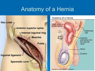
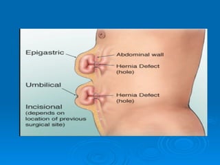
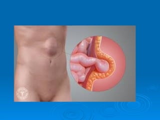
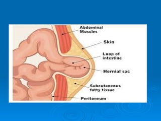
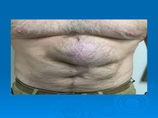
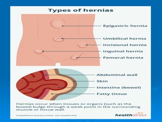
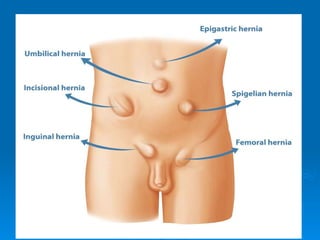
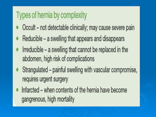
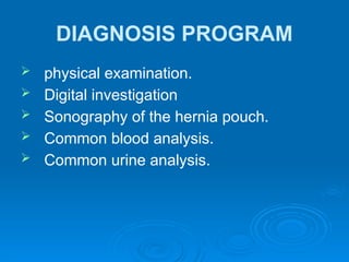
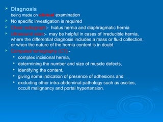
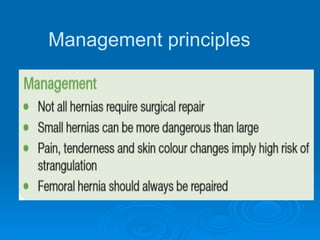
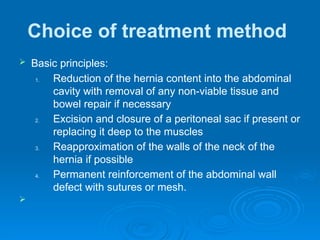
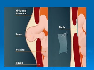
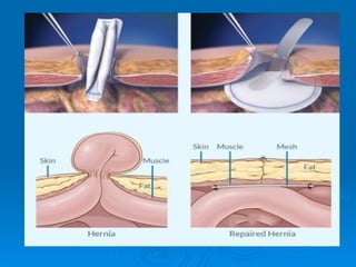
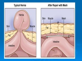
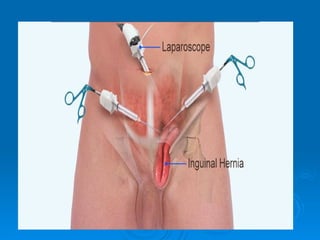
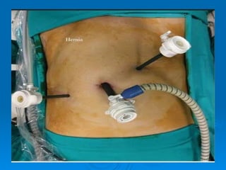
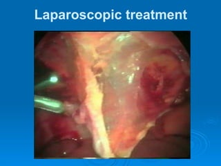
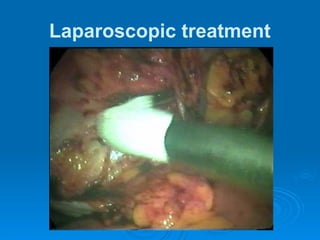
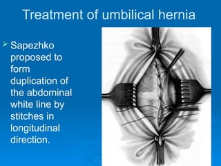
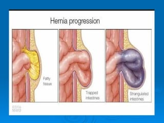
HERNIA
HERNIA
Hernia of the abdominal wall or
external hernia is the bulging of part
of the contents of the abdominal
cavity through a weakness in the
abdominal wall.
 Internal hernia is such disease, visceral
organs hit the peritoneum pouch. It formed
in the place of natural peritoneum fold or
recess and generally kept in the
abdominal cavity.
Anatomy of a Hernia
Anatomical causes of abdominal w
herniation
The Walls Of The Inguinal Canal
ANTERIOR WALL
 laterally - muscles fibers of the external oblique
 medially - aponeurosis of the external oblique
 most medially there is not wall but instead there is a deficiency called the
superficial inguinal ring.
SUPERIOR -- arching fibers of the internal oblique and sometimes transverse
abdominis. These fibers start anterior and lateral, pass over the spermatic
cord and the medially forms part of the posterior wall of the canal.
POSTERIOR -- lateral the posterior wall is deficient at the deep inguinal ring.
Medially the posterior wall is made up of the fused aponeuroses of the
internal oblique and transverse abdominis, called the conjoined tendon X.
INFERIOR (or floor) -- inguinal ligament. Medially, some of the fibers of the
inguinal ligament curve under the spermatic cord and fasten into the
pectineal line of the pubis, this is the lacunar ligament which forms part of
the floor of the inguinal canal.
PATHOMORPHOLOGY
 Each abdominal hernia consists of hernia
gate, hernia sac and hernia contents.
Hernia sac forms by outpouching of
parietal peritoneum and can contain small
intestine and omentum. Sometimes it
containes other organs: large intestine,
urinary bladder, ovary, and appendix.
 The main parts of the hernia pouch are
neck, body and fundus.
CLASSIFICATION
 1) Depends on anatomical localization: inguinal
(indirect and direct), midline hernia, epigastric
hernia, femoral hernia, lumbar hernia, sciatic
hernia, incisional hernia, umbilical hernia .
 2) depends on etiology: congenital and acquired
 3) Depends on clinical presentations: complete
and incomplete, reducible and nonreducible,
traumatic and postoperative, complicated and
noncomplicated.
Examination for hernia
 Examined on lying down initially and then
standing.
 Asked to cough
 Divarification is best seen by asking a supine
patient to simply lift his head off the pillow.
 The overlying skin is usually of normal colour.
 If bruising is present this may suggest venous
engorgement of the content.
 If there is overlying cellulitis then hernia content
is strangulating.
 In most cases a cough impulse is felt
DIAGNOSIS PROGRAM
 physical examination.
 Digital investigation
 Sonography of the hernia pouch.
 Common blood analysis.
 Common urine analysis.
 Diagnosis
being made on clinical examination
 No specific investigation is required
 Chest radiograph:- hiatus hernia and diaphragmatic hernia
 Ultrasound scan:- may be helpful in cases of irreducible hernia,
where the differential diagnosis includes a mass or fluid collection,
or when the nature of the hernia content is in doubt.
 Computed tomography (CT):-

complex incisional hernia,

determining the number and size of muscle defects,

identifying the content,

giving some indication of presence of adhesions and

excluding other intra-abdominal pathology such as ascites,
occult malignancy and portal hypertension.
Management principles
Choice of treatment method
 Basic principles:
1. Reduction of the hernia content into the abdominal
cavity with removal of any non-viable tissue and
bowel repair if necessary
2. Excision and closure of a peritoneal sac if present or
replacing it deep to the muscles
3. Reapproximation of the walls of the neck of the
hernia if possible
4. Permanent reinforcement of the abdominal wall
defect with sutures or mesh.

Treatment of the femoral hernia
 The term ‘mesh’ refers to prosthetic material,
either a net or a flat sheet, which is used to
strengthen a hernia repair.
 Mesh can be used:

to bridge a defect: the mesh is simply fixed
over the defect as a tension-free patch;

to plug a defect: a plug of mesh is pushed into
the defect;

to augment a repair: the defect is closed with
sutures and the mesh added for
reinforcement.
 A well-placed mesh should have good overlap around all
margins of the defect, at least 2 cm but up to 5 cm if
possible.
 Suturing a mesh edge to edge into the defect (inlay),
with no overlap, is not recommended.
 Mesh plug repairs have gained some popularity in small
defects especially where overlap is hard to achieve.
 Plug operations are fast but plugs can form a dense
‘meshoma’ of plug and collagen.
 Other complications include migration, erosion into
adjacent organs, fistula formation and chronic pain.
Bassini repair
Girard repair
Laparoscopic treatment
Treatment of umbilical hernia
 The Lexer operation is most widespread. It
performed by imposition of sutures on an
umbilical ring
Treatment of umbilical hernia
 After the Meyo
method defect
of anterior
abdominal wall
in the umbilical
ring is sutured
by U-shaped
stitches in
transversal
direction
Treatment of umbilical hernia
 Sapezhko
proposed to
form
duplication of
the abdominal
white line by
stitches in
longitudinal
direction.
INCARCERATED HERNIA
 Incarcerated hernia is sudden pressing of
hernia contents in a hernia orifice.
Incarceration is the most frequent and
most dangerous complication of hernia
diseases.
Etiology and pathogenesis
 At the elastic incarceration, after increasing
intraabdominal pressure, one or a few organs
relocated from an abdominal cavity to the hernia
sack, where it is compressed with following
ischemia and necrosis in the area of hernia gate.
 At the fecal incarceration in the intestinal
loop which is in a hernia sack, plenty of
excrement passed quickly. Proximal part
of loop is overfilled, and distal is
compressed in a hernia gate.
Classification of the incarcerated
hernia
 complete
 Incomplete
 partial (the Richter’s hernia)
 retrograde
 without the destructive changes of hernia
contents
 with the phlegmon of hernia sack
Retrograde incarceration
Diagnosis program
 examination.
 Physical examination.
 Blood analysis and urine analysis.
 Digital investigation of the rectum.
 Survey X-Ray of abdominal cavity
organs.
Treatment
 Approximately 70% of incarcerated hernia
can be reduced manually.
 in the case of failure, surgery should be
immediately performed for incarcerated
hernia.
abdominal hernias for nursing students.pptx

abdominal hernias for nursing students.pptx

  • 1.
  • 2.
    HERNIA Hernia of theabdominal wall or external hernia is the bulging of part of the contents of the abdominal cavity through a weakness in the abdominal wall.
  • 3.
     Internal herniais such disease, visceral organs hit the peritoneum pouch. It formed in the place of natural peritoneum fold or recess and generally kept in the abdominal cavity.
  • 4.
  • 10.
    Anatomical causes ofabdominal w herniation
  • 12.
    The Walls OfThe Inguinal Canal ANTERIOR WALL  laterally - muscles fibers of the external oblique  medially - aponeurosis of the external oblique  most medially there is not wall but instead there is a deficiency called the superficial inguinal ring. SUPERIOR -- arching fibers of the internal oblique and sometimes transverse abdominis. These fibers start anterior and lateral, pass over the spermatic cord and the medially forms part of the posterior wall of the canal. POSTERIOR -- lateral the posterior wall is deficient at the deep inguinal ring. Medially the posterior wall is made up of the fused aponeuroses of the internal oblique and transverse abdominis, called the conjoined tendon X. INFERIOR (or floor) -- inguinal ligament. Medially, some of the fibers of the inguinal ligament curve under the spermatic cord and fasten into the pectineal line of the pubis, this is the lacunar ligament which forms part of the floor of the inguinal canal.
  • 13.
    PATHOMORPHOLOGY  Each abdominalhernia consists of hernia gate, hernia sac and hernia contents. Hernia sac forms by outpouching of parietal peritoneum and can contain small intestine and omentum. Sometimes it containes other organs: large intestine, urinary bladder, ovary, and appendix.  The main parts of the hernia pouch are neck, body and fundus.
  • 14.
    CLASSIFICATION  1) Dependson anatomical localization: inguinal (indirect and direct), midline hernia, epigastric hernia, femoral hernia, lumbar hernia, sciatic hernia, incisional hernia, umbilical hernia .  2) depends on etiology: congenital and acquired  3) Depends on clinical presentations: complete and incomplete, reducible and nonreducible, traumatic and postoperative, complicated and noncomplicated.
  • 16.
    Examination for hernia Examined on lying down initially and then standing.  Asked to cough  Divarification is best seen by asking a supine patient to simply lift his head off the pillow.  The overlying skin is usually of normal colour.  If bruising is present this may suggest venous engorgement of the content.  If there is overlying cellulitis then hernia content is strangulating.  In most cases a cough impulse is felt
  • 19.
    DIAGNOSIS PROGRAM  physicalexamination.  Digital investigation  Sonography of the hernia pouch.  Common blood analysis.  Common urine analysis.
  • 20.
     Diagnosis being madeon clinical examination  No specific investigation is required  Chest radiograph:- hiatus hernia and diaphragmatic hernia  Ultrasound scan:- may be helpful in cases of irreducible hernia, where the differential diagnosis includes a mass or fluid collection, or when the nature of the hernia content is in doubt.  Computed tomography (CT):-  complex incisional hernia,  determining the number and size of muscle defects,  identifying the content,  giving some indication of presence of adhesions and  excluding other intra-abdominal pathology such as ascites, occult malignancy and portal hypertension.
  • 21.
  • 22.
    Choice of treatmentmethod  Basic principles: 1. Reduction of the hernia content into the abdominal cavity with removal of any non-viable tissue and bowel repair if necessary 2. Excision and closure of a peritoneal sac if present or replacing it deep to the muscles 3. Reapproximation of the walls of the neck of the hernia if possible 4. Permanent reinforcement of the abdominal wall defect with sutures or mesh. 
  • 23.
    Treatment of thefemoral hernia  The term ‘mesh’ refers to prosthetic material, either a net or a flat sheet, which is used to strengthen a hernia repair.  Mesh can be used:  to bridge a defect: the mesh is simply fixed over the defect as a tension-free patch;  to plug a defect: a plug of mesh is pushed into the defect;  to augment a repair: the defect is closed with sutures and the mesh added for reinforcement.
  • 27.
     A well-placedmesh should have good overlap around all margins of the defect, at least 2 cm but up to 5 cm if possible.  Suturing a mesh edge to edge into the defect (inlay), with no overlap, is not recommended.  Mesh plug repairs have gained some popularity in small defects especially where overlap is hard to achieve.  Plug operations are fast but plugs can form a dense ‘meshoma’ of plug and collagen.  Other complications include migration, erosion into adjacent organs, fistula formation and chronic pain.
  • 28.
  • 29.
  • 32.
  • 33.
  • 34.
    Treatment of umbilicalhernia  The Lexer operation is most widespread. It performed by imposition of sutures on an umbilical ring
  • 35.
    Treatment of umbilicalhernia  After the Meyo method defect of anterior abdominal wall in the umbilical ring is sutured by U-shaped stitches in transversal direction
  • 36.
    Treatment of umbilicalhernia  Sapezhko proposed to form duplication of the abdominal white line by stitches in longitudinal direction.
  • 37.
    INCARCERATED HERNIA  Incarceratedhernia is sudden pressing of hernia contents in a hernia orifice. Incarceration is the most frequent and most dangerous complication of hernia diseases.
  • 39.
    Etiology and pathogenesis At the elastic incarceration, after increasing intraabdominal pressure, one or a few organs relocated from an abdominal cavity to the hernia sack, where it is compressed with following ischemia and necrosis in the area of hernia gate.
  • 40.
     At thefecal incarceration in the intestinal loop which is in a hernia sack, plenty of excrement passed quickly. Proximal part of loop is overfilled, and distal is compressed in a hernia gate.
  • 41.
    Classification of theincarcerated hernia  complete  Incomplete  partial (the Richter’s hernia)  retrograde  without the destructive changes of hernia contents  with the phlegmon of hernia sack
  • 42.
  • 43.
  • 44.
    Diagnosis program  examination. Physical examination.  Blood analysis and urine analysis.  Digital investigation of the rectum.  Survey X-Ray of abdominal cavity organs.
  • 45.
    Treatment  Approximately 70%of incarcerated hernia can be reduced manually.  in the case of failure, surgery should be immediately performed for incarcerated hernia.