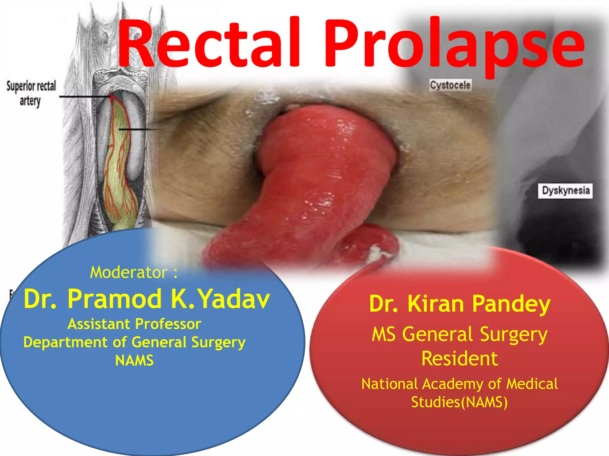
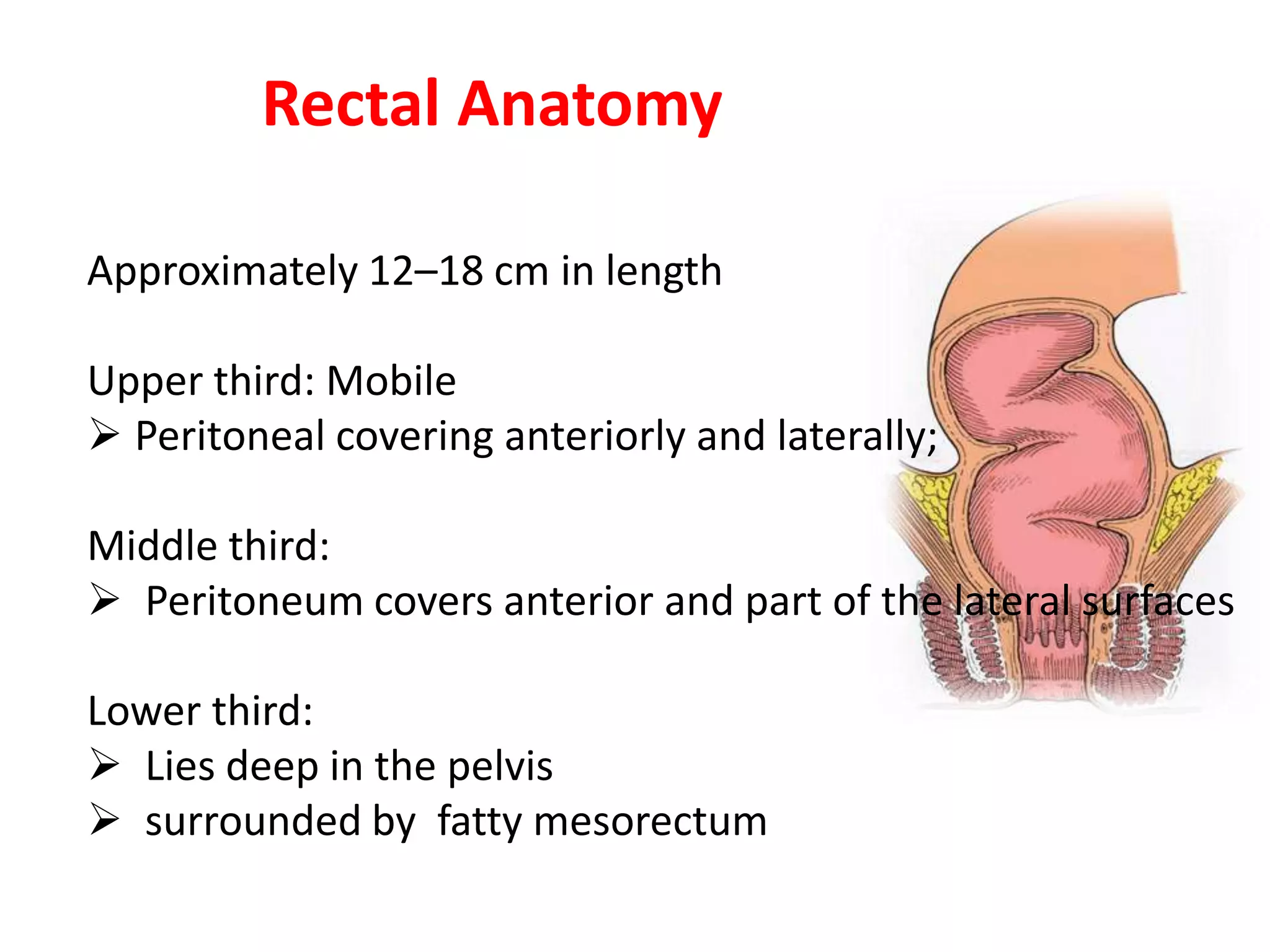
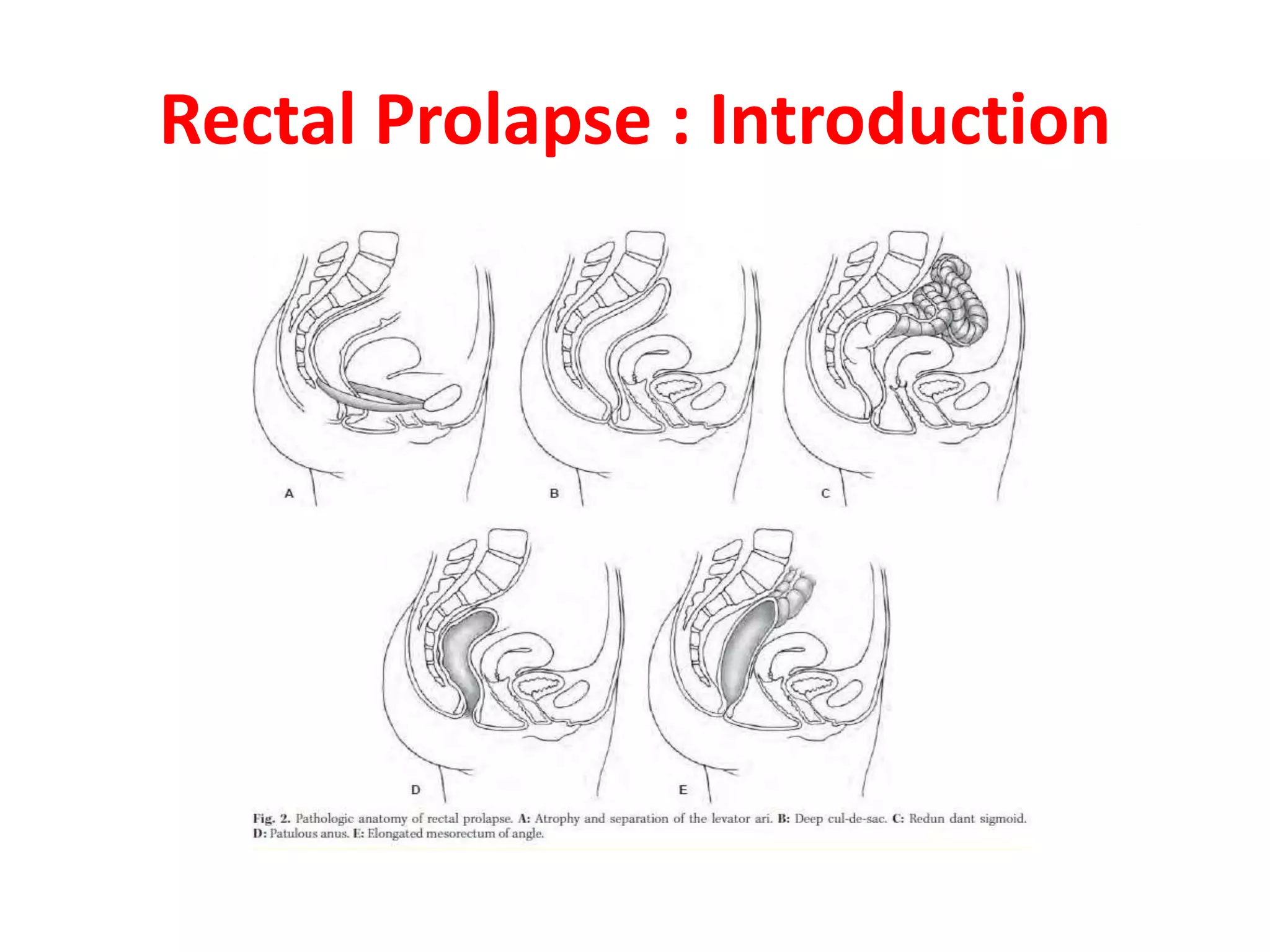
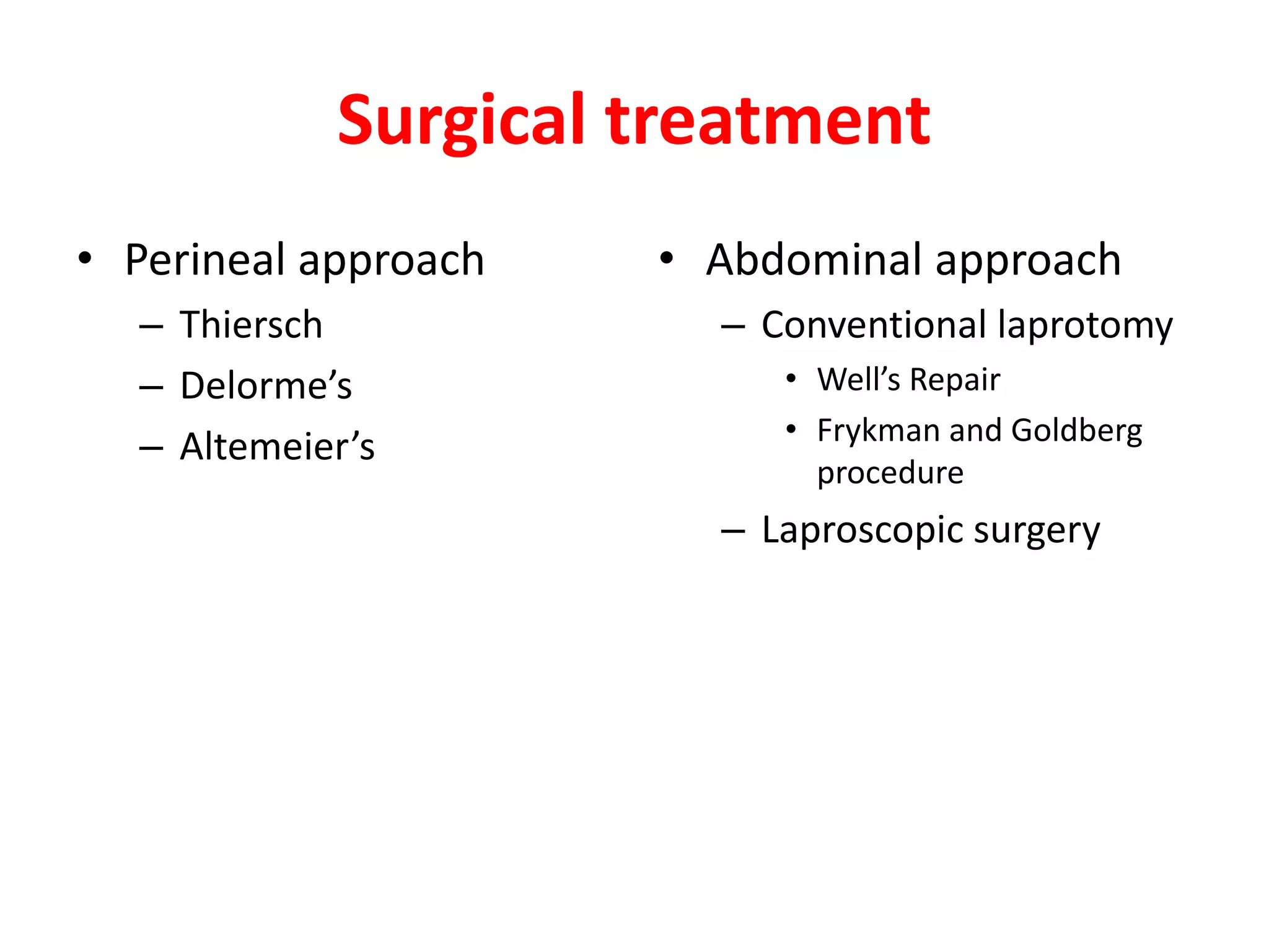
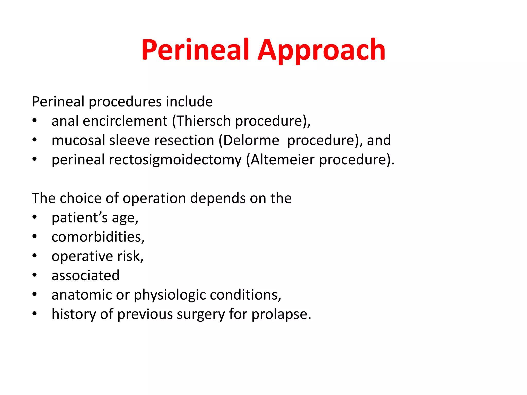
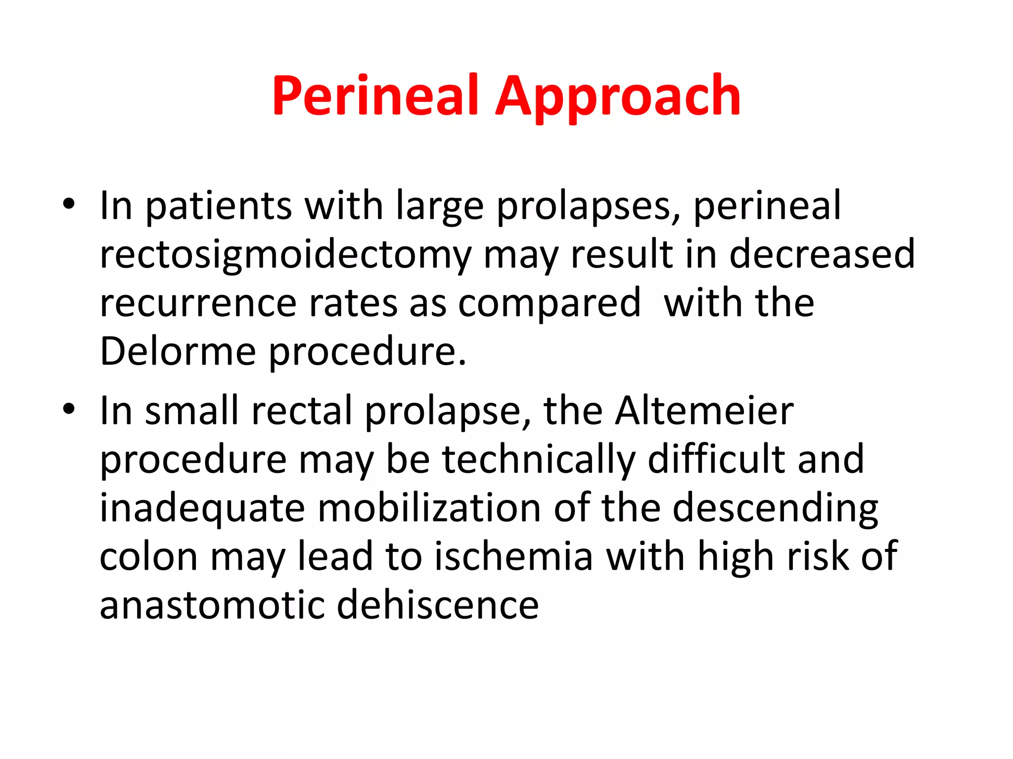
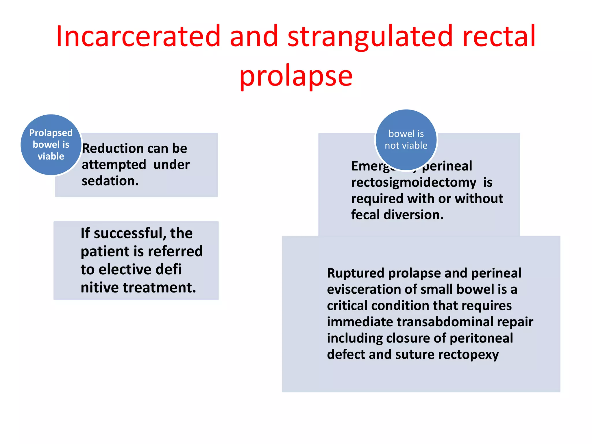
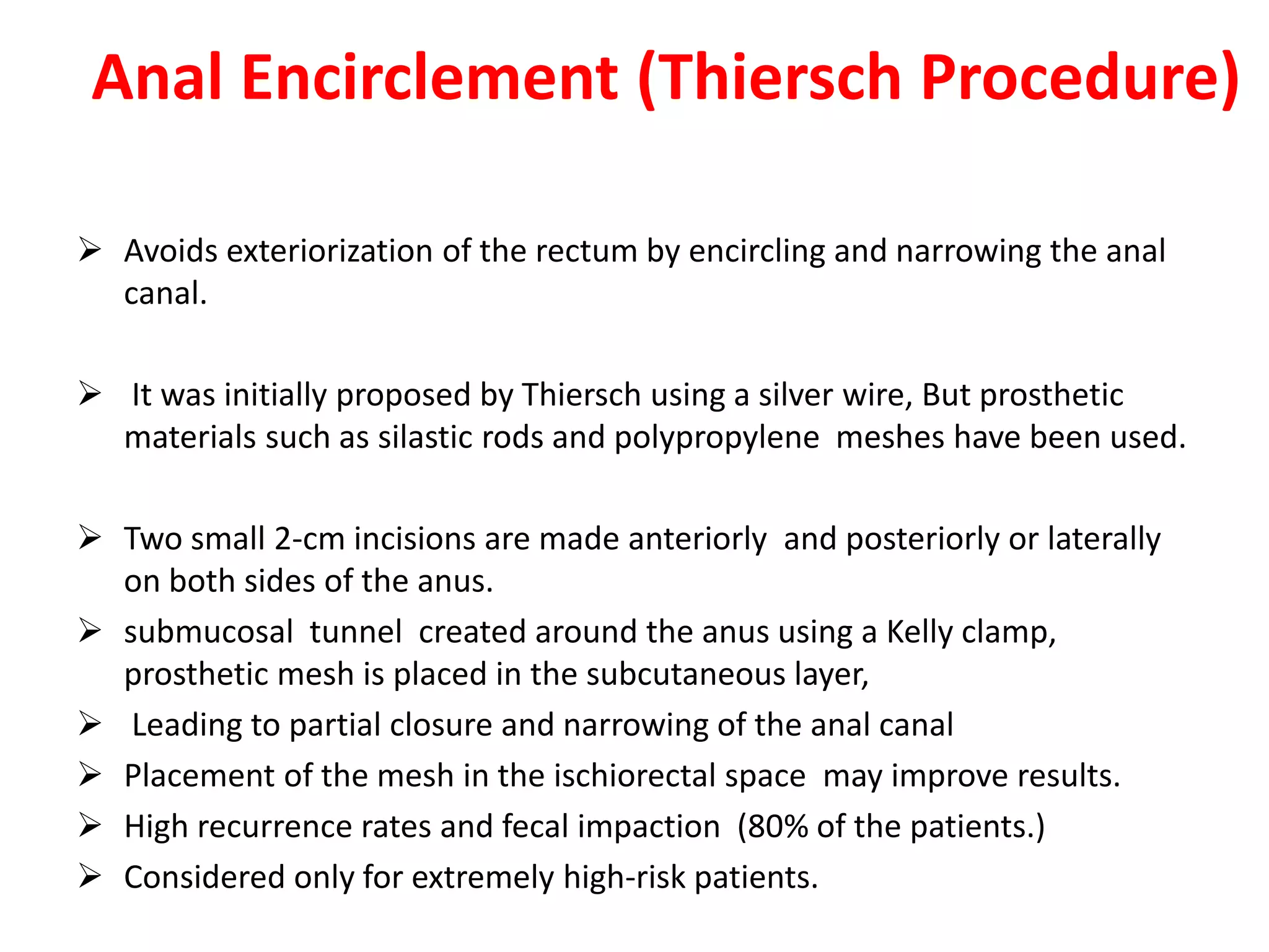
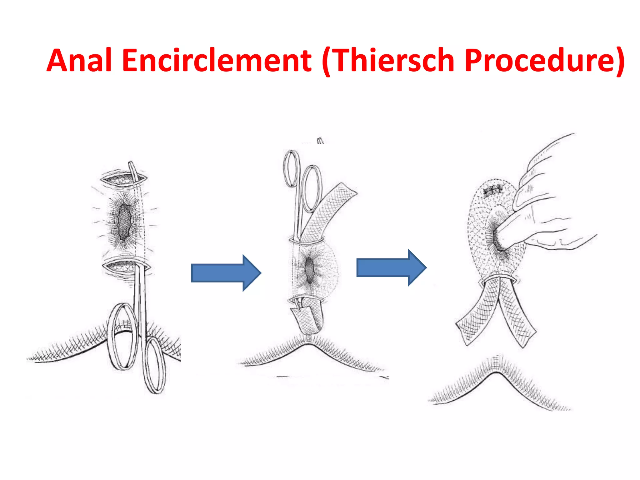
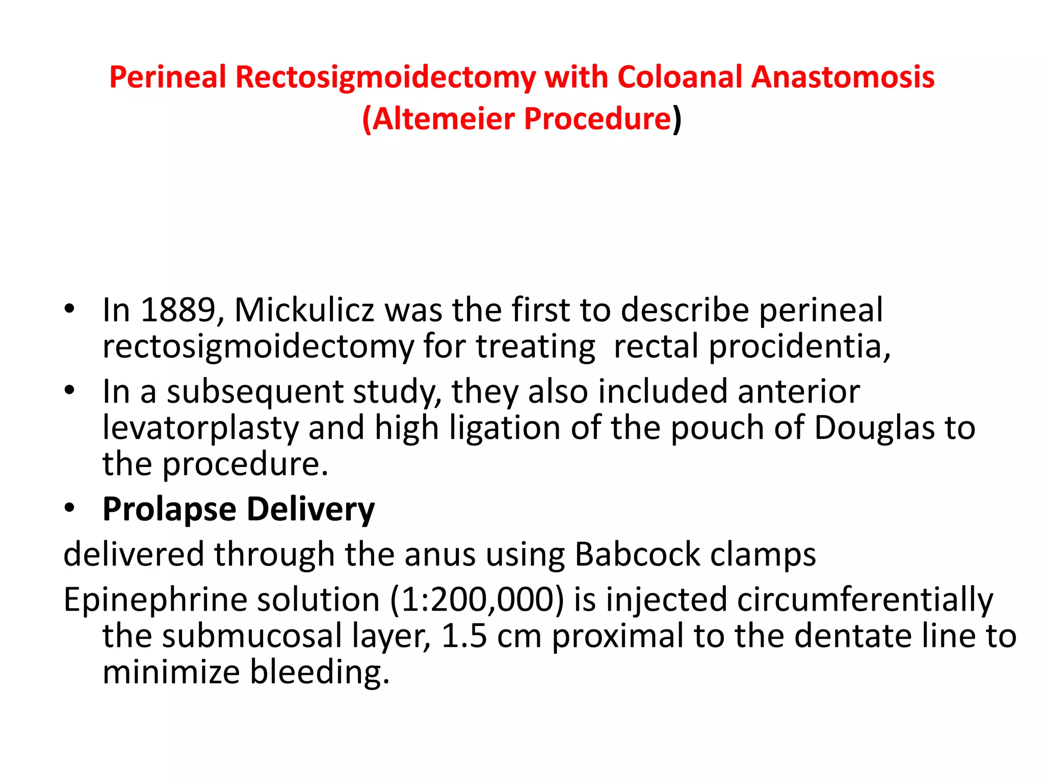
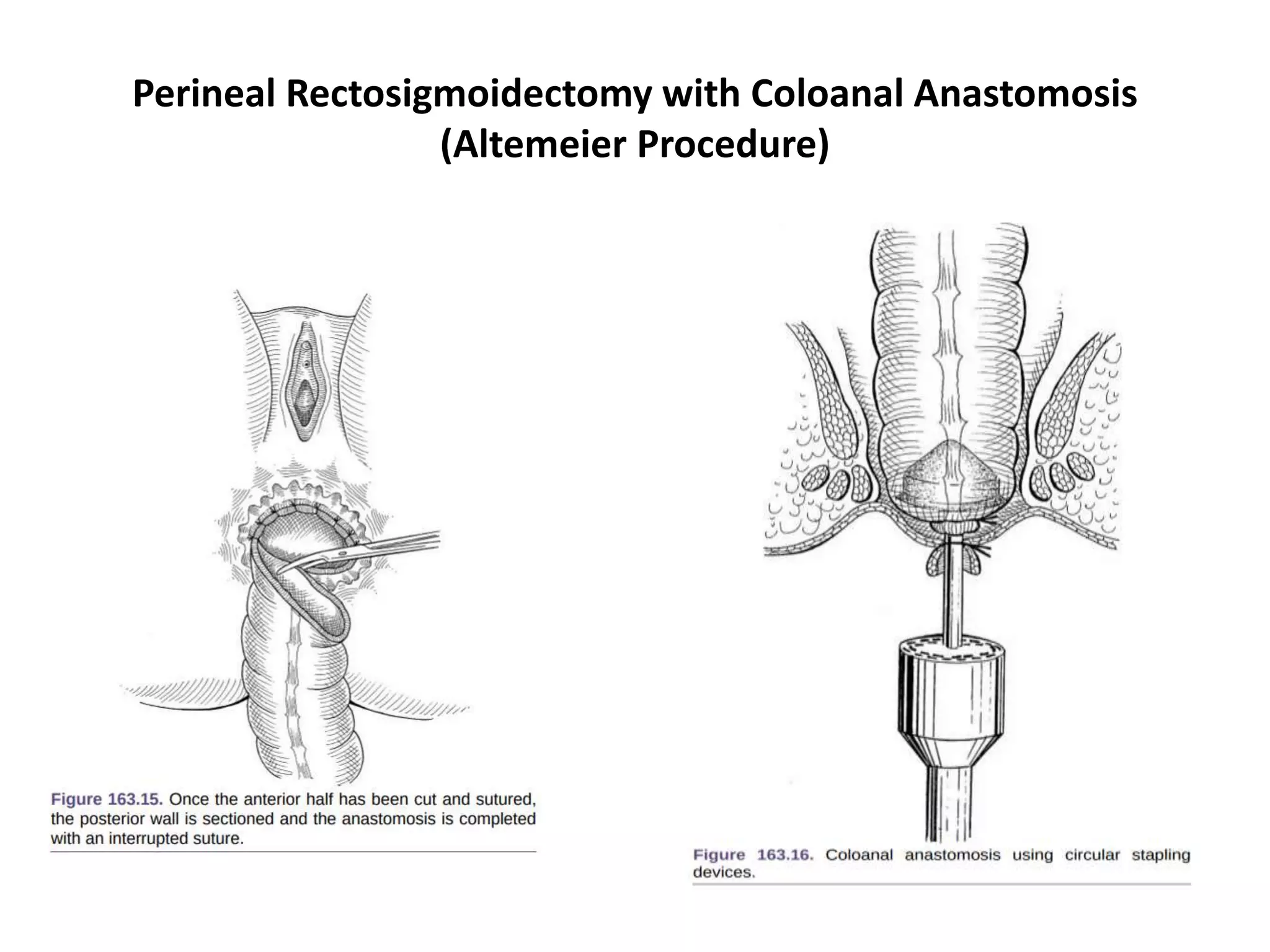
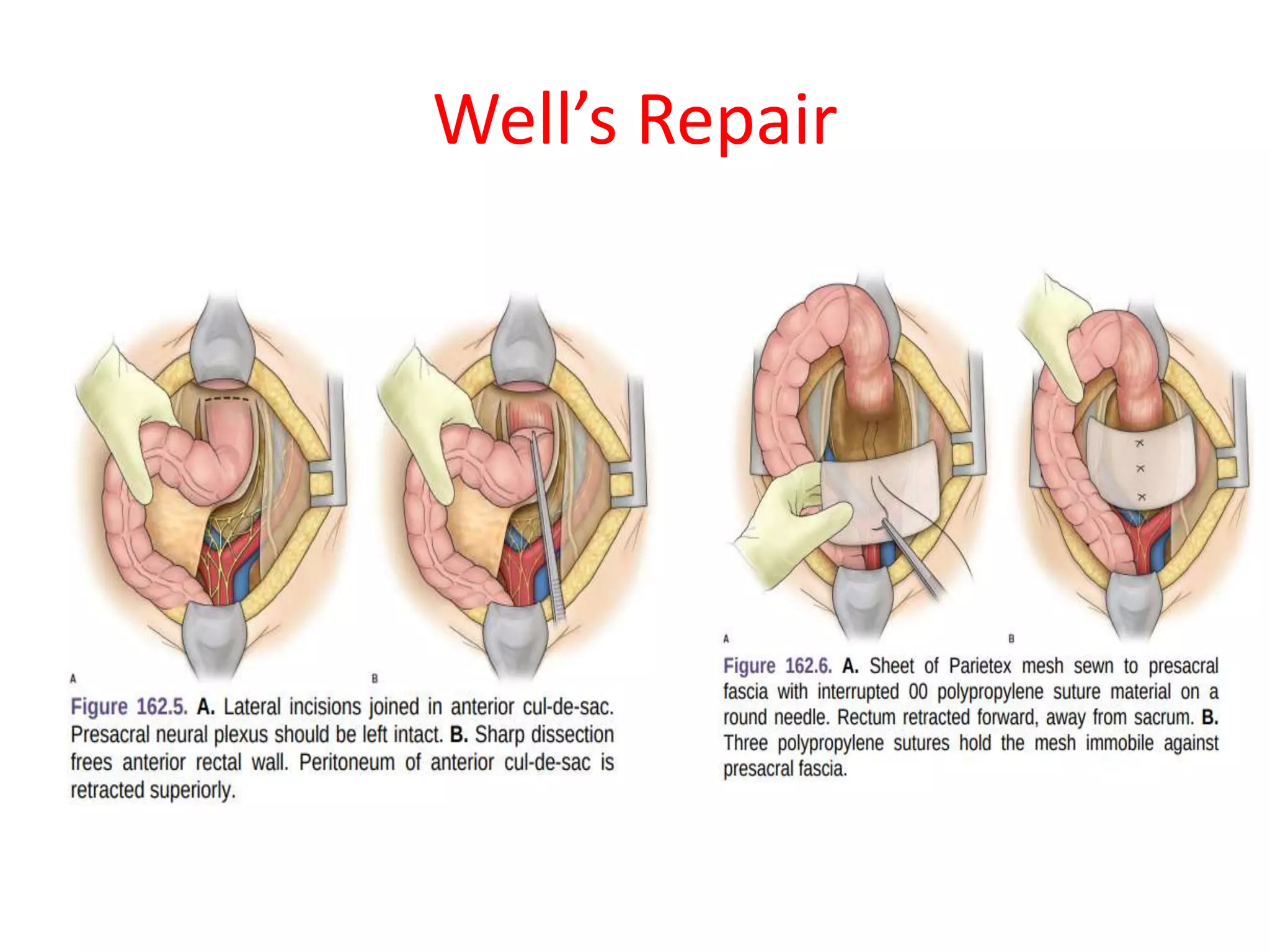
This document provides information about rectal prolapse including its anatomy, causes, clinical presentation, and surgical treatment options. It begins with a description of the rectal anatomy including its blood supply, lymphatic drainage, and curves. It then discusses the causes and types of rectal prolapse and explains how factors like pelvic floor weakness can lead to its development. Common signs and symptoms are outlined. Both perineal and abdominal surgical approaches are described in detail including the Thiersch, Delorme, Altemeier, and Wells procedures. Postoperative care is also reviewed. The document provides a comprehensive overview of rectal prolapse.
Rectal prolapse: Do we really have a perfect surgical solution? pptx copyDr Amit Dangi
Ventral rectopexy has gained worldwide acceptance for surgical correction of rectal prolapse and high-grade internal rectal intussusception. The technique is based on correcting the descent of the posterior and middle compartments combined with reinforcement of the vaginal septum and elevation of the pelvic floor. anterior mobilization of the distal rectum and mesh suspension performed during VR can correct full-thickness rectal prolapse, rectoceles, and internal rec- tal prolapse and can be combined with vaginal prolapse procedures, such as sacrocolpopexy, in patients with multicompartment pelvic floor defects.
Rectal prolapse (Surgical anatomy of rectum, pathology and management0sunil kumar daha
Please find the powerpoint on Rectal prolapse. I tried present it on understandable way and all the contents are reviewed by experts and from very reliable references.
LAPAROSCOPIC INGUINAL HERNIA REPAIR- OPERATIVE SURGERY
#surgicaleducator #operativesurgery #laparascopicinguinalherniarepair #usmle #babysurgeon #surgicaltutor
Dear viewers,
• Greetings from “Surgical Educator”
• Because of the popular demand by viewers of the YouTube channel “Surgical Educator”, I have decided to create and upload videos on common surgeries.
• I have already uploaded videos on open and Laparoscopic Appendicectomy, Thyroidectomy, Modified Radical Mastectomy and open and
Laparoscopic Cholecystectomy and Open inguinal hernia repair
• In this video today, I have discussed Laparoscopic Inguinal Hernia Repair- both TAPP and TEP approaches.
• However, these videos are not real surgeries but the theoretical aspect of operative surgery like going through an atlas of operative surgery.
• Along with these videos, I recommend you to watch real operative surgery videos as well and I will give a link for each surgery in the end of the video as end-cards, which I think will be very useful.
• This will give a very good opportunity for the surgical trainees to mentally rehearse various surgical steps in a sequential manner prior to actual surgery. You can watch the video in the following links:
• surgicaleducator.blogspot.com
• youtube.com/c/surgicaleducator
• Thank you for watching the videos.
In this presentation, I have shown how to do open anterior resection both high and low varieties in a step-by-step manner with clear pictures as if reading an atlas of operative surgery.
Approximately 75% of abdominal wall hernias occur in the groin.
The lifetime risk of inguinal hernia is 27% in men and 3% in women.
And hence Of inguinal hernia repairs, 90% are performed in men, and 10% are performed in women.
The incidence of inguinal hernia in men has a distribution, with peaks before the first year of life and after age 40.
Indirect inguinal and femoral hernias occur more commonly on the right side.
This is attributed to a delay in atrophy of the processus vaginalis after the normal slower descent of the right testis to the scrotum during fetal development.
The predominance of right-sided femoral hernias is thought to be caused by the tamponading effect of the sigmoid colon on the left femoral canal
The prevalence of hernias increases and the likelihood of strangulation and need for hospitalization increase with aging.
New Directions in Targeted Therapeutic Approaches for Older Adults With Mantl...i3 Health
i3 Health is pleased to make the speaker slides from this activity available for use as a non-accredited self-study or teaching resource.
This slide deck presented by Dr. Kami Maddocks, Professor-Clinical in the Division of Hematology and
Associate Division Director for Ambulatory Operations
The Ohio State University Comprehensive Cancer Center, will provide insight into new directions in targeted therapeutic approaches for older adults with mantle cell lymphoma.
STATEMENT OF NEED
Mantle cell lymphoma (MCL) is a rare, aggressive B-cell non-Hodgkin lymphoma (NHL) accounting for 5% to 7% of all lymphomas. Its prognosis ranges from indolent disease that does not require treatment for years to very aggressive disease, which is associated with poor survival (Silkenstedt et al, 2021). Typically, MCL is diagnosed at advanced stage and in older patients who cannot tolerate intensive therapy (NCCN, 2022). Although recent advances have slightly increased remission rates, recurrence and relapse remain very common, leading to a median overall survival between 3 and 6 years (LLS, 2021). Though there are several effective options, progress is still needed towards establishing an accepted frontline approach for MCL (Castellino et al, 2022). Treatment selection and management of MCL are complicated by the heterogeneity of prognosis, advanced age and comorbidities of patients, and lack of an established standard approach for treatment, making it vital that clinicians be familiar with the latest research and advances in this area. In this activity chaired by Michael Wang, MD, Professor in the Department of Lymphoma & Myeloma at MD Anderson Cancer Center, expert faculty will discuss prognostic factors informing treatment, the promising results of recent trials in new therapeutic approaches, and the implications of treatment resistance in therapeutic selection for MCL.
Target Audience
Hematology/oncology fellows, attending faculty, and other health care professionals involved in the treatment of patients with mantle cell lymphoma (MCL).
Learning Objectives
1.) Identify clinical and biological prognostic factors that can guide treatment decision making for older adults with MCL
2.) Evaluate emerging data on targeted therapeutic approaches for treatment-naive and relapsed/refractory MCL and their applicability to older adults
3.) Assess mechanisms of resistance to targeted therapies for MCL and their implications for treatment selection
Couples presenting to the infertility clinic- Do they really have infertility...Sujoy Dasgupta
Dr Sujoy Dasgupta presented the study on "Couples presenting to the infertility clinic- Do they really have infertility? – The unexplored stories of non-consummation" in the 13th Congress of the Asia Pacific Initiative on Reproduction (ASPIRE 2024) at Manila on 24 May, 2024.
Anti ulcer drugs and their Advance pharmacology ||
Anti-ulcer drugs are medications used to prevent and treat ulcers in the stomach and upper part of the small intestine (duodenal ulcers). These ulcers are often caused by an imbalance between stomach acid and the mucosal lining, which protects the stomach lining.
||Scope: Overview of various classes of anti-ulcer drugs, their mechanisms of action, indications, side effects, and clinical considerations.
HOT NEW PRODUCT! BIG SALES FAST SHIPPING NOW FROM CHINA!! EU KU DB BK substit...GL Anaacs
Contact us if you are interested:
Email / Skype : kefaya1771@gmail.com
Threema: PXHY5PDH
New BATCH Ku !!! MUCH IN DEMAND FAST SALE EVERY BATCH HAPPY GOOD EFFECT BIG BATCH !
Contact me on Threema or skype to start big business!!
Hot-sale products:
NEW HOT EUTYLONE WHITE CRYSTAL!!
5cl-adba precursor (semi finished )
5cl-adba raw materials
ADBB precursor (semi finished )
ADBB raw materials
APVP powder
5fadb/4f-adb
Jwh018 / Jwh210
Eutylone crystal
Protonitazene (hydrochloride) CAS: 119276-01-6
Flubrotizolam CAS: 57801-95-3
Metonitazene CAS: 14680-51-4
Payment terms: Western Union,MoneyGram,Bitcoin or USDT.
Deliver Time: Usually 7-15days
Shipping method: FedEx, TNT, DHL,UPS etc.Our deliveries are 100% safe, fast, reliable and discreet.
Samples will be sent for your evaluation!If you are interested in, please contact me, let's talk details.
We specializes in exporting high quality Research chemical, medical intermediate, Pharmaceutical chemicals and so on. Products are exported to USA, Canada, France, Korea, Japan,Russia, Southeast Asia and other countries.
Title: Sense of Taste
Presenter: Dr. Faiza, Assistant Professor of Physiology
Qualifications:
MBBS (Best Graduate, AIMC Lahore)
FCPS Physiology
ICMT, CHPE, DHPE (STMU)
MPH (GC University, Faisalabad)
MBA (Virtual University of Pakistan)
Learning Objectives:
Describe the structure and function of taste buds.
Describe the relationship between the taste threshold and taste index of common substances.
Explain the chemical basis and signal transduction of taste perception for each type of primary taste sensation.
Recognize different abnormalities of taste perception and their causes.
Key Topics:
Significance of Taste Sensation:
Differentiation between pleasant and harmful food
Influence on behavior
Selection of food based on metabolic needs
Receptors of Taste:
Taste buds on the tongue
Influence of sense of smell, texture of food, and pain stimulation (e.g., by pepper)
Primary and Secondary Taste Sensations:
Primary taste sensations: Sweet, Sour, Salty, Bitter, Umami
Chemical basis and signal transduction mechanisms for each taste
Taste Threshold and Index:
Taste threshold values for Sweet (sucrose), Salty (NaCl), Sour (HCl), and Bitter (Quinine)
Taste index relationship: Inversely proportional to taste threshold
Taste Blindness:
Inability to taste certain substances, particularly thiourea compounds
Example: Phenylthiocarbamide
Structure and Function of Taste Buds:
Composition: Epithelial cells, Sustentacular/Supporting cells, Taste cells, Basal cells
Features: Taste pores, Taste hairs/microvilli, and Taste nerve fibers
Location of Taste Buds:
Found in papillae of the tongue (Fungiform, Circumvallate, Foliate)
Also present on the palate, tonsillar pillars, epiglottis, and proximal esophagus
Mechanism of Taste Stimulation:
Interaction of taste substances with receptors on microvilli
Signal transduction pathways for Umami, Sweet, Bitter, Sour, and Salty tastes
Taste Sensitivity and Adaptation:
Decrease in sensitivity with age
Rapid adaptation of taste sensation
Role of Saliva in Taste:
Dissolution of tastants to reach receptors
Washing away the stimulus
Taste Preferences and Aversions:
Mechanisms behind taste preference and aversion
Influence of receptors and neural pathways
Impact of Sensory Nerve Damage:
Degeneration of taste buds if the sensory nerve fiber is cut
Abnormalities of Taste Detection:
Conditions: Ageusia, Hypogeusia, Dysgeusia (parageusia)
Causes: Nerve damage, neurological disorders, infections, poor oral hygiene, adverse drug effects, deficiencies, aging, tobacco use, altered neurotransmitter levels
Neurotransmitters and Taste Threshold:
Effects of serotonin (5-HT) and norepinephrine (NE) on taste sensitivity
Supertasters:
25% of the population with heightened sensitivity to taste, especially bitterness
Increased number of fungiform papillae
These simplified slides by Dr. Sidra Arshad present an overview of the non-respiratory functions of the respiratory tract.
Learning objectives:
1. Enlist the non-respiratory functions of the respiratory tract
2. Briefly explain how these functions are carried out
3. Discuss the significance of dead space
4. Differentiate between minute ventilation and alveolar ventilation
5. Describe the cough and sneeze reflexes
Study Resources:
1. Chapter 39, Guyton and Hall Textbook of Medical Physiology, 14th edition
2. Chapter 34, Ganong’s Review of Medical Physiology, 26th edition
3. Chapter 17, Human Physiology by Lauralee Sherwood, 9th edition
4. Non-respiratory functions of the lungs https://academic.oup.com/bjaed/article/13/3/98/278874
The prostate is an exocrine gland of the male mammalian reproductive system
It is a walnut-sized gland that forms part of the male reproductive system and is located in front of the rectum and just below the urinary bladder
Function is to store and secrete a clear, slightly alkaline fluid that constitutes 10-30% of the volume of the seminal fluid that along with the spermatozoa, constitutes semen
A healthy human prostate measures (4cm-vertical, by 3cm-horizontal, 2cm ant-post ).
It surrounds the urethra just below the urinary bladder. It has anterior, median, posterior and two lateral lobes
It’s work is regulated by androgens which are responsible for male sex characteristics
Generalised disease of the prostate due to hormonal derangement which leads to non malignant enlargement of the gland (increase in the number of epithelial cells and stromal tissue)to cause compression of the urethra leading to symptoms (LUTS
ARTIFICIAL INTELLIGENCE IN HEALTHCARE.pdfAnujkumaranit
Artificial intelligence (AI) refers to the simulation of human intelligence processes by machines, especially computer systems. It encompasses tasks such as learning, reasoning, problem-solving, perception, and language understanding. AI technologies are revolutionizing various fields, from healthcare to finance, by enabling machines to perform tasks that typically require human intelligence.
MANAGEMENT OF ATRIOVENTRICULAR CONDUCTION BLOCK.pdfJim Jacob Roy
Cardiac conduction defects can occur due to various causes.
Atrioventricular conduction blocks ( AV blocks ) are classified into 3 types.
This document describes the acute management of AV block.
micro teaching on communication m.sc nursing.pdfAnurag Sharma
Microteaching is a unique model of practice teaching. It is a viable instrument for the. desired change in the teaching behavior or the behavior potential which, in specified types of real. classroom situations, tends to facilitate the achievement of specified types of objectives.
1. Dr. Kiran Pandey
MS General Surgery
Resident
National Academy of Medical
Studies(NAMS)
Rectal Prolapse
Moderator :
Dr. Pramod K.Yadav
Assistant Professor
Department of General Surgery
NAMS
3. Approximately 12–18 cm in length
Upper third: Mobile
Peritoneal covering anteriorly and laterally;
Middle third:
Peritoneum covers anterior and part of the lateral surfaces
Lower third:
Lies deep in the pelvis
surrounded by fatty mesorectum
Rectal Anatomy
4. Rectal Anatomy
Begins at sacral promentry
Taenia coli joins to form a continuous
outer longitudinal muscle layer
Follows the curve of the sacrum
Ends at the anorectal junction.
The puborectalis muscle encircles the posterior and lateral aspects of the
junction, creating th anorectal angle (normally 120°).
5. Rectal Anatomy
The rectum has three lateral curvatures:
Upper and Lower: convex to the right
Middle : Convex to the left.
On the luminal
Three curves are marked by
semicircular folds(Houston’s valves)
Separated by adjacent structures by fascial layers.
Denonvilliers’ fascia seperates from the prostate/vagina
Waldeyer’s fascia seperates from the coccyx and lower two sacral
vertebrae
They are a barrier to malignant invasion.
7. Rectal Anatomy
Lymphatic drainage
• The lymphatics of the mucosal lining communicate with muscular layers.
• The usual flow is upwards
• Surgical clearance of malignant disease
Concentrates mainly wide resection of proximal lymph nodes.
8. Rectal Prolapse : Introduction
Defined as drooping of the rectum or Falling down of hind gut.
• First described by Papyrus in 1500 BC
• Types:
Occult (internal) rectal prolapse :
Rectal wall has prolapsed but does not protrude
through the anus.
Mucosal prolapse
only of the rectal and anal mucosa
Complete rectal prolapse or rectal procidentia:
The prolapse is delivered through the
anal canal leading to a protrusion of all
layers of rectum
9. Rectal Prolapse : Introduction
Factors associated
Sliding hernia through a defect in the pelvic fascia
Intussusception
Complex, redundant rectosigmoid colon
Deep pouch of doglus
Loose rectal fixation
Pelvic fascia defect, resulting in a sliding hernia.
Constipation and difficulty in defecation,
Pelvic floor atony
Patulous anus,
Excessive inhibition reflex of pelvic floor.
Pudendal neuropathy
Abnormal hindgut motility associated with
Reduced high- amplitude propagated contractions,
Increased resting colonic pressure,
Slow colonic transit
11. Rectal Prolapse : Introduction
Factors preventing prolapse:
Curvature of sacrum
Tilt of pelvis
Serpentine course of rectum
Levator ani muscles- fixes rectum
Puborectalis sling-Tilt and elevate lower
end of rectum
12. Rectal Prolapse : Introduction
• Bimodal distribution
– one peak occurs in children within the first 3 years of
life
– second peak occurs after the seventh decade
• In the elderly, rectal prolapse is more common in
women (80% to 90%), and the prevalence of this
abnormality increases with age
• Institutionalized patients who have neurologic or
psychiatric comorbidities (15%)
13. Clinical features
• Mass coming out of anal canal during
straining/coughing
• Mucous leakage
• Rectal bleeding
• Dull perineal pain or pressure
• Tenesmus
• Difficult defecation
• Fecal incontinence
14. Assessment
• Detailed history and examination
• Flexible Sigmoidoscope/ total colonoscopy
• Defecating Proctography
• Electromyography and measurement of the
pudendal nerve terminal motor latency
15. Treatment of Mucosal Prolapse
In infants and young children
Digital repositioning.
The parents are taught to replace the protrusion, and any underlying
causes are addressed.
Submucosal injections.
If digital repositioning fails after 6 weeks’ trial, injections of 5 per cent
phenol in almond oil are carried out under general anaesthetic.
Aseptic inflammation following these injections,
the mucous membrane becomes tethered to the muscle coat.
Surgery.
Occasionally, surgery is required and, in such cases,
The child is placed in the prone jack-knife position, the retrorectal space is
entered, and the rectum is sutured to the sacrum.
16. Treatment of Mucosal Prolapse
In Adults:
Local treatments.
Submucosal Injections of phenol
In almond oil or the application of rubber bands
Excision of the prolapsed mucosa.
17. Surgical Approach
• Surgery definitive treatment
• Therapeutic options
– narrowing of anal orifice
– obliteration of pouch of douglas
– restoration of pelvic floor
– resection of redundant bowel
– Suspension / fixation of rectum to sacrum
Garely et.al., Rectal prolapse. In: Cameron JL, Cameron AM (editors): Current surgical therapy. Elsevier Saunders: Philadelphia; 2014
Rickert et al. Laparoscopic surgery for rectal prolapse and pelvic floor disorders. World J Gastrointest Endosc 2015
19. Perineal Approach
Perineal procedures include
• anal encirclement (Thiersch procedure),
• mucosal sleeve resection (Delorme procedure), and
• perineal rectosigmoidectomy (Altemeier procedure).
The choice of operation depends on the
• patient’s age,
• comorbidities,
• operative risk,
• associated
• anatomic or physiologic conditions,
• history of previous surgery for prolapse.
20. Perineal Approach
Since affeccted patients are ,numerous comorbidities and
high operative risk, Perineal approach is excellent
treatment strategy.
Perineal procedures allow the
use of spinal or locoregional anesthesia
associated with decreased perioperative morbidity rates,
Shorter hospital stay and recovery to normal activity,
less pain, avoidance of peritoneal adhesions,
lower risk of injury to the pelvic nerves, when
compared with abdominal approaches
21. Perineal Approach
• In patients with large prolapses, perineal
rectosigmoidectomy may result in decreased
recurrence rates as compared with the
Delorme procedure.
• In small rectal prolapse, the Altemeier
procedure may be technically difficult and
inadequate mobilization of the descending
colon may lead to ischemia with high risk of
anastomotic dehiscence
22. Incarcerated and strangulated rectal
prolapse
Reduction can be
attempted under
sedation.
If successful, the
patient is referred
to elective defi
nitive treatment.
Prolapsed
bowel is
viable
Emergency perineal
rectosigmoidectomy is
required with or without
fecal diversion.
Ruptured prolapse and perineal
evisceration of small bowel is a
critical condition that requires
immediate transabdominal repair
including closure of peritoneal
defect and suture rectopexy
bowel is
not viable
23. Anal Encirclement (Thiersch Procedure)
Avoids exteriorization of the rectum by encircling and narrowing the anal
canal.
It was initially proposed by Thiersch using a silver wire, But prosthetic
materials such as silastic rods and polypropylene meshes have been used.
Two small 2-cm incisions are made anteriorly and posteriorly or laterally
on both sides of the anus.
submucosal tunnel created around the anus using a Kelly clamp,
prosthetic mesh is placed in the subcutaneous layer,
Leading to partial closure and narrowing of the anal canal
Placement of the mesh in the ischiorectal space may improve results.
High recurrence rates and fecal impaction (80% of the patients.)
Considered only for extremely high-risk patients.
25. Mucosal Sleeve Resection
(Delorme Procedure)
fi rst described by Edmond Delorme in 1900 and
Involves rectal muscle layer plication and mucosal layer resection
relaps rates ranging from 7% to 23%
Sphincteroplasty can be added to the Delorme procedure in patients with
fecal incontinence to improve results.
Complications may include bleeding, leakage, stricture, fecal
incontinence, and recurrence
Incision
Th e rectum is pulled down to the full extent of the prolapse using
Babcock clamps.
Injection of an epinephrine solution (1:200,000) in the submucosal layer
facilitates dissection and decreases bleeding. A
circumferential incision is made with electrocautery, 2 cm proximal to the
dentate line to preserve anorectal sensorial area, exposing the circular
muscular layer.
27. Mucosal Sleeve Resection
(Delorme Procedure)
Mucosal Sleeve Resection
Muscular Plication
• After the mucosal sleeve is stripped from the muscularis, nonabsorbable
longitudinal sutures are placed in the muscular layer to createm an
accordion eff ect.
• Th is plication begins at the proximal end of the mucosectomy and ends at
the distal edge of the internal sphincter close to the dentate line
Mucosal Anastomosis
Once muscular plication is finished, the stripped mucosa is resected and a
mucosal anastomosis is performed using 4-0 absorbable sutures,
beginning with four sutures placed in the four quadrants
Once the anastomosis is complete it usually is pulled back spontaneously into
the anal canal
30. Perineal Rectosigmoidectomy with Coloanal Anastomosis
(Altemeier Procedure)
• In 1889, Mickulicz was the first to describe perineal
rectosigmoidectomy for treating rectal procidentia,
• In a subsequent study, they also included anterior
levatorplasty and high ligation of the pouch of Douglas to
the procedure.
• Prolapse Delivery
delivered through the anus using Babcock clamps
Epinephrine solution (1:200,000) is injected circumferentially
the submucosal layer, 1.5 cm proximal to the dentate line to
minimize bleeding.
31. Perineal Rectosigmoidectomy with Coloanal Anastomosis
(Altemeier Procedure)
• Incision
• A circumferential incision is initiated at the
• posterior outer rectal wall, 1.5 to 2 cm above
• the dentate line, preserving the entire transitional zone
of the anal canal.
• The incision is made through all layers of the outer
rectal wall and deepened until the perirectal fat is
encountered and the mesorectum identifi ed.
• Care should be taken not to damage the mesorectal
vessels and to avoid simultaneous transection of the
inner rectal wall
Rectal Mobilization
• Mobilization of the rectum proceeds cranially with
careful isolation and ligation of mesorectal vessels close
to the intestinal wall.
Levatorplasty
• Posterior plication of the levator muscles is
• one modifi cation to the Altemeier procedure
• proposed to include the treatment of fecal incontinence
by restoring the angles of the pelvic floor.
Resection and Anastomosis
• Before sectioning the proximal colon, arterial
• patency is tested by dividing the terminal branches
before routinely perform in all intestinal anastomoses.
• Anastomosis can also be performed using circular
staples
• Th e anastomosis should be located above the pectinate
line, thus preservin the columns of Morgagni that play
an importan role in preserving fi ne anal continence
and decreasing the risk of postoperative stenosis.
clamping them, as we cilitate sigmoid mo-
35. COMPLICATIONS
Deaths related to perineal rectosigmoidectomy rare.
In the literature vary from 0% to 32%.
They may occur in the early or immediate
Postoperative period and include pelvic hematomas,
Anastomotic dehiscence, abscesses,
and bleeding. The most frequently
observed late postoperative complications
are anal strictures, which may be managed
by anal dilation.
36. Open abdominal approach
• Anterior Mesh Sling Or Ripstein Repair
• Posterior Mesh Sling Or Wells Repair
• Sigmoid Resection And Suture Proctopexy
37. BOWEL PREPARATION
A bowel devoid of stool is easier to handle
especially if a resection is anticipated.
At present, two sachets of oral Pico-Salax are
used the day prior to operation.
Antibiotics are also administered preoperatively
and our current choice is
Timentin 3.1-g i.v. within 1 hour of incision and
two postoperative doses q8h.
38. WELL’S REPAIR
Anterior mesh sling or Ripstein repair : most popular repair for rectal prolapse
Ripstein himself abandoned it, for posterior sling d/t complication
Complication reduced by half, if the sling were placed posterior to the rectum
leaving the anterior one-fourth to one third of the circumference of the
rectum free to expand, a principle expounded by Wells.
valon sponge (polyvinyl alcohol), (polypropylene) and Teflon are the most
common materials used.
Th e abdomen entered by infraumbilical transverse incision.
Care is taken to protect the ureters
39. The retropresacral space is entered at the level of the sacral
promontory, and the rectum is gently mobilized from the sacral hollow
Ensure that the mesorectum is anterior to the plane of dissection or
the intestine may be devascularized and there may be annoying
bleeding.
At the same time, one should avoid the presacral nerves.
Dissection in the presacral plane should be performed with cautery
taking care not to injure the rectal wall.
The rectum is fully mobilized to the level of the coccyx.
Anterior mobilization is begun by continuing the lateral peritoneal
incisions distally until they meet in the deepest portion of the cul-de-
sac.
43. Postoperative Care
• Intravenous fl uids are given until the postoperative ileus has resolved,
usually in 2 or 3 days.
• Nasogastric suction is ordinarily not necessary.
• Th e Foley catheter is removed on the second day.
• ensure that the patient does not strain at defecation.
• oral intake, a bulk-forming agent in the form of a psyllium seed
preparation is given two or three times a day,
• supplemented by milk of magnesia as necessary.
44. SIGMOID RESECTION AND
SUTURE PROCTOPEXY
Described by Frykman , performed through a transverse or midline abdominal incision.
Foley catheter is inserted into the urinary bladder.
The head of the operating room table is tilted down 30 to 40 degrees, depending
on the shape of the pelvis.
The entire small bowel is packed to the upper abdomen with moist lap pads.
Mobilization of Sigmoid Colon and Rectum
Th e lateral peritoneal reflection of the sigmoid colon (white line of Toldt) is incised by
The gonadal vessels and left ureter are identified and swept posteriorly.
The peritoneal incision is continued to the left side of the rectum, about 1 cm lateral to
the rectal wall, and curves anteriorly to the rectovesical sulcus.
Similarly, the peritoneum at the base of the sigmoid mesentery is incised by cautery and
continued to the right side of the rectum to unite with the incision on the left side
45. SIGMOID RESECTION AND
SUTURE PROCTOPEXY
Entering the Retrorectal Space (Posterior Mobilization).
Sweeping the Ureters and Pelvic Nerves
Anterior Mobilization .
Resection of the Redundant Sigmoid and Anastomosis
Proctopexy Sutures
46. COMPLICATIONS: THEIR PREVENTION AND MANAGEMENT
Th is review revealed a recurrence rate of 2.3%, and complications directly
related to placement of the sling occurred in 16.5% of the patients.
The overall reoperation rate was 4.1%
Indications for reoperation included
fecal impaction,
small-bowel obstruction,
stricture,
pelvic abscess,
rectal erosion, and
hemorrhage.
Presacral bleeding may range from nuisance bleeding to hemorrhages of
terrifying proportions.
Strictures were common, but only those that required further operation,
either division or removal of the sling or resection of the segment of
bowel, were included.
50. Laparoscopic approach
Goals of surgery
correct anatomic abnormalities associated with prolapse
provide functional improvement of his or her symptoms.
Laparoscopic surgical options include
rectopexy with or without mesh,
sigmoid resection alone, or
combination of the two.
51. Laparoscopic approach
• None operation is universal for prolapse patients
• Must be tailored to the patient and his or her individual symptoms.
A resection with rectopexy is indicated in
sigmoid diverticular disease,
a sigmoidocele,
a redundant sigmoid colon,
slow transit time, and/or constipation.
52. Laparoscopic approach
• A rectopexy relatively contraindicated in constipation
(high rates of persistence or worsening of constipation following this
procedure. )
Absolute contraindication to a laparoscopic repair
patient’s inability to tolerate general anesthesia
Relative contraindications
prior abdominal surgery
irreducible prolapse
53. Preoperative Preparation
• Clear liquid diet 2 days before surgery.
• On the day prior to surgery,
Two doses of an phosphosoda preparation.
1 g metronidazole and 1 g neomycin at 2 p.m., 3 p.m.,
and 11 p.m.
• NPO the night before surgery.
• Preoperative antibiotics prior induction of general anesthesia.
• Positioned supine on split-leg table and sequential venous
compression stockings are placed on both lower extremities.
54. Patient Positioning and Port Placement
• Surgeon s on the patient’s right side and the assistant on the left.
• Two monitors are positioned at the foot of the bed
• The patient is then placed in steep Trendelenburg position secured with
bean bag.
Port Placement
• Either a Veress needle or the Hassan technique to access the peritoneal
cavity at or superior to the umbilicus.
• A carbon dioxide pneumoperitoneum of 15 mm Hg is created, a 30-degree
laparoscope is inserted, and the abdominal cavity is inspected.
55. Patient Positioning and Port
Placement
5-mm port just lateral to the rectus sheath cephalad to thecamera port
12-mm port in the right iliac fossa at the McBurney point.
(used in resection cases for the introduction of stapling devices and in
rectopexy cases for intracorporeal suturing.)
5-mm port is placed on the left that mirrors the right upper port
location.
56.
57. Rectopexy without Mesh
A complete mobilization of the rectum from the sacral promontory to the coccyx
In the course of developing this plane, the right and left ureters are identified, and
the hypogastric nerves are kept with the retroperitoneal tissue posterior to the
rectum.
Preservation of the lateral rectal ligaments to prevent disruption of the
parasympathetic supply to the rectum,
(high rate of constipation after rectopexy alone may be caused by disruption of
these nerves.
58. Rectopexy without Mesh
Once the rectum has been fully mobilized, the surgeon performs the rectopexy.
Rectum is placed on cephalad tension, two or three nonabsorbable stitches are
used to secure it to the presacral fascia at the level of the sacral promontory.
These lateral rectal stitches are placed on both sides and should include the
submucosa but not the mucosa.
The presacral stitches should engage the fascia without piercing the presacral
venous plexus.
After the pelvis is examined for hemostasis, the ports are removed, the
pneumoperitoneum is evacuated, and the incisions are closed
59. Rectopexy with Mesh
After rectal mobilization, sacral fixation can be achieved with
Mesh placed anteriorly (per Ripstein) or
Mesh placed anteriorly posteriorly (per Wells).
Ripstein procedure
Anterior, circumferential fixation of the rectum by a piece of polypropylene mesh.
Most surgeons have abandoned
postoperative fibrotic reaction is often dense and leads to varying degrees of obstruction.
Wells procedure
posterior, partial mesh wrap.
posterior rectopexy with the creation of a T-shaped or rectangulat piece of mesh.
The long part of the “T” is sutured with nonabsorbable suture to the presacral with junction of
“T” is at the level of the sacral promontory.
The “arms” of the “T” are sewn to the lateral rectal walls with two or three nonabsorbable
stitches.
Th e arms do not completely encircle the rectum, and the final product resembles a hotdog
(rectum) in a bun (mesh).
60. Sigmoid Resection with Rectopexy
Sigmoid resections without rectopexy have been abandoned in favor of
sigmoid resection with fixation.
After complete rectal mobilization, sigmoid colon dissected free from its
lateral attachments.
Left ureter is identifid and a proximal margin of resection is chosen based
on the ability of the colon to easily reach the rectum (which is placed on
tension to eliminate redundancy).
An articulating, linear, endoscopic stapler is used to divide the proximal
rectum at the distal line of resection. The distal end of the specimen is
eviscerated via a left lower quadrant muscle–splitting incision and the
proximal line of resection is divided extracorporeally.
61. Sigmoid Resection with Rectopexy
A mesh rectopexy is not recommended
After removing the stapler from the rectum and inspecting the
anastomotic rings for completeness, the pelvis is fi lled with irrigation
fluid, and a flexible sigmoidoscope is used to inspect the anastomosis for
hemostasis and integrity.
If there are no air bubbles present upon insuffl ation of the rectum, the air
is evacuated and the scope is withdrawn.
A single absorbable suture is used to close the right lower quadrant 12-
mm port site using a suture passer device.
After a final inspection of the abdominal cavity, the 5-mm ports are
removed as the pneumoperitoneum is evacuated.
The skin is closed.
62. Postoperative Management
• Early postoperative pain is controlled with parenteral narcotics and
parenteral NSAIDs
• On the day of surgery, patients are encouraged to ambulate and are
allowed clear liquids.
• The average length of stay for rectopexy patients is 1 to 2 days, but with
resection remain hospitalized for longer.
• Most patients begin to mobilize third-spaced fluids between postoperative
day 1 and 2 and their diet is advanced with the passage of flatus.
• The parenteral narcotics are replaced on postoperative day 2 with an oral
narcotic/acetaminophen combination.
• Most patients are discharged on postoperative day 3.
• Resolution of incontinence may take as long as 6 months, so patients may
need reassurance
63. Complications
Lap. Rectopexy with or without mesh has been safe as open rectopexy.
The complication rates range from 0% to 30% in most series.
leak rates remain in the 1% to 2% range.
Anal incontinence rates decrease by 40% to 60%, and constipation improves in
60% to 90% of patients after resection rectopexy.
Reoperation
• bleeding (1% to 2%)
• fecal impaction (1%).
Rectopexy alone is associated with a slightly lower reported rate in improvement
in constipation than resection rectopexy.
Surgical treatment for Internal rectal prolapse, or rectal intussusceptio has been
ineffective (posterior rectopexy) or thought to be too risky (sigmoid resection with
anastomosis) for a condition without a clear natural history toward full, external
rectal prolapse.
64. Lap. Rectopexy Vs Rectopexy with
resection
Resection with rectopexy has become the preferred approach for rectAal
prolapse repair,
In relatively young and healthy and suff er from constipation.
Resection with rectopexy is as effi cacious as rectopexy alone in resolving
incontinence, but has decreased postoperative constipation rates.
Eliminate the creation of an acute angle at the rectosigmoid junction by
removing a redundant colon,
Preserves the rectal reservoir and avoids damage to sphincters as opposed
to the perineal approach.
Reported prolapse recurrence rates of rectopexy both with and without
resection are low and range from 0% to 7% over 2 to 10 years of follow-up.
65. Open Vs Laparoscopic Approach
Laparoscopic procedures have been shown to be equivalent to the open
variants in terms of safety, effectiveness, and recurrence rates.
In centers with the necessary expertise in laparoscopic pelvic surgery,
recurrences can be treated laparoscopically as well otherwise open
approach.
Laproscopic surgical procedures have following benefits over open
• Minimal surgical stress
• Decreased lengths of stay,
• Minimal Postoperative pain
• Decreased use of narcotics
• High patient satisfaction.
66. Postoperative rates of morbidity,
mortality and recurrence
.
Hori et. al. Surgical options for full-thickness rectal prolapse: current status and institutional choice. Annals of
gastroenterology. 2018
67.
68. Recent Advances
Robotic Modified Orr Loygue
• Involves standard ventral mesh with narrow mesh
posteriorly.
• Robotic dissection allows a long narrow tube to be
dissected posterior to rectum in between hypogastric
nerve down to rectal tube and waldeyers fascia
allowing the mesh to be tied to the rectal muscle. Not
known if it improves the outcome.
Disadvantage :cost, prolong surgery and off sight injury
69. References
Fischer’s mastery of surgery, 6th edition
Bailey and love’s short practice of surgery,
27th edition
Sabiston textbook of surgery, 20th edition
Surgical options for full-thickness rectal
prolapse: current status and institutional
choice, Hori et. al.
Editor's Notes
The mucous membrane and submucosa of the rectum protrude outside the anus for approximately 1–4 cm. When the
prolapsed mucosa is palpated between the finger and the thumb, it is evident that it is composed of no more than a double layer of mucous membrane. In infants The direct downward course of the rectum, due to the as-yet undeveloped sacral curve, predisposes infants to this condition .In children, Mucosal prolapse often commences after an attack of diarrhoea, or from loss of weight and consequent loss of fat in
the ischiorectal fossa. It may also be associated with cystic fibrosis and should be treated conservatively .In the adult often associated with third-degree
haemorrhoids, when it is referred to as mucohaemorrhoidal prolapse . In the female, a torn perineum, and in the male straining from urethral obstruction, predispose to mucosal prolapse. In old age, both mucosal and full-thickness prolapse are associated with weakness of the sphincter mechanism.
It is possible that chronic constipation and straining to defecate may be a contributing factor
Benign disease : into their underwear and around the anus that causes variable degrees of perineal skin irritation.
By the time the patients seek medical consultation, about 50% of them already have
In a review of 32 young adults (<30 years), Sun et al. found 41% had chronic psychiatric disease requiring treatment and these patients experienced significantly more constipation than nonpsychiatric patients (83% vs. 50%)
All patients in whom rectalprolapse is suspected should be examined while straining on the toilet. The diagnosis of rectal prolapse is easy if the protrusion comes through the anus
to rule out associated abnormalities, especially polyps, cancer, and inflammatory bowel disease. Occasionally, a solitary rectal ulcer is found
are useful to confirm the nerve damage to the external anal sphincter and the pelvic floor muscles. Patients with damage to the pudendal nerves usually continue to have fecal incontinence after successful repair of the rectal
prolapse
Abdominal procedures are preferred by most surgeons, because they are more effective and are associated with lower recurrence rates, even in older
patients . Perineal approaches are less invasive and potentially advantageous.