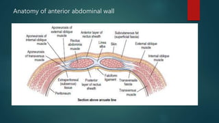
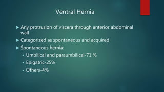
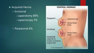
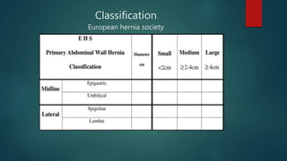
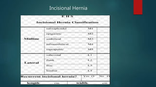
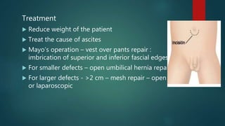
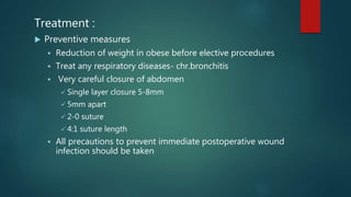
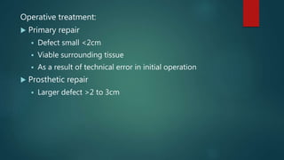
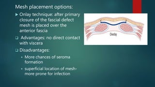
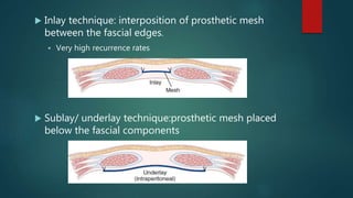
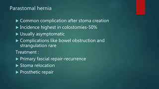
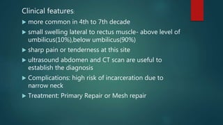
Ventral Hernia Management
DR MANJIL MALLA
1ST YEAR RESIDENT.,GENERAL SURGERY
PAHS
Anatomy of anterior abdominal wall
Ventral Hernia
 Any protrusion of viscera through anterior abdominal
wall
 Categorized as spontaneous and acquired
 Spontaneous hernia:
 Umbilical and paraumbilical-71 %
 Epigatric-25%
 Others-4%
 Acquired Hernia
 Incisional
o Laparotomy 89%
o Laparoscopy 5%
 Parastomal 6%
Classification
European hernia society
Incisional Hernia
Umbilical Hernia:
 Umbilical hernia occurs when the umbilical
scar closes incompletely in the child or fails
and stretches in later years in the adult
patient
 In infants
 Congenital and common
 Closes spontaneously by 2 years of age
 If persist after 5 years –surgical repair
 In adults
 Largely acquired
 Female>male
 hernia does not protrude through umbilical cicatrix
 protrusion through the linea alba just above the
the umbilicus -supraumbilical
 occasionally below the umbilicus (infraumbilical) –
so called as paraumbilical hernia
Etiology
 multifactorial, commonly found in association with
processes that increase intraabdominal pressure
 pregnancy
 obesity
 ascites
 persistent or repetitive abdominal distention in
bowel obstruction or peritoneal dialysis
Clinical Features
 Pain and swelling are the main symptoms
 Pain increases on prolonged standing or heavy
exercise
 Content: mostly omentum
Treatment
 Reduce weight of the patient
 Treat the cause of ascites
 Mayo’s operation – vest over pants repair :
imbrication of superior and inferior fascial edges
 For smaller defects – open umbilical hernia repair
 For larger defects - >2 cm – mesh repair – open
or laparoscopic
Epigastric Hernia :
 Hernia protruding through interlacing fibres of the linea
alba anywhere between umbilicus and xiphisternum
 protrusion of extraperitoneal fat - fatty hernia of linea alba
 Multiple in up to 20% of patients and approximately 80%
are in midline
Etiology:
Sudden strain leading to tearing of interlacing
fibers of linea alba
Clinical Features:
 Symptomless
 Painful- in partial strangulation of fat
 Referred dyspepsia
 On palpation – feels firm, no cough impulse and
cannot be reduced
Treatment:
 Midline defect is usually elliptical in nature with the
long axis oriented transversely
 Hernia will often not be seen on laparoscopy owing
to the lack of peritoneal involvement through the
hernia defect
 Open repair – excision of incarcerated preperitoneal
tissue and simple closure of defect
Incisional Hernia
 It is herniation through a weak abdominal scar (scar of
previous surgery)
 Etiology:
 Factors related to patients:
1. Obesity – due to fat encroaching in between the muscle layers
2. Advanced age
3. Multiparity,malnutrition,peritoneal
dialysis,jaundice,hypoproteinemia,anaemia, malignant diseases
4. Coughing,vomting and overzealous ventilation in early postoperative
period
5. Steroids and chemotherapy
6.Smoking in postoperative period.
7.Causes which increases the intra-abdominal pressure(BPH, straining,
stricture urethra or rectum, ascites)
 Factors related to procedure:
o Vertical incision higher chances of incisional hernia than horizontal
incision
o Layered closure of the abdomen has got higher chance than single
layer
o Continuous closure has got higher chances than interrupted closure
o Using absorbable suture material
o Emergency surgical wound has higher chances than elective
surgical wound
o Laparotomy wound
Clinical Features:
 Pain and swelling in the vicinity of previous scar
 Bulging more prominent on standing and coughing,reduces
spontaneously on lying down
 Attacks of subacute intestinal obstruction
 Expansile impulse on cough
 skin over the hernia is thin and atrophic
Treatment :
 Preventive measures
 Reduction of weight in obese before elective procedures
 Treat any respiratory diseases- chr.bronchitis
 Very careful closure of abdomen
 Single layer closure 5-8mm
 5mm apart
 2-0 suture
 4:1 suture length
 All precautions to prevent immediate postoperative wound
infection should be taken
Operative treatment:
 Primary repair
 Defect small <2cm
 Viable surrounding tissue
 As a result of technical error in initial operation
 Prosthetic repair
 Larger defect >2 to 3cm
Mesh placement options:
 Onlay technique: after primary
closure of the fascial defect
mesh is placed over the
anterior fascia
 Advantages: no direct contact
with viscera
 Disadvantages:
 More chances of seroma
formation
 superficial location of mesh-
more prone for infection
 Inlay technique: interposition of prosthetic mesh
between the fascial edges.
 Very high recurrence rates
 Sublay/ underlay technique:prosthetic mesh placed
below the fascial components
 Retromuscular technique:
 also called as Rives-Stoppa-Wantz Retrorectus Repair
 placement of mesh under the rectus muscle & above the
posterior rectus sheath
 Advantage
 intraabdominal forces hold the prosthesis against the muscles.
 Forces that created the hernia now are used to prevent its
recurrence
Component separation technique
skin and subcutaneous fat dissected
free
from the anterior sheath of the
rectus abdominis muscle and the
aponeurosis of the external oblique
muscle.
external abdominal oblique is
incised 1 to 2 cm lateral to the
rectus abdominis muscle.
• external oblique separated
from the internal oblique
Dissection carried to posterior
axillary wall
Additional length can be achieved by incising post rectus sheath
above the arcuate line
Laproscopic surgical repair
 IPOM(intraperitoneal onlay mesh)
 IPOM plus:
 defect >2cm
 6 to 10 cm difficult to close
 >10 cm: laproscopic TAR or Robotic TAR
Lap vs open
 Whenever feasible laproscopic repair is preferred.
Advantages of LAP
 Lower rate of wound infection
 Shorter hospital stay
 Quicker return to work
 Presence of swiss cheese defect can be seen
Disadvantages
 Higher operating cost
 Expertise needed
 More serious complication
Parastomal hernia
 Common complication after stoma creation
 Incidence highest in colostomies-50%
 Usually asymptomatic
 Complications like bowel obstruction and
strangulation rare
Treatment :
 Primary fascial repair-recurrence
 Stoma relocation
 Prosthetic repair
Spigelian hernia
 Unusual hernia
 Occurs through the
Spigelian fascia –composed
of aponeurotic layer
between rectus muscle
medially and semilunar line
laterally
 Often interparietal
Clinical features:
 more common in 4th to 7th decade
 small swelling lateral to rectus muscle- above level of
umbilicus(10%),below umbilicus(90%)
 sharp pain or tenderness at this site
 ultrasound abdomen and CT scan are useful to
establish the diagnosis
 Complications: high risk of incarceration due to
narrow neck
 Treatment: Primary Repair or Mesh repair
 References:
 sabiston textbook of surgery,south asia edition
 Schwartz’s principles of surgery,10th edition
THANK YOU

ventralherniamanagement-190502154429.pdf

  • 1.
    Ventral Hernia Management DRMANJIL MALLA 1ST YEAR RESIDENT.,GENERAL SURGERY PAHS
  • 2.
    Anatomy of anteriorabdominal wall
  • 4.
    Ventral Hernia  Anyprotrusion of viscera through anterior abdominal wall  Categorized as spontaneous and acquired  Spontaneous hernia:  Umbilical and paraumbilical-71 %  Epigatric-25%  Others-4%
  • 5.
     Acquired Hernia Incisional o Laparotomy 89% o Laparoscopy 5%  Parastomal 6%
  • 6.
  • 7.
  • 9.
    Umbilical Hernia:  Umbilicalhernia occurs when the umbilical scar closes incompletely in the child or fails and stretches in later years in the adult patient  In infants  Congenital and common  Closes spontaneously by 2 years of age  If persist after 5 years –surgical repair
  • 10.
     In adults Largely acquired  Female>male  hernia does not protrude through umbilical cicatrix  protrusion through the linea alba just above the the umbilicus -supraumbilical  occasionally below the umbilicus (infraumbilical) – so called as paraumbilical hernia
  • 11.
    Etiology  multifactorial, commonlyfound in association with processes that increase intraabdominal pressure  pregnancy  obesity  ascites  persistent or repetitive abdominal distention in bowel obstruction or peritoneal dialysis
  • 12.
    Clinical Features  Painand swelling are the main symptoms  Pain increases on prolonged standing or heavy exercise  Content: mostly omentum
  • 13.
    Treatment  Reduce weightof the patient  Treat the cause of ascites  Mayo’s operation – vest over pants repair : imbrication of superior and inferior fascial edges  For smaller defects – open umbilical hernia repair  For larger defects - >2 cm – mesh repair – open or laparoscopic
  • 14.
    Epigastric Hernia : Hernia protruding through interlacing fibres of the linea alba anywhere between umbilicus and xiphisternum  protrusion of extraperitoneal fat - fatty hernia of linea alba  Multiple in up to 20% of patients and approximately 80% are in midline
  • 15.
    Etiology: Sudden strain leadingto tearing of interlacing fibers of linea alba Clinical Features:  Symptomless  Painful- in partial strangulation of fat  Referred dyspepsia  On palpation – feels firm, no cough impulse and cannot be reduced
  • 16.
    Treatment:  Midline defectis usually elliptical in nature with the long axis oriented transversely  Hernia will often not be seen on laparoscopy owing to the lack of peritoneal involvement through the hernia defect  Open repair – excision of incarcerated preperitoneal tissue and simple closure of defect
  • 17.
    Incisional Hernia  Itis herniation through a weak abdominal scar (scar of previous surgery)  Etiology:  Factors related to patients: 1. Obesity – due to fat encroaching in between the muscle layers 2. Advanced age 3. Multiparity,malnutrition,peritoneal dialysis,jaundice,hypoproteinemia,anaemia, malignant diseases 4. Coughing,vomting and overzealous ventilation in early postoperative period 5. Steroids and chemotherapy
  • 18.
    6.Smoking in postoperativeperiod. 7.Causes which increases the intra-abdominal pressure(BPH, straining, stricture urethra or rectum, ascites)  Factors related to procedure: o Vertical incision higher chances of incisional hernia than horizontal incision o Layered closure of the abdomen has got higher chance than single layer o Continuous closure has got higher chances than interrupted closure
  • 19.
    o Using absorbablesuture material o Emergency surgical wound has higher chances than elective surgical wound o Laparotomy wound
  • 20.
    Clinical Features:  Painand swelling in the vicinity of previous scar  Bulging more prominent on standing and coughing,reduces spontaneously on lying down  Attacks of subacute intestinal obstruction  Expansile impulse on cough  skin over the hernia is thin and atrophic
  • 21.
    Treatment :  Preventivemeasures  Reduction of weight in obese before elective procedures  Treat any respiratory diseases- chr.bronchitis  Very careful closure of abdomen  Single layer closure 5-8mm  5mm apart  2-0 suture  4:1 suture length  All precautions to prevent immediate postoperative wound infection should be taken
  • 22.
    Operative treatment:  Primaryrepair  Defect small <2cm  Viable surrounding tissue  As a result of technical error in initial operation  Prosthetic repair  Larger defect >2 to 3cm
  • 23.
    Mesh placement options: Onlay technique: after primary closure of the fascial defect mesh is placed over the anterior fascia  Advantages: no direct contact with viscera  Disadvantages:  More chances of seroma formation  superficial location of mesh- more prone for infection
  • 24.
     Inlay technique:interposition of prosthetic mesh between the fascial edges.  Very high recurrence rates  Sublay/ underlay technique:prosthetic mesh placed below the fascial components
  • 25.
     Retromuscular technique: also called as Rives-Stoppa-Wantz Retrorectus Repair  placement of mesh under the rectus muscle & above the posterior rectus sheath  Advantage  intraabdominal forces hold the prosthesis against the muscles.  Forces that created the hernia now are used to prevent its recurrence
  • 26.
    Component separation technique skinand subcutaneous fat dissected free from the anterior sheath of the rectus abdominis muscle and the aponeurosis of the external oblique muscle. external abdominal oblique is incised 1 to 2 cm lateral to the rectus abdominis muscle.
  • 27.
    • external obliqueseparated from the internal oblique Dissection carried to posterior axillary wall Additional length can be achieved by incising post rectus sheath above the arcuate line
  • 28.
    Laproscopic surgical repair IPOM(intraperitoneal onlay mesh)  IPOM plus:  defect >2cm  6 to 10 cm difficult to close  >10 cm: laproscopic TAR or Robotic TAR
  • 29.
    Lap vs open Whenever feasible laproscopic repair is preferred.
  • 30.
    Advantages of LAP Lower rate of wound infection  Shorter hospital stay  Quicker return to work  Presence of swiss cheese defect can be seen
  • 31.
    Disadvantages  Higher operatingcost  Expertise needed  More serious complication
  • 32.
    Parastomal hernia  Commoncomplication after stoma creation  Incidence highest in colostomies-50%  Usually asymptomatic  Complications like bowel obstruction and strangulation rare Treatment :  Primary fascial repair-recurrence  Stoma relocation  Prosthetic repair
  • 33.
    Spigelian hernia  Unusualhernia  Occurs through the Spigelian fascia –composed of aponeurotic layer between rectus muscle medially and semilunar line laterally  Often interparietal
  • 34.
    Clinical features:  morecommon in 4th to 7th decade  small swelling lateral to rectus muscle- above level of umbilicus(10%),below umbilicus(90%)  sharp pain or tenderness at this site  ultrasound abdomen and CT scan are useful to establish the diagnosis  Complications: high risk of incarceration due to narrow neck  Treatment: Primary Repair or Mesh repair
  • 35.
     References:  sabistontextbook of surgery,south asia edition  Schwartz’s principles of surgery,10th edition
  • 36.