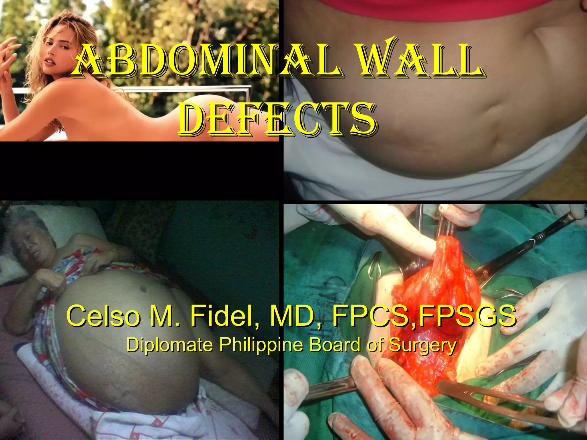
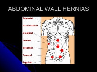
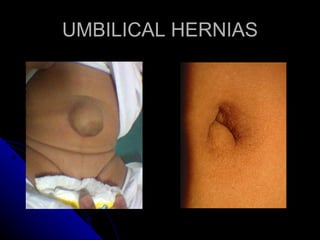
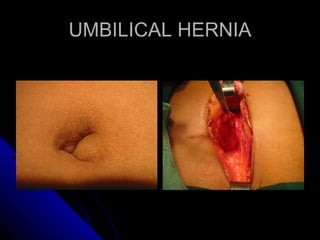
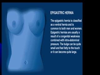
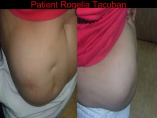
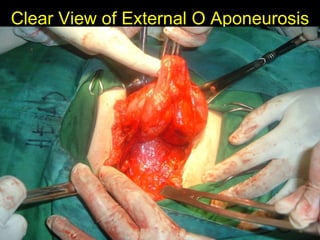
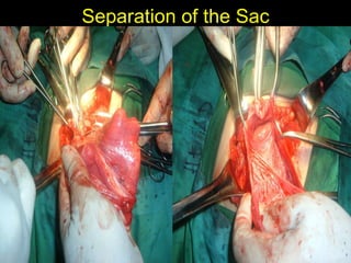
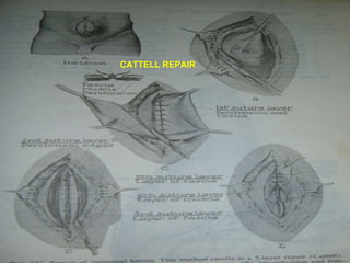
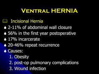
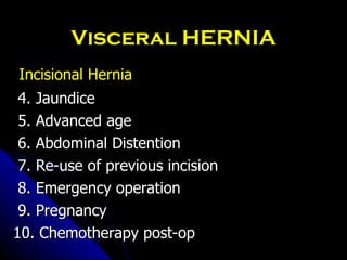
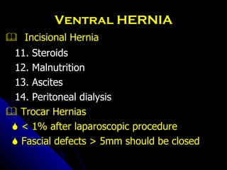
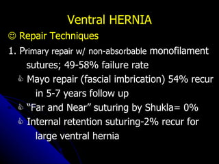
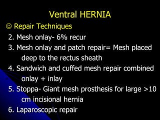
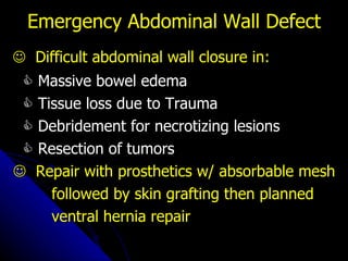
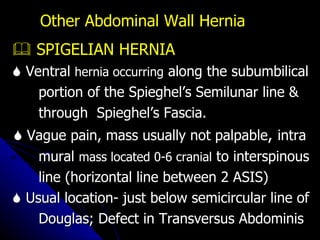
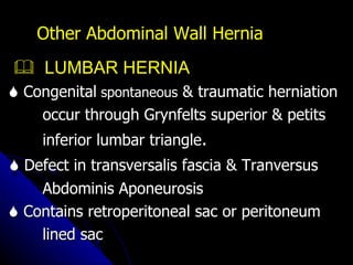
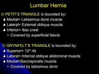
ABDOMINAL WALL DEFECTS Celso M. Fidel, MD, FPCS,FPSGS Diplomate Philippine Board of Surgery
Introduction ABDOMINAL WALL     Complex musculo-aponeurotic structure     Attached to the :    Vertebral column posteriorly     Ribs superiorly     Bones of the pelvis inferiorly    Derived embryonically in a  segmental,  metameric manner, and is reflected  in blood supply and innervation.
Introduction ABDOMINAL WALL    Protects and restrains the abdominal viscera,  and its musculature    Acts indirectly to flex the vertebral column.    Integrity is essential to the prevention of  hernias, whether they be:    Congenital    Acquired     Iatrogenic
Introduction ABDOMINAL WALL It is the repository of the panniculus adiposus  May reach considerable proportions in some members of the species afflicted with morbid obesity .
Introduction   ABDOMINAL WALL Variety of pathology  difficult to assess on  physical examination. Computed tomography (CT) often delineates  these abnormalities
GENERAL CONSIDERATIONS    VENTRAL HERNIA    UMBILICAL HERNIAS    EPIGASTRIC HERNIA    INCISIONAL HERNIA    TROCAR HERNIA    Emergency Abdominal wall Defects    Difficult Abdominal Wall Closure
ABDOMINAL WALL HERNIAS
UMBILICAL HERNIAS
UMBILICAL HERNIA
 
GENERAL CONSIDERATIONS    Other Abdominal Wall Hernia    Spigelian Hernia    Lumbar Hernia 1. Petit’s or inferior triangle hernia 2. Grynfelt’s or sup. Triangle hernia    Pelvic Floor Hernia 1. Obturator Hernia 2. Perineal Hernia 3. Sciatic Hernia
GENERAL CONSIDERATIONS    Other Abdominal Wall Hernia 4. Parastomal Hernia 5. Internal hernia  (a) Normal Orifice (b) Abnormal Orifice (c) Iatrogenic ( post-operative)
GENERAL CONSIDERATIONS    Other Abdominal Wall Hernia 6. Co ngenital Abdominal  Wall defect (a) Gastroschisis (b) Omphalocele 7. C ongenital  Diaphragmatic Hernia (a) Bochdalek   (b) Morgagni
Abdominal Wall Defects      Ventral Hernia Defect in the abdominal wall with intestines  or preperitoneal fat thru fascial defect On PE fascial defect usually palpable in  obese patients  Ultrasound or CT scan for the diagnosis Same principle of management as groin hernia
Ventral HERNIA    Umbilical Hernia    Occur more frequently in females; 10-30%  live birth    Obesity and repeated pregnancies precludes  this problem    In infants aponeurotic defect of 1.5 cm or less  would close spontaneously    Repair for children present by the age of  three or four & infants whose defect is 2 cm
Ventral HERNIA    Umbilical Hernia    MAYO HERNIOPLASTY    Vest over pants imbrication of the superior  & inferior aponeurotic fascia layer    EPIGASTRIC HERNIA    Protrusion of properitoneal fat & peritoneum  through the dicussating fibers of the rectus  sheath in  the midline (linea alba) between  the xiphoid.
Ventral HERNIA    Epigastric Hernia    Diastasis Recti    Wide gap between the medial borders of the rectus sheath     Diffuse bulge at upper midline of abdomen    Not a fascial defect, hence repaired for cosmetic purposes    Incisional Hernia
Patient Rogelia Tacuban
INCISIONAL HERNIOPLASTY    Anatomic reconstruction of the  abdominal  wall and Includes;    Closure of the parietal defect Restoration of normal intra-abdominal  pressure Tendinous reinforcement of the lateral abdominal muscles.
Clear View of External O Aponeurosis
Separation of the Sac
CATTELL REPAIR
Ventral HERNIA    Incisional Hernia    2-11% of abdominal wall closure    56% in the first year postoperative    17% incarcerate    20-46% repeat recurrence    Causes: 1. Obesity 2. post-op pulmonary complications 3. Wound infection
Visceral HERNIA Incisional Hernia 4. Jaundice 5. Advanced age 6. Abdominal Distention 7. Re-use of previous incision 8. Emergency operation 9. Pregnancy 10. Chemotherapy post-op
Ventral HERNIA    Incisional Hernia 11. Steroids 12. Malnutrition 13. Ascites 14. Peritoneal dialysis    Trocar Hernias    < 1% after laparoscopic procedure    Fascial defects > 5mm should be closed
Ventral HERNIA    Repair Techniques 1. P rimary repair w/   non-absorbable  monofilament sutures; 49-58% failure rate    Mayo repair (fascial imbrication) 54% recur in 5-7 years follow up   “ Far and Near” suturing by Shukla= 0%    Internal retention suturing-2% recur for large ventral hernia
Ventral HERNIA    Repair Techniques 2. Mesh onlay- 6% recur 3. Mesh onlay and patch repair= Mesh placed deep to the rectus sheath 4. Sandwich and cuffed mesh repair combined onlay + inlay 5. Stoppa- Giant mesh prosthesis for large >10 cm incisional hernia 6. Laparoscopic repair
Emergency Abdominal Wall Defect    Difficult abdominal wall closure in:    Massive bowel edema    Tissue loss due to Trauma    Debridement for necrotizing lesions    Resection of tumors    Repair with prosthetics w/ absorbable mesh followed by skin grafting then planned ventral hernia repair
Other Abdominal Wall Hernia    SPIGELIAN HERNIA    Ventral  hernia occurring  along the subumbilical  portion of the Spieghel’s Semilunar line &  through  Spieghel’s Fascia.    Vague pain, mass usually not palpable ,  intra mural  mass located 0-6 cranial  to interspinous line (horizontal line between 2 ASIS)    Usual location- just below semicircular line of Douglas; Defect in Transversus Abdominis
Other Abdominal Wall Hernia    LUMBAR HERNIA    Congenital  spontaneous  & traumatic herniation  occur through Grynfelts superior & petits  inferior lumbar triangle .    Defect in transversalis fascia & Tranversus Abdominis Aponeurosis    Contains retroperitoneal sac or peritoneum lined sac
Lumbar Hernia    PETIT’S TRIANGLE  is bounded by:    Medial= Latissimus dorsi muscle    Lateral= External oblique muscle    Inferior= Iliac crest      Covered by superficial fascia    GRYNFELT’S TRIANGLE  is bounded by:    Superior= 12 th  rib    Lateral= Internal oblique abdominal muscle    Medial=Sacrospinalis muscle      Covered by latissimus dorsi
   PELVIC HERNIA-  occurs in cachetic, elderly patients in the, Obturator fossa, Perineum & Greater and lesser sciatic foramina 1. Obturator Hernia    50% with Howship-Romberg Sign Pain in the region of the hip, and of the knee  and on the inner aspect of the thigh because  of pressure on the obturator  nerve by an  obturator hernia. Other Abdominal Wall Hernia
   Usually in emaciated females in late 70’s on the right side    Often with either large or small bowel  incarceration or strangulation    Rarely with a mass at the anteromedial thigh or a bulge on rectal or pelvic examination    Diagnosis by CT scan    Repair by midline approach to take care of bowel problem too. Other Abdominal Wall Hernia
2. Perineal Hernia    Occur spontaneously or after APR or pelvic exenteration 1. Anterior- defect in urogenital diaphragm; mass in labia majora 2. Po sterior-   defect in the levator  ani between the urinary bladder and rectum    Repair= Transperineal or transabdominal  primary repair or with mesh   Other Abdominal Wall Hernia
3. Sciatic Hernia    Rarest  of all hernias;     Occurs in the greater or lesser sciatic  foramen or thru a defect in the pyriformis muscle    Presents as sciatic nerve palsy and a mass or simply intestinal obstruction    Repair= G luteal   or Transabdominal  approach Other Abdominal Wall Hernia
   PARASTOMAL HERNIA    Occurs thru defects adjacent to ostomy site    Incidence: 12-32% paracolostomy < 10% paraileostomy    Prevention: Small fascial incision, avoid maturing thru the abdominal incision    Complications:  1. Obstruction; 2. Incarceration 3. Poor Appliance fit 4. Local pain Other Abdominal Wall Hernia
   PARASTOMAL HERNIA    Repair: Primary fascial or prosthetic repair or relocation of stoma    Symptoms generally well tolerated    All repairs associated  with: 1.  significant morbidity 2. high recurrence  Other Abdominal Wall Hernia
   INTERNAL HERNIA    Abdominal contents protrude thru  normal or abnormal intra-abdominal  orifice 3. Iatrogenic (Post operative) (a) Defect in Mesentery or Omentum Peterson Hernia=thru Roux limb    CONGENITAL ABDOMINAL WALL DEFECTS    Gastroschisis 1. Herniation of abdominal viscera without a sac, intact umbilical cord Other Abdominal Wall Hernia
   CONGENITAL ABDOMINAL WALL DEFECTS    Omphalocele 1. Herniation of abdominal viscera into the umbilical cord, hence lined by internally by peritoneal sac and externally by amnion 2. Associated anomalies: (a) Cloacal exstrophy (b) Chromosomal abnormality in 50% Other Abdominal Wall Hernia
   CONGENITAL ABDOMINAL WALL DEFECTS    Gastroschisis 2. Two times (2X) more common than omphalocele 3. Associated anomalies: Intestinal Atresia  10% 4. Eviscerated Bowels are: (a) Edematous (b) Shortened with fibrinous adhesions 5. < 10% mortality Other Abdominal Wall Hernia
   CONGENITAL DIAPHRAGMATIC HERNIA    Bochdalek Hernia (a) Postero-lateral, most common at costal and spinal diaphragmatic attachment (b) Associated with malrotation, pulmonary hypoplasia (c)  4 x more common in the left side (d) Only 10-20% have a sac; 80% mortality by the first year of life Other Abdominal Wall Hernia
   CONGENITAL DIAPHRAGMATIC HERNIA    Bochdalek Hernia (e) Better repaired after a few days to weeks when the child stabilizes; higher mortality if repaired at birth (f) Repair: Transabdominal with the Ladd procedure for the malrotation    Morgagni  (a) Between sternal & costal diaphragmatic m argin either retrosternal  or parasternal Other Abdominal Wall Hernia
THANK YOU!!!

abdominal wall

  • 1.
    ABDOMINAL WALL DEFECTSCelso M. Fidel, MD, FPCS,FPSGS Diplomate Philippine Board of Surgery
  • 2.
    Introduction ABDOMINAL WALL  Complex musculo-aponeurotic structure  Attached to the :  Vertebral column posteriorly  Ribs superiorly  Bones of the pelvis inferiorly  Derived embryonically in a segmental, metameric manner, and is reflected in blood supply and innervation.
  • 3.
    Introduction ABDOMINAL WALL Protects and restrains the abdominal viscera, and its musculature  Acts indirectly to flex the vertebral column.  Integrity is essential to the prevention of hernias, whether they be:  Congenital  Acquired  Iatrogenic
  • 4.
    Introduction ABDOMINAL WALLIt is the repository of the panniculus adiposus May reach considerable proportions in some members of the species afflicted with morbid obesity .
  • 5.
    Introduction ABDOMINAL WALL Variety of pathology difficult to assess on physical examination. Computed tomography (CT) often delineates these abnormalities
  • 6.
    GENERAL CONSIDERATIONS  VENTRAL HERNIA  UMBILICAL HERNIAS  EPIGASTRIC HERNIA  INCISIONAL HERNIA  TROCAR HERNIA  Emergency Abdominal wall Defects  Difficult Abdominal Wall Closure
  • 7.
  • 8.
  • 9.
  • 10.
  • 11.
    GENERAL CONSIDERATIONS  Other Abdominal Wall Hernia  Spigelian Hernia  Lumbar Hernia 1. Petit’s or inferior triangle hernia 2. Grynfelt’s or sup. Triangle hernia  Pelvic Floor Hernia 1. Obturator Hernia 2. Perineal Hernia 3. Sciatic Hernia
  • 12.
    GENERAL CONSIDERATIONS  Other Abdominal Wall Hernia 4. Parastomal Hernia 5. Internal hernia (a) Normal Orifice (b) Abnormal Orifice (c) Iatrogenic ( post-operative)
  • 13.
    GENERAL CONSIDERATIONS  Other Abdominal Wall Hernia 6. Co ngenital Abdominal Wall defect (a) Gastroschisis (b) Omphalocele 7. C ongenital Diaphragmatic Hernia (a) Bochdalek (b) Morgagni
  • 14.
    Abdominal Wall Defects  Ventral Hernia Defect in the abdominal wall with intestines or preperitoneal fat thru fascial defect On PE fascial defect usually palpable in obese patients Ultrasound or CT scan for the diagnosis Same principle of management as groin hernia
  • 15.
    Ventral HERNIA  Umbilical Hernia  Occur more frequently in females; 10-30% live birth  Obesity and repeated pregnancies precludes this problem  In infants aponeurotic defect of 1.5 cm or less would close spontaneously  Repair for children present by the age of three or four & infants whose defect is 2 cm
  • 16.
    Ventral HERNIA  Umbilical Hernia  MAYO HERNIOPLASTY  Vest over pants imbrication of the superior & inferior aponeurotic fascia layer  EPIGASTRIC HERNIA  Protrusion of properitoneal fat & peritoneum through the dicussating fibers of the rectus sheath in the midline (linea alba) between the xiphoid.
  • 17.
    Ventral HERNIA  Epigastric Hernia  Diastasis Recti  Wide gap between the medial borders of the rectus sheath  Diffuse bulge at upper midline of abdomen  Not a fascial defect, hence repaired for cosmetic purposes  Incisional Hernia
  • 18.
  • 19.
    INCISIONAL HERNIOPLASTY  Anatomic reconstruction of the abdominal wall and Includes;  Closure of the parietal defect Restoration of normal intra-abdominal pressure Tendinous reinforcement of the lateral abdominal muscles.
  • 20.
    Clear View ofExternal O Aponeurosis
  • 21.
  • 22.
  • 23.
    Ventral HERNIA  Incisional Hernia  2-11% of abdominal wall closure  56% in the first year postoperative  17% incarcerate  20-46% repeat recurrence  Causes: 1. Obesity 2. post-op pulmonary complications 3. Wound infection
  • 24.
    Visceral HERNIA IncisionalHernia 4. Jaundice 5. Advanced age 6. Abdominal Distention 7. Re-use of previous incision 8. Emergency operation 9. Pregnancy 10. Chemotherapy post-op
  • 25.
    Ventral HERNIA  Incisional Hernia 11. Steroids 12. Malnutrition 13. Ascites 14. Peritoneal dialysis  Trocar Hernias  < 1% after laparoscopic procedure  Fascial defects > 5mm should be closed
  • 26.
    Ventral HERNIA  Repair Techniques 1. P rimary repair w/ non-absorbable monofilament sutures; 49-58% failure rate  Mayo repair (fascial imbrication) 54% recur in 5-7 years follow up  “ Far and Near” suturing by Shukla= 0%  Internal retention suturing-2% recur for large ventral hernia
  • 27.
    Ventral HERNIA  Repair Techniques 2. Mesh onlay- 6% recur 3. Mesh onlay and patch repair= Mesh placed deep to the rectus sheath 4. Sandwich and cuffed mesh repair combined onlay + inlay 5. Stoppa- Giant mesh prosthesis for large >10 cm incisional hernia 6. Laparoscopic repair
  • 28.
    Emergency Abdominal WallDefect  Difficult abdominal wall closure in:  Massive bowel edema  Tissue loss due to Trauma  Debridement for necrotizing lesions  Resection of tumors  Repair with prosthetics w/ absorbable mesh followed by skin grafting then planned ventral hernia repair
  • 29.
    Other Abdominal WallHernia  SPIGELIAN HERNIA  Ventral hernia occurring along the subumbilical portion of the Spieghel’s Semilunar line & through Spieghel’s Fascia.  Vague pain, mass usually not palpable , intra mural mass located 0-6 cranial to interspinous line (horizontal line between 2 ASIS)  Usual location- just below semicircular line of Douglas; Defect in Transversus Abdominis
  • 30.
    Other Abdominal WallHernia  LUMBAR HERNIA  Congenital spontaneous & traumatic herniation occur through Grynfelts superior & petits inferior lumbar triangle .  Defect in transversalis fascia & Tranversus Abdominis Aponeurosis  Contains retroperitoneal sac or peritoneum lined sac
  • 31.
    Lumbar Hernia  PETIT’S TRIANGLE is bounded by:  Medial= Latissimus dorsi muscle  Lateral= External oblique muscle  Inferior= Iliac crest  Covered by superficial fascia  GRYNFELT’S TRIANGLE is bounded by:  Superior= 12 th rib  Lateral= Internal oblique abdominal muscle  Medial=Sacrospinalis muscle  Covered by latissimus dorsi
  • 32.
    PELVIC HERNIA- occurs in cachetic, elderly patients in the, Obturator fossa, Perineum & Greater and lesser sciatic foramina 1. Obturator Hernia  50% with Howship-Romberg Sign Pain in the region of the hip, and of the knee and on the inner aspect of the thigh because of pressure on the obturator nerve by an obturator hernia. Other Abdominal Wall Hernia
  • 33.
    Usually in emaciated females in late 70’s on the right side  Often with either large or small bowel incarceration or strangulation  Rarely with a mass at the anteromedial thigh or a bulge on rectal or pelvic examination  Diagnosis by CT scan  Repair by midline approach to take care of bowel problem too. Other Abdominal Wall Hernia
  • 34.
    2. Perineal Hernia Occur spontaneously or after APR or pelvic exenteration 1. Anterior- defect in urogenital diaphragm; mass in labia majora 2. Po sterior- defect in the levator ani between the urinary bladder and rectum  Repair= Transperineal or transabdominal primary repair or with mesh Other Abdominal Wall Hernia
  • 35.
    3. Sciatic Hernia Rarest of all hernias;  Occurs in the greater or lesser sciatic foramen or thru a defect in the pyriformis muscle  Presents as sciatic nerve palsy and a mass or simply intestinal obstruction  Repair= G luteal or Transabdominal approach Other Abdominal Wall Hernia
  • 36.
    PARASTOMAL HERNIA  Occurs thru defects adjacent to ostomy site  Incidence: 12-32% paracolostomy < 10% paraileostomy  Prevention: Small fascial incision, avoid maturing thru the abdominal incision  Complications: 1. Obstruction; 2. Incarceration 3. Poor Appliance fit 4. Local pain Other Abdominal Wall Hernia
  • 37.
    PARASTOMAL HERNIA  Repair: Primary fascial or prosthetic repair or relocation of stoma  Symptoms generally well tolerated  All repairs associated with: 1. significant morbidity 2. high recurrence Other Abdominal Wall Hernia
  • 38.
    INTERNAL HERNIA  Abdominal contents protrude thru normal or abnormal intra-abdominal orifice 3. Iatrogenic (Post operative) (a) Defect in Mesentery or Omentum Peterson Hernia=thru Roux limb  CONGENITAL ABDOMINAL WALL DEFECTS  Gastroschisis 1. Herniation of abdominal viscera without a sac, intact umbilical cord Other Abdominal Wall Hernia
  • 39.
    CONGENITAL ABDOMINAL WALL DEFECTS  Omphalocele 1. Herniation of abdominal viscera into the umbilical cord, hence lined by internally by peritoneal sac and externally by amnion 2. Associated anomalies: (a) Cloacal exstrophy (b) Chromosomal abnormality in 50% Other Abdominal Wall Hernia
  • 40.
    CONGENITAL ABDOMINAL WALL DEFECTS  Gastroschisis 2. Two times (2X) more common than omphalocele 3. Associated anomalies: Intestinal Atresia 10% 4. Eviscerated Bowels are: (a) Edematous (b) Shortened with fibrinous adhesions 5. < 10% mortality Other Abdominal Wall Hernia
  • 41.
    CONGENITAL DIAPHRAGMATIC HERNIA  Bochdalek Hernia (a) Postero-lateral, most common at costal and spinal diaphragmatic attachment (b) Associated with malrotation, pulmonary hypoplasia (c) 4 x more common in the left side (d) Only 10-20% have a sac; 80% mortality by the first year of life Other Abdominal Wall Hernia
  • 42.
    CONGENITAL DIAPHRAGMATIC HERNIA  Bochdalek Hernia (e) Better repaired after a few days to weeks when the child stabilizes; higher mortality if repaired at birth (f) Repair: Transabdominal with the Ladd procedure for the malrotation  Morgagni (a) Between sternal & costal diaphragmatic m argin either retrosternal or parasternal Other Abdominal Wall Hernia
  • 43.