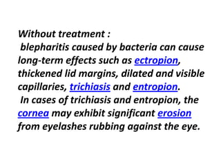
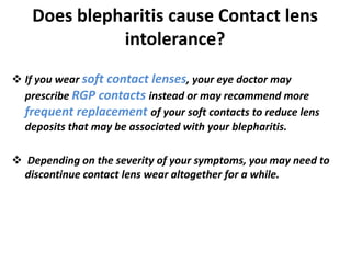
In The Name of God
A . Doostdar Razi
Member of TUMS
asgar_doostdar@yahoo.com
Blepharitis @ Contact lenses
Introduction
• Blepharitis is inflammation or infection of the
eyelid margins .
• Blepharitis is one the most common
ophthalmological complications as well as one
of the most difficult conditions to treat.
Blepharitis
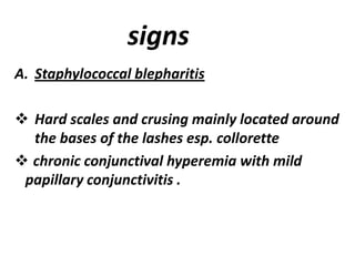
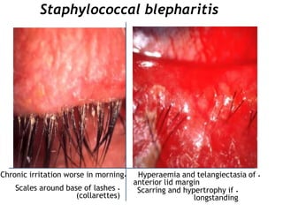
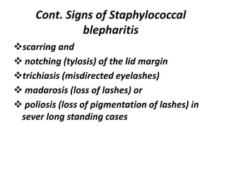
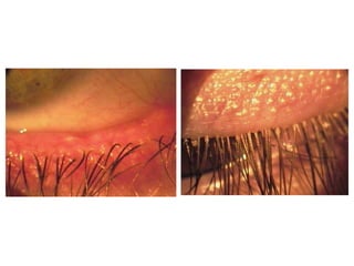
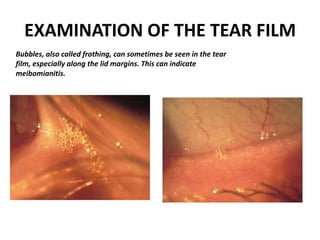
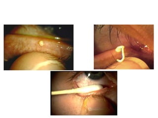
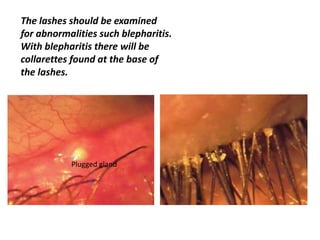
Blepharitis is a common eyelid inflammation
that sometimes is associated with a bacterial
or certaindry eyeseye infection, symptoms of
.rosaceaacnetypes of skin conditions such as
Blepharitis is one of the most frequently observed
conditions among eye care practitioners, yet
remains largely misunderstood.
SLIT-LAMP BIOMICROSCOPY
The slit-lamp biomicroscope is an
important diagnostic instrument for
the examination of the anterior eye
structures as well as the contact lens.
Being able to proficiently use the
instrument is important. It use
critical in the fitting of contact
lenses.
CLASSIFICATION
I will discuss blepharitis according to
its predominant anatomic location,
as :
 anterior
 posterior
1. Anterior
• Staphylococcal
• Seborrhoeic
• Meibomianitis
• Meibomian seborrhoea
2. Posterior Staph
Blepharitis
Anterior blepharitis
Anterior blepharitis is characterized by inflammation at
the base of the eyelashes .
Patients with anterior blepharitis, compared to those
with posterior blepharitis, are more likely to be female
and younger *
Two variants of anterior blepharitis are
identified: staphylococcal and seborrheic.
In staphylococcal anterior blepharitis,
colonization of the eyelids by staphylococci leads
to formation of fibrinous scales and crust around
the eyelashes.
The seborrheic variant is characterized by
dandruff-like skin changes around the base of
the eyelids, resulting in greasy scales around the
eyelashes.
PATHOPHYSIOLOGY
The pathophysiology of blepharitis is not completely
understood. A role for lid-colonizing staphylococcal
bacteria was first noted in 1946 . Several mechanisms by
which staphylococci may alter meibomian gland secretion
and cause blepharitis are supported by many studies .
Direct infection of the lids
Evoke reaction to staphylococcal exotoxin
Provoke allergic response to staphylococcal antigens .

It is likely that a combination of these is responsible for the
clinical manifestations of staphylococcal blepharitis.
CLINICAL PRESENTATION
symptoms
 burning , grittiness and mild photophobia with
remission and exacerbation is characteristic .
 Symptoms are usually worse in the morning ,
although in patients with dry eye they may
increase during the day .
signs
A. Staphylococcal blepharitis
 Hard scales and crusing mainly located around
the bases of the lashes esp. collorette
 chronic conjunctival hyperemia with mild
papillary conjunctivitis .
Staphylococcal blepharitis
•Hyperaemia and telangiectasia of
anterior lid margin
•Scarring and hypertrophy if
longstanding
•Scales around base of lashes
(collarettes)
•Chronic irritation worse in morning
Cont. Signs of Staphylococcal
blepharitis
scarring and
 notching (tylosis) of the lid margin
trichiasis (misdirected eyelashes)
 madarosis (loss of lashes) or
 poliosis (loss of pigmentation of lashes) in
sever long standing cases
Cont. Signs of Staphylococcal
blepharitis
secondary changes include :
 stye formation
 marginal keratitis and occasionally
phlyctenulosis (Corneal nodulesthat developed
near the limbus and then spread onto the
cornea, carrying behind them a leash of vessels)
 associated with tear film instability and dry eye
.
Cont. Signs of Staphylococcal blepharitis
 The diagnosis of blepharitis is clinical, based on
the patient's history and physical examination
findings. There are no confirmatory diagnostic
tests or laboratory investigations.
The history should include questions about
symptom duration, smoking, allergens, contact
lenses, and use of retinoids which may provoke
or exacerbate symptoms. A history of acne,
rosacea, or eczema should be evaluated .
DIAGNOSIS
EXAMINATION OF THE TEAR FILM
Bubbles, also called frothing, can sometimes be seen in the tear
film, especially along the lid margins. This can indicate
meibomianitis.
The lashes should be examined
for abnormalities such blepharitis.
With blepharitis there will be
collarettes found at the base of
the lashes.
Plugged gland
• Burning
• Flaking
• Crusting
• Tearing
• Irritation
• Itching
• redness
• Foreign body sensation
• bulbar and palpebral
hyperemia
Treatment for Blepharitis
Anterior Blepharitis
• staphylococcal blepharitis
more severe
loss of eyelashes
eyelid cleansing and hygiene
antibiotic ointment for eyelids
Viruses and other types of
bacteria besides
staphylococcus also can cause
anterior blepharitis
• Seborrheic blepharitis
seborrheic dermatitis
Regular cleansing with eyelid
scrubs and gentle, non-
detergent shampoos
Without treatment :
blepharitis caused by bacteria can cause
,ectropionterm effects such as-long
thickened lid margins, dilated and visible
.entropionandtrichiasiscapillaries,
In cases of trichiasis and entropion, the
erosionmay exhibit significantcornea
from eyelashes rubbing against the eye.
Eyelid Hygiene Relieves Blepharitis
Symptoms
 Blepharitis can be difficult to manage because it is often chronic,
meaning that it never warm compress goes away completely.
Your eye doctor may recommend one or more of these steps
involving good eyelid hygiene and massage:
 Apply a such as a washcloth to the outer eyelids.
 Cleanse the eyelids with a commercial lid scrub or other
recommended product.
 Gently massage the outer eyelids.
 A warm compress loosens the crust on your eyelids and eyelashes
before you clean them. The warmth also can loosen any blocked
residue in the oil-secreting meibomian glands in your eyelids
Does blepharitis cause Contact lens
intolerance?
 If you wear soft contact lenses, your eye doctor may
prescribe RGP contacts instead or may recommend more
frequent replacement of your soft contacts to reduce lens
deposits that may be associated with your blepharitis.
 Depending on the severity of your symptoms, you may need to
discontinue contact lens wear altogether for a while.
According to recent research
People With Blepharitis More Likely to Have Certain
 Inflammatory Diseases
 Psychological Issues
 Cardiovascular Diseases and More
Thank you

Blepharitis

  • 1.
    In The Nameof God A . Doostdar Razi Member of TUMS asgar_doostdar@yahoo.com
  • 2.
  • 3.
    Introduction • Blepharitis isinflammation or infection of the eyelid margins . • Blepharitis is one the most common ophthalmological complications as well as one of the most difficult conditions to treat.
  • 4.
    Blepharitis Blepharitis is acommon eyelid inflammation that sometimes is associated with a bacterial or certaindry eyeseye infection, symptoms of .rosaceaacnetypes of skin conditions such as Blepharitis is one of the most frequently observed conditions among eye care practitioners, yet remains largely misunderstood.
  • 5.
    SLIT-LAMP BIOMICROSCOPY The slit-lampbiomicroscope is an important diagnostic instrument for the examination of the anterior eye structures as well as the contact lens. Being able to proficiently use the instrument is important. It use critical in the fitting of contact lenses.
  • 7.
    CLASSIFICATION I will discussblepharitis according to its predominant anatomic location, as :  anterior  posterior
  • 8.
    1. Anterior • Staphylococcal •Seborrhoeic • Meibomianitis • Meibomian seborrhoea 2. Posterior Staph Blepharitis
  • 9.
    Anterior blepharitis Anterior blepharitisis characterized by inflammation at the base of the eyelashes . Patients with anterior blepharitis, compared to those with posterior blepharitis, are more likely to be female and younger *
  • 10.
    Two variants ofanterior blepharitis are identified: staphylococcal and seborrheic. In staphylococcal anterior blepharitis, colonization of the eyelids by staphylococci leads to formation of fibrinous scales and crust around the eyelashes. The seborrheic variant is characterized by dandruff-like skin changes around the base of the eyelids, resulting in greasy scales around the eyelashes.
  • 11.
    PATHOPHYSIOLOGY The pathophysiology ofblepharitis is not completely understood. A role for lid-colonizing staphylococcal bacteria was first noted in 1946 . Several mechanisms by which staphylococci may alter meibomian gland secretion and cause blepharitis are supported by many studies . Direct infection of the lids Evoke reaction to staphylococcal exotoxin Provoke allergic response to staphylococcal antigens .  It is likely that a combination of these is responsible for the clinical manifestations of staphylococcal blepharitis.
  • 12.
    CLINICAL PRESENTATION symptoms  burning, grittiness and mild photophobia with remission and exacerbation is characteristic .  Symptoms are usually worse in the morning , although in patients with dry eye they may increase during the day .
  • 13.
    signs A. Staphylococcal blepharitis Hard scales and crusing mainly located around the bases of the lashes esp. collorette  chronic conjunctival hyperemia with mild papillary conjunctivitis .
  • 14.
    Staphylococcal blepharitis •Hyperaemia andtelangiectasia of anterior lid margin •Scarring and hypertrophy if longstanding •Scales around base of lashes (collarettes) •Chronic irritation worse in morning
  • 15.
    Cont. Signs ofStaphylococcal blepharitis scarring and  notching (tylosis) of the lid margin trichiasis (misdirected eyelashes)  madarosis (loss of lashes) or  poliosis (loss of pigmentation of lashes) in sever long standing cases
  • 16.
    Cont. Signs ofStaphylococcal blepharitis
  • 17.
    secondary changes include:  stye formation  marginal keratitis and occasionally phlyctenulosis (Corneal nodulesthat developed near the limbus and then spread onto the cornea, carrying behind them a leash of vessels)  associated with tear film instability and dry eye . Cont. Signs of Staphylococcal blepharitis
  • 18.
     The diagnosisof blepharitis is clinical, based on the patient's history and physical examination findings. There are no confirmatory diagnostic tests or laboratory investigations. The history should include questions about symptom duration, smoking, allergens, contact lenses, and use of retinoids which may provoke or exacerbate symptoms. A history of acne, rosacea, or eczema should be evaluated . DIAGNOSIS
  • 20.
    EXAMINATION OF THETEAR FILM Bubbles, also called frothing, can sometimes be seen in the tear film, especially along the lid margins. This can indicate meibomianitis.
  • 22.
    The lashes shouldbe examined for abnormalities such blepharitis. With blepharitis there will be collarettes found at the base of the lashes. Plugged gland
  • 23.
    • Burning • Flaking •Crusting • Tearing • Irritation • Itching • redness • Foreign body sensation • bulbar and palpebral hyperemia
  • 24.
    Treatment for Blepharitis AnteriorBlepharitis • staphylococcal blepharitis more severe loss of eyelashes eyelid cleansing and hygiene antibiotic ointment for eyelids Viruses and other types of bacteria besides staphylococcus also can cause anterior blepharitis • Seborrheic blepharitis seborrheic dermatitis Regular cleansing with eyelid scrubs and gentle, non- detergent shampoos
  • 25.
    Without treatment : blepharitiscaused by bacteria can cause ,ectropionterm effects such as-long thickened lid margins, dilated and visible .entropionandtrichiasiscapillaries, In cases of trichiasis and entropion, the erosionmay exhibit significantcornea from eyelashes rubbing against the eye.
  • 26.
    Eyelid Hygiene RelievesBlepharitis Symptoms  Blepharitis can be difficult to manage because it is often chronic, meaning that it never warm compress goes away completely. Your eye doctor may recommend one or more of these steps involving good eyelid hygiene and massage:  Apply a such as a washcloth to the outer eyelids.  Cleanse the eyelids with a commercial lid scrub or other recommended product.  Gently massage the outer eyelids.  A warm compress loosens the crust on your eyelids and eyelashes before you clean them. The warmth also can loosen any blocked residue in the oil-secreting meibomian glands in your eyelids
  • 27.
    Does blepharitis causeContact lens intolerance?  If you wear soft contact lenses, your eye doctor may prescribe RGP contacts instead or may recommend more frequent replacement of your soft contacts to reduce lens deposits that may be associated with your blepharitis.  Depending on the severity of your symptoms, you may need to discontinue contact lens wear altogether for a while.
  • 28.
    According to recentresearch People With Blepharitis More Likely to Have Certain  Inflammatory Diseases  Psychological Issues  Cardiovascular Diseases and More
  • 29.