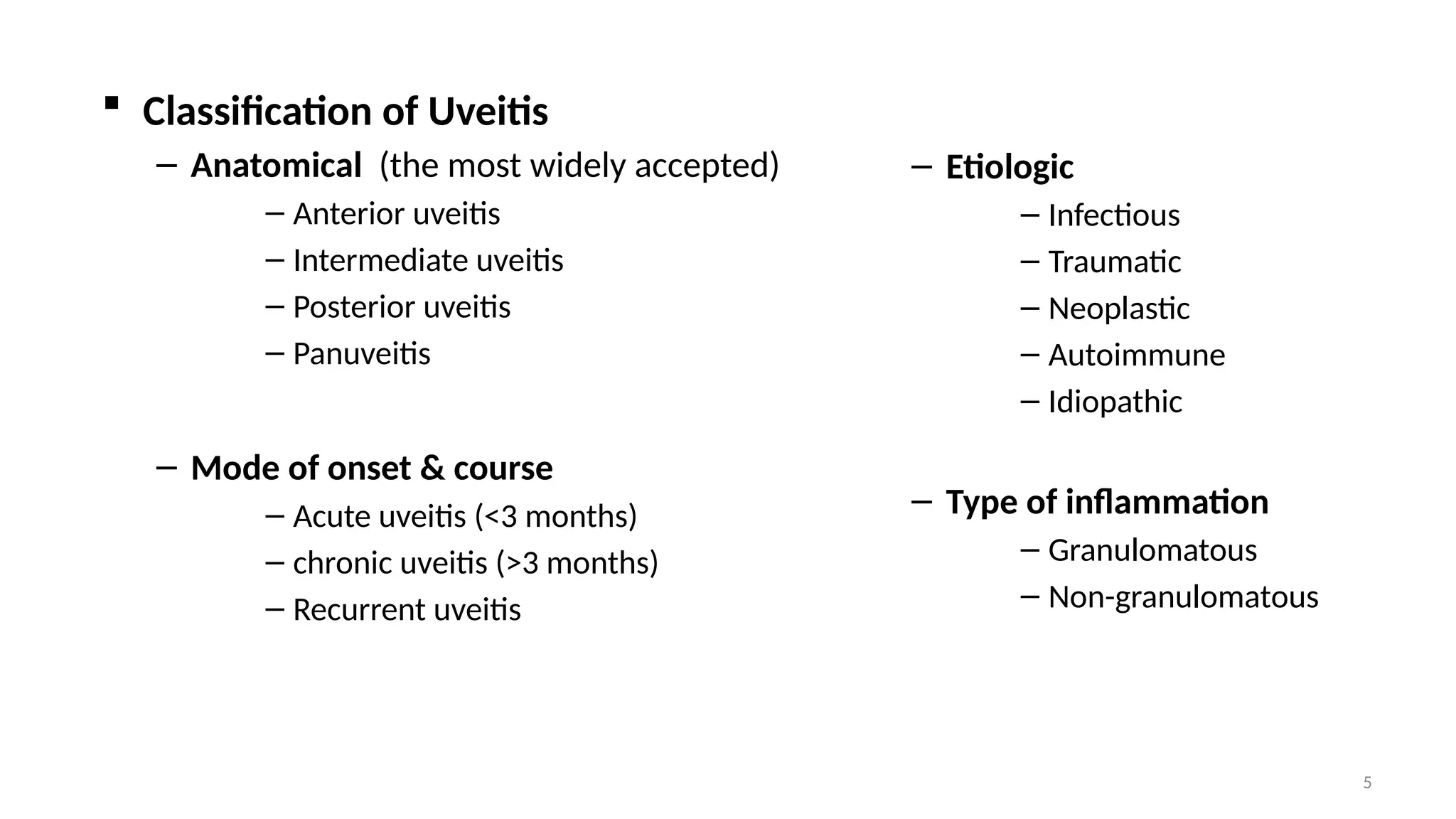
1
Uveitis
By Bekuma Jima (MD)
Ophthalmology Resident- (R4)
2
 Introduction
– “Uvea” = “Grape” (Latin).
– Uveal Tract = Three parts --- Iris + Ciliary body + Choroid.
3
– Ciliary body --- two parts:
– Pars plicata --- anterior part.
– Pars plana --- posterior part.
4
 Uveitis
– It is an inflammation of the uvea --- and, it can affect:
– Any part of the uveal tract:
• Iris ---> iritis.
• Choroid ---> choroiditis.
• Ciliary body --- cyclitis.
• Pars plicata ---> anterior cyclitis.
• Pars plana ---> pars planitis (intermediate uveitis).
– More than one part of the uveal tract:
• Iris and ciliary body ---> iridocyclitis.
• Whole uveal tract ---> panuveitis.
5
 Classification of Uveitis
– Anatomical (the most widely accepted)
– Anterior uveitis
– Intermediate uveitis
– Posterior uveitis
– Panuveitis
– Mode of onset & course
– Acute uveitis (<3 months)
– chronic uveitis (>3 months)
– Recurrent uveitis
– Etiologic
– Infectious
– Traumatic
– Neoplastic
– Autoimmune
– Idiopathic
– Type of inflammation
– Granulomatous
– Non-granulomatous
6
Anatomical classification of uveitis
• Anterior Uveitis
– The inflammation involves the iris
and anterior part of the ciliary
body.
» Iritis
» Iridocyclitis
» Keratouveitis
– Anterior uveitis is further divided
into acute and chronic.
7
– Causes of anterior uveitis:
– Arthritis --- JRA, ankylosing spondylitis, Reiter’s syndrome, psoriasis
– Sarcoidosis
– Behcet’s disease
– Infections ---bacterial, viral, fungal, parasitic, others.
» HSV, HZV, Syphilis, TB, Lyme disease
– Trauma
– Surgery --- esp. associated with lens
– Idiopathic (unknown cause)
8
– Acute Anterior Uveitis --- AAU
– Has classic presentation
• Rapid onset of unilateral pain, visual loss, photophobia, redness and
watery discharge
• sometimes preceding mild ocular discomfort for a few days.
– Signs
• Conjunctival injection
• Perilimbal (ciliary flush) --- in early cases
• Diffuse --- in severe cases
9
• Miosis due to pupillary sphincter spasm
• Endothelial dusting or Keratic precipitates (KP’s)
• Inflamm. cells and flare (protein influx) in A/C
• Sometimes inflamm. membrane covering pupil
• Posterior synechiae
10
11
12
– Chronic Anterior Uveitis --- CAU
– Gradual onset, persistent inflammation, lasts > 3months.
– Symptoms:
• Variable --- redness, discomfort, photophobia.
• Sometimes --- asymptomatic until complications develop --- E.g.,
cataract.
13
– Signs:
• Aqueous cells and flare
• Old KP’s (endothelial aggregates of inflammatory cells)
• Posterior synechiae
• Iris atrophy or nodules
14
• Intermediate Uveitis
– Inflammation of the middle portion of the uvea.
– Presentation:
– Insidious onset of blurred vision and floaters.
– Externally, the eye looks quiet and normal.
– Anterior vitreous cells.
– Snow ball --- aggregation of inflammatory cells in
the anterior part of vitreous.
– Snow banking --- grey-white fibrovascular plaque in
the inferior peripheral retina
15
• Posterior Uveitis
– Inflammation affecting the choroid, retina, and/or retinal vessels.
– Clinical Presentations:
– The eye may look quiet or may have AC inflammation.
– Symptoms --- blurry / loss of vision, scotoma, floaters.
– Signs --- infiltrates within the vitreous, retina or choroid; signs of RD.
16
• Panuveitis --- Diffuse Uveitis
– Inflammation of the entire inner eye.
– Presentation --- findings of the anterior and posterior uveitis.
– Endophthalmitis
– A type of panuveitis which is of infectious cause --- usually unilateral.
17
 Investigation for Uveitis
– Diagnosis often made on clinical grounds --- as in:
– Mild unilateral acute anterior uveitis.
– Systemic diagnosis already made --- E.g., sarcoidosis.
– Distinct features --- E.g., toxoplasmosis, CMV retinitis, sympathetic ophthalmia.
– When investigations is needed, it should be done based on the most likely
cause clinically.
18
– General investigations:
– CBC, ESR, CRP
– Serology for syphilis --- VDRL, rapid plasma reagin (RPR),
– Chest x-ray
– Specific workups:
– Infectious workup --- HIV test, toxoplasma IgG/IgM, Sputum AFB
– ANA --- for children with arthritis
– Serum ACE, lysozyme
– Biopsy --- from conj., aqueous, vitreous, retina, choroid
– Imaging tests --- Ultrasound, Fluorescein angiography, OCT, CT scan, MRI
19
 Treatment of Uveitis
– Steps of management:
– Proper workup and diagnosis.
• Especially, differentiate infectious from the non infectious causes.
– Treatment of the underlying cause, if any.
– Supportive management.
20
– Treatment of underlying causes:
– TB --- initiate proper anti-TB
– CMV retinitis --- Gancyclovir, Foscarnet
– HSV --- Acyclovir
– Toxoplasmosis:
• 1st
line --- pyrimethamine, sulfadiazine, folinic acid, and prednisolone.
• 2nd
Line --- Clindamycin.
• 3rd Line --- trimethoprim and sulfamethoxazole.
– Systemic inflammatory d’ses:
• Systemic corticosteroids
• Immunomodulatory agents
21
– Supportive treatment:
– Important for both infectious & non infectious causes.
– Include:
• Cycloplegics
• Uses --- relieve pain, prevent synechiae.
• Tropicamide, cyclopentolate, atropine.
• Corticosteroids --- to control inflammation.
• Immunosuppressive drugs --- indications being:
• Sight threatening uveitis despite steroid use.
• Steroid resistant or steroid dependent cases.
• Intolerable side effects of steroids.
• If corticosteroid use is contraindicated.
22
– Possible routes for corticosteroids:
– Topical --- for treatment of anterior uveitis.
– Sub-Tenon injection --- for intermediate or posterior uveitis.
– Intravitreal --- as injection or implant --- for posterior uveitis.
– Systemic --- for:
• Vision threatening uveitis --- posterior uveitis, panuveitis.
• Simultaneous treatment of underlying systemic inflammatory d’ses.
23
Thank you!

5.opththalmology Uveitis.over view pptx

  • 1.
    1 Uveitis By Bekuma Jima(MD) Ophthalmology Resident- (R4)
  • 2.
    2  Introduction – “Uvea”= “Grape” (Latin). – Uveal Tract = Three parts --- Iris + Ciliary body + Choroid.
  • 3.
    3 – Ciliary body--- two parts: – Pars plicata --- anterior part. – Pars plana --- posterior part.
  • 4.
    4  Uveitis – Itis an inflammation of the uvea --- and, it can affect: – Any part of the uveal tract: • Iris ---> iritis. • Choroid ---> choroiditis. • Ciliary body --- cyclitis. • Pars plicata ---> anterior cyclitis. • Pars plana ---> pars planitis (intermediate uveitis). – More than one part of the uveal tract: • Iris and ciliary body ---> iridocyclitis. • Whole uveal tract ---> panuveitis.
  • 5.
    5  Classification ofUveitis – Anatomical (the most widely accepted) – Anterior uveitis – Intermediate uveitis – Posterior uveitis – Panuveitis – Mode of onset & course – Acute uveitis (<3 months) – chronic uveitis (>3 months) – Recurrent uveitis – Etiologic – Infectious – Traumatic – Neoplastic – Autoimmune – Idiopathic – Type of inflammation – Granulomatous – Non-granulomatous
  • 6.
    6 Anatomical classification ofuveitis • Anterior Uveitis – The inflammation involves the iris and anterior part of the ciliary body. » Iritis » Iridocyclitis » Keratouveitis – Anterior uveitis is further divided into acute and chronic.
  • 7.
    7 – Causes ofanterior uveitis: – Arthritis --- JRA, ankylosing spondylitis, Reiter’s syndrome, psoriasis – Sarcoidosis – Behcet’s disease – Infections ---bacterial, viral, fungal, parasitic, others. » HSV, HZV, Syphilis, TB, Lyme disease – Trauma – Surgery --- esp. associated with lens – Idiopathic (unknown cause)
  • 8.
    8 – Acute AnteriorUveitis --- AAU – Has classic presentation • Rapid onset of unilateral pain, visual loss, photophobia, redness and watery discharge • sometimes preceding mild ocular discomfort for a few days. – Signs • Conjunctival injection • Perilimbal (ciliary flush) --- in early cases • Diffuse --- in severe cases
  • 9.
    9 • Miosis dueto pupillary sphincter spasm • Endothelial dusting or Keratic precipitates (KP’s) • Inflamm. cells and flare (protein influx) in A/C • Sometimes inflamm. membrane covering pupil • Posterior synechiae
  • 10.
  • 11.
  • 12.
    12 – Chronic AnteriorUveitis --- CAU – Gradual onset, persistent inflammation, lasts > 3months. – Symptoms: • Variable --- redness, discomfort, photophobia. • Sometimes --- asymptomatic until complications develop --- E.g., cataract.
  • 13.
    13 – Signs: • Aqueouscells and flare • Old KP’s (endothelial aggregates of inflammatory cells) • Posterior synechiae • Iris atrophy or nodules
  • 14.
    14 • Intermediate Uveitis –Inflammation of the middle portion of the uvea. – Presentation: – Insidious onset of blurred vision and floaters. – Externally, the eye looks quiet and normal. – Anterior vitreous cells. – Snow ball --- aggregation of inflammatory cells in the anterior part of vitreous. – Snow banking --- grey-white fibrovascular plaque in the inferior peripheral retina
  • 15.
    15 • Posterior Uveitis –Inflammation affecting the choroid, retina, and/or retinal vessels. – Clinical Presentations: – The eye may look quiet or may have AC inflammation. – Symptoms --- blurry / loss of vision, scotoma, floaters. – Signs --- infiltrates within the vitreous, retina or choroid; signs of RD.
  • 16.
    16 • Panuveitis ---Diffuse Uveitis – Inflammation of the entire inner eye. – Presentation --- findings of the anterior and posterior uveitis. – Endophthalmitis – A type of panuveitis which is of infectious cause --- usually unilateral.
  • 17.
    17  Investigation forUveitis – Diagnosis often made on clinical grounds --- as in: – Mild unilateral acute anterior uveitis. – Systemic diagnosis already made --- E.g., sarcoidosis. – Distinct features --- E.g., toxoplasmosis, CMV retinitis, sympathetic ophthalmia. – When investigations is needed, it should be done based on the most likely cause clinically.
  • 18.
    18 – General investigations: –CBC, ESR, CRP – Serology for syphilis --- VDRL, rapid plasma reagin (RPR), – Chest x-ray – Specific workups: – Infectious workup --- HIV test, toxoplasma IgG/IgM, Sputum AFB – ANA --- for children with arthritis – Serum ACE, lysozyme – Biopsy --- from conj., aqueous, vitreous, retina, choroid – Imaging tests --- Ultrasound, Fluorescein angiography, OCT, CT scan, MRI
  • 19.
    19  Treatment ofUveitis – Steps of management: – Proper workup and diagnosis. • Especially, differentiate infectious from the non infectious causes. – Treatment of the underlying cause, if any. – Supportive management.
  • 20.
    20 – Treatment ofunderlying causes: – TB --- initiate proper anti-TB – CMV retinitis --- Gancyclovir, Foscarnet – HSV --- Acyclovir – Toxoplasmosis: • 1st line --- pyrimethamine, sulfadiazine, folinic acid, and prednisolone. • 2nd Line --- Clindamycin. • 3rd Line --- trimethoprim and sulfamethoxazole. – Systemic inflammatory d’ses: • Systemic corticosteroids • Immunomodulatory agents
  • 21.
    21 – Supportive treatment: –Important for both infectious & non infectious causes. – Include: • Cycloplegics • Uses --- relieve pain, prevent synechiae. • Tropicamide, cyclopentolate, atropine. • Corticosteroids --- to control inflammation. • Immunosuppressive drugs --- indications being: • Sight threatening uveitis despite steroid use. • Steroid resistant or steroid dependent cases. • Intolerable side effects of steroids. • If corticosteroid use is contraindicated.
  • 22.
    22 – Possible routesfor corticosteroids: – Topical --- for treatment of anterior uveitis. – Sub-Tenon injection --- for intermediate or posterior uveitis. – Intravitreal --- as injection or implant --- for posterior uveitis. – Systemic --- for: • Vision threatening uveitis --- posterior uveitis, panuveitis. • Simultaneous treatment of underlying systemic inflammatory d’ses.
  • 23.