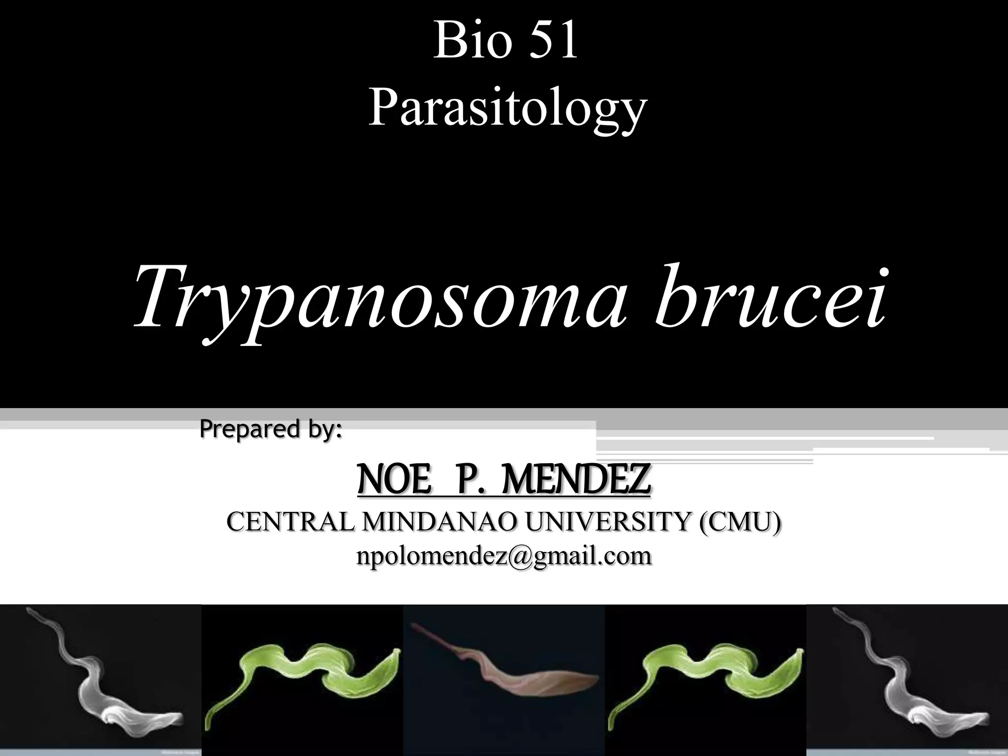
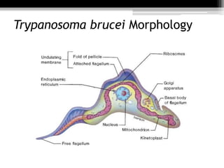
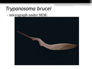
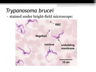
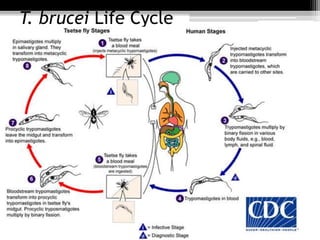
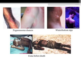
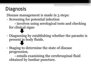
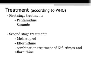
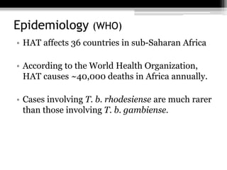
Bio 51
Parasitology
Trypanosoma brucei
Prepared by:
NOE P. MENDEZ
CENTRAL MINDANAO UNIVERSITY (CMU)
npolomendez@gmail.com
References
• Neva, F. A. & Brown, H. W. 1994. Basic clinical parasitology.
6th Ed. Appilton & Lange Paramount Publishing Business
and Professional Group: USA.
• Steverding, D. 2008. The history of African trypanosomiasis.
Biomed Central. Accessed on Dec. 1, 2015 at http://www.
parasitesandvectors.com/content/pdf /1756-3305-1-3.pdf
• Trypanosomiasis. World Health Organization. Accessed on
Dec. 1, 2015 at http://www.who.int/ mediacentre/factsheets/
fs259/en/
• Parasites – African trypanosomiasis. CDC. Accessed on Dec.
1, 2015 at http://www.cdc.gov/parasites/sleepingsickness/
biology.html
Scientific Classification
• Domain: Eukarya
• Kindom: Protista
• Phylum: Sarcomastigphora
• Class: Zoomastogophorea
• Order: Kinetoplastida
• Family: Trypanosomatidae
• Genus: Trypanosoma
• Species: T. brucei
• Sub-species: T. b. brucei, T. b. gambiense, T. b. rhodesiense
Trypanosoma brucei Morphology
Trypanosoma brucei
• micrograph under SEM:
Trypanosoma brucei
• stained under bright-field microscope:
Trypanosoma brucei
• T. brucei causes Human African Trypanosomiasis
(HAT) or sleeping sickness
• T. brucei is not killed by the immune system
because it has a glycoprotein (VSG) coating.
Trypanosoma brucei subspecies
• Two subspecies that are morphologically
indistinguishable cause distinct disease patterns
in humans:
1) T. b. gambiense causes West African
trypanosomiasis
2) T. b. rhodesiense causes East African
trypanosomiasis
• A third member, T. b. brucei, under normal
conditions does not infect humans.
Vector of T. brucei
• Tsetse fly belongs to the genus Glossina
Geographical Distribution of African
Trypanosomiasis
G.palpalis G.morsitans
In West Africa In East Africa
T. brucei Life Cycle
Stages of Infection
• In the first stage, the trypanosomes multiply in
subcutaneous tissues, blood and lymph. This is
also called haemo-lymphatic stage.
• In the second stage, the parasites cross the
blood-brain barrier to infect the central nervous
system. This is known as the neurological or
meningo-encephalic stage.
Pathogenesis
• Incubation period: 2 weeks
• Trypanosomal chancre will develop at the site of
bite
• Via lymphatics: enlarged lymph nodes especially
posterior cervical region (Winterbottom’s sign)
• Via blood stream: headache, fever, muscle &
joint pain, irregular erythematous rash
Pathogenesis
• Invasion of bone marrow (hypoplastic anemia)
• Enlarged liver & spleen
• Invasion of the CNS: severe headache, mental
apathy, slow speech, deep sleep, coma & death
Winterbottom signTrypanosoma chancre
Coma before death
Diagnosis
Disease management is made in 3 steps:
• Screening for potential infection
- involves using serological tests and checking
for clinical signs
• Diagnosing by establishing whether the parasite is
present in body fluids.
• Staging to determine the state of disease
progression.
- entails examining the cerebrospinal fluid
obtained by lumbar puncture.
Treatment (according to WHO)
• First stage treatment:
- Pentamidine
- Suramin
• Second stage treatment:
- Melarsoprol
- Eflornithine
- combination treatment of Nifurtimox and
Eflornithine
Epidemiology (WHO)
• HAT affects 36 countries in sub-Saharan Africa
• According to the World Health Organization,
HAT causes ~40,000 deaths in Africa annually.
• Cases involving T. b. rhodesiense are much rarer
than those involving T. b. gambiense.
Epidemiology (Steverding, 2008)
• The total amount of reported HAT cases has
decreased substantially over time.
• 1998: ~40,000 reported cases; >250,000
actual cases
• 2004: ~18,000 reported cases; between
50,000 and 70,000 actual cases
• 2010: ~7,000 reported cases; ~30,000 actual
cases
End of Report

Trypanosoma brucei

  • 1.
    Bio 51 Parasitology Trypanosoma brucei Preparedby: NOE P. MENDEZ CENTRAL MINDANAO UNIVERSITY (CMU) npolomendez@gmail.com
  • 2.
    References • Neva, F.A. & Brown, H. W. 1994. Basic clinical parasitology. 6th Ed. Appilton & Lange Paramount Publishing Business and Professional Group: USA. • Steverding, D. 2008. The history of African trypanosomiasis. Biomed Central. Accessed on Dec. 1, 2015 at http://www. parasitesandvectors.com/content/pdf /1756-3305-1-3.pdf • Trypanosomiasis. World Health Organization. Accessed on Dec. 1, 2015 at http://www.who.int/ mediacentre/factsheets/ fs259/en/ • Parasites – African trypanosomiasis. CDC. Accessed on Dec. 1, 2015 at http://www.cdc.gov/parasites/sleepingsickness/ biology.html
  • 3.
    Scientific Classification • Domain:Eukarya • Kindom: Protista • Phylum: Sarcomastigphora • Class: Zoomastogophorea • Order: Kinetoplastida • Family: Trypanosomatidae • Genus: Trypanosoma • Species: T. brucei • Sub-species: T. b. brucei, T. b. gambiense, T. b. rhodesiense
  • 4.
  • 5.
  • 6.
    Trypanosoma brucei • stainedunder bright-field microscope:
  • 7.
    Trypanosoma brucei • T.brucei causes Human African Trypanosomiasis (HAT) or sleeping sickness • T. brucei is not killed by the immune system because it has a glycoprotein (VSG) coating.
  • 8.
    Trypanosoma brucei subspecies •Two subspecies that are morphologically indistinguishable cause distinct disease patterns in humans: 1) T. b. gambiense causes West African trypanosomiasis 2) T. b. rhodesiense causes East African trypanosomiasis • A third member, T. b. brucei, under normal conditions does not infect humans.
  • 9.
    Vector of T.brucei • Tsetse fly belongs to the genus Glossina
  • 10.
    Geographical Distribution ofAfrican Trypanosomiasis G.palpalis G.morsitans In West Africa In East Africa
  • 12.
  • 13.
    Stages of Infection •In the first stage, the trypanosomes multiply in subcutaneous tissues, blood and lymph. This is also called haemo-lymphatic stage. • In the second stage, the parasites cross the blood-brain barrier to infect the central nervous system. This is known as the neurological or meningo-encephalic stage.
  • 14.
    Pathogenesis • Incubation period:2 weeks • Trypanosomal chancre will develop at the site of bite • Via lymphatics: enlarged lymph nodes especially posterior cervical region (Winterbottom’s sign) • Via blood stream: headache, fever, muscle & joint pain, irregular erythematous rash
  • 15.
    Pathogenesis • Invasion ofbone marrow (hypoplastic anemia) • Enlarged liver & spleen • Invasion of the CNS: severe headache, mental apathy, slow speech, deep sleep, coma & death
  • 16.
  • 17.
    Diagnosis Disease management ismade in 3 steps: • Screening for potential infection - involves using serological tests and checking for clinical signs • Diagnosing by establishing whether the parasite is present in body fluids. • Staging to determine the state of disease progression. - entails examining the cerebrospinal fluid obtained by lumbar puncture.
  • 18.
    Treatment (according toWHO) • First stage treatment: - Pentamidine - Suramin • Second stage treatment: - Melarsoprol - Eflornithine - combination treatment of Nifurtimox and Eflornithine
  • 19.
    Epidemiology (WHO) • HATaffects 36 countries in sub-Saharan Africa • According to the World Health Organization, HAT causes ~40,000 deaths in Africa annually. • Cases involving T. b. rhodesiense are much rarer than those involving T. b. gambiense.
  • 20.
    Epidemiology (Steverding, 2008) •The total amount of reported HAT cases has decreased substantially over time. • 1998: ~40,000 reported cases; >250,000 actual cases • 2004: ~18,000 reported cases; between 50,000 and 70,000 actual cases • 2010: ~7,000 reported cases; ~30,000 actual cases
  • 21.

Editor's Notes

  • #5 Kinetoplasts are a network of circular DNA found inside the mitochondrion adjacent to the basal body of the flagellum. They have unique replication.
  • #8 *Variable Surface Glycoprotein for antigenic variation
  • #13 http://www.dpd.cdc.gov/dpdx/HTML/TrypanosomiasisAfrican.htm
  • #20 Center for Disease Control and Prevention
  • #21 Steverding, Dietmar. 2008. The history of African trypanosomiasis. Biomed Central. Accessed on Dec. 1, 2015 at http://www.parasitesandvectors.com/content/pdf/1756-3305-1-3.pdf