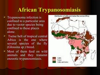
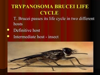
Trypanosomiasis is caused by pathogenic Trypanosoma and is endemic in Africa and South America. It is transmitted between hosts by blood-sucking insects. Trypanosoma brucei causes African trypanosomiasis (sleeping sickness) and exists in the vertebrate host as trypomastigotes, passing between hosts via the tsetse fly vector. The disease occurs in two stages with initial symptoms of fever and swelling followed by neurological involvement if untreated. Diagnosis involves blood, lymph node aspirate, or CSF examination to detect the parasites.
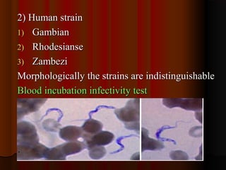
Helminthology is such an important topic not only in India but worldwide. Here is an introduction to the medically important parasites causing diseases to man.
LUMEN DWELLING FLAGELLATES - GIARDIA
REFS:
INTERNATIONALLY ACCEPTED BOOK OF MEDICAL PARASITOLOGY BY K. D. CHATTERJEE
TEXT BOOK OF MEDICAL PARASITOLOGY BY PANIKER
IMAGE SOURCES : FROM INTERNET
This is the presentation on Trypanosomiasis that covers classification and diseases caused by Trypanosoma, its life cycle, Geographical distribution, Transmission, diagnosis and treatment and finally its scenario in India.
Some flow charts have been taken from published articles, that can be searched directly from net.
A lecture Leish & trypanosoma Parasites.pdfssuser868036
بالطبع! يبدو أنك تبحث عن معلومات حول الأمراض المعروفة باسم Leishmaniasis و Trypanosomiasis. إليك نبذة عن كل منهما بالإنجليزية:
1. **Leishmaniasis:**
Leishmaniasis is a vector-borne disease caused by protozoan parasites of the Leishmania genus. It is transmitted through the bite of infected sandflies. The disease manifests in three main forms: cutaneous, mucocutaneous, and visceral leishmaniasis. Cutaneous leishmaniasis presents with skin sores, while mucocutaneous leishmaniasis affects the mucous membranes of the nose, mouth, and throat. Visceral leishmaniasis, also known as kala-azar, is the most severe form, affecting internal organs such as the spleen, liver, and bone marrow. Symptoms include prolonged fever, weight loss, and enlargement of the spleen and liver. Leishmaniasis is endemic in many tropical and subtropical regions, particularly in countries with poor socioeconomic conditions.
2. **Trypanosomiasis:**
Trypanosomiasis, also known as sleeping sickness in humans and nagana in animals, is a parasitic disease caused by protozoan parasites of the Trypanosoma genus. There are two main types of the disease: African trypanosomiasis and American trypanosomiasis, also called Chagas disease. African trypanosomiasis is transmitted to humans through the bite of infected tsetse flies, while Chagas disease is transmitted by triatomine bugs, also known as kissing bugs. The symptoms of African trypanosomiasis progress in two stages: an early stage characterized by fever, headaches, joint pains, and itching, followed by a late stage where the parasites invade the central nervous system, leading to neurological symptoms such as sleep disturbances, confusion, and poor coordination. Chagas disease can cause acute symptoms such as fever and swelling at the site of infection, followed by chronic symptoms affecting the heart, digestive system, and nervous system. Trypanosomiasis is endemic in parts of sub-Saharan Africa and Central and South America.
هل تحتاج إلى مزيد من التفاصيل حول أي من هذه الأمراض؟
TEST BANK for Operations Management, 14th Edition by William J. Stevenson, Ve...kevinkariuki227
TEST BANK for Operations Management, 14th Edition by William J. Stevenson, Verified Chapters 1 - 19, Complete Newest Version.pdf
The prostate is an exocrine gland of the male mammalian reproductive system
It is a walnut-sized gland that forms part of the male reproductive system and is located in front of the rectum and just below the urinary bladder
Function is to store and secrete a clear, slightly alkaline fluid that constitutes 10-30% of the volume of the seminal fluid that along with the spermatozoa, constitutes semen
A healthy human prostate measures (4cm-vertical, by 3cm-horizontal, 2cm ant-post ).
It surrounds the urethra just below the urinary bladder. It has anterior, median, posterior and two lateral lobes
It’s work is regulated by androgens which are responsible for male sex characteristics
Generalised disease of the prostate due to hormonal derangement which leads to non malignant enlargement of the gland (increase in the number of epithelial cells and stromal tissue)to cause compression of the urethra leading to symptoms (LUTS
These simplified slides by Dr. Sidra Arshad present an overview of the non-respiratory functions of the respiratory tract.
Learning objectives:
1. Enlist the non-respiratory functions of the respiratory tract
2. Briefly explain how these functions are carried out
3. Discuss the significance of dead space
4. Differentiate between minute ventilation and alveolar ventilation
5. Describe the cough and sneeze reflexes
Study Resources:
1. Chapter 39, Guyton and Hall Textbook of Medical Physiology, 14th edition
2. Chapter 34, Ganong’s Review of Medical Physiology, 26th edition
3. Chapter 17, Human Physiology by Lauralee Sherwood, 9th edition
4. Non-respiratory functions of the lungs https://academic.oup.com/bjaed/article/13/3/98/278874
- Video recording of this lecture in English language: https://youtu.be/lK81BzxMqdo
- Video recording of this lecture in Arabic language: https://youtu.be/Ve4P0COk9OI
- Link to download the book free: https://nephrotube.blogspot.com/p/nephrotube-nephrology-books.html
- Link to NephroTube website: www.NephroTube.com
- Link to NephroTube social media accounts: https://nephrotube.blogspot.com/p/join-nephrotube-on-social-media.html
Ozempic: Preoperative Management of Patients on GLP-1 Receptor Agonists Saeid Safari
Preoperative Management of Patients on GLP-1 Receptor Agonists like Ozempic and Semiglutide
ASA GUIDELINE
NYSORA Guideline
2 Case Reports of Gastric Ultrasound
Tom Selleck Health: A Comprehensive Look at the Iconic Actor’s Wellness Journeygreendigital
Tom Selleck, an enduring figure in Hollywood. has captivated audiences for decades with his rugged charm, iconic moustache. and memorable roles in television and film. From his breakout role as Thomas Magnum in Magnum P.I. to his current portrayal of Frank Reagan in Blue Bloods. Selleck's career has spanned over 50 years. But beyond his professional achievements. fans have often been curious about Tom Selleck Health. especially as he has aged in the public eye.
Follow us on: Pinterest
Introduction
Many have been interested in Tom Selleck health. not only because of his enduring presence on screen but also because of the challenges. and lifestyle choices he has faced and made over the years. This article delves into the various aspects of Tom Selleck health. exploring his fitness regimen, diet, mental health. and the challenges he has encountered as he ages. We'll look at how he maintains his well-being. the health issues he has faced, and his approach to ageing .
Early Life and Career
Childhood and Athletic Beginnings
Tom Selleck was born on January 29, 1945, in Detroit, Michigan, and grew up in Sherman Oaks, California. From an early age, he was involved in sports, particularly basketball. which played a significant role in his physical development. His athletic pursuits continued into college. where he attended the University of Southern California (USC) on a basketball scholarship. This early involvement in sports laid a strong foundation for his physical health and disciplined lifestyle.
Transition to Acting
Selleck's transition from an athlete to an actor came with its physical demands. His first significant role in "Magnum P.I." required him to perform various stunts and maintain a fit appearance. This role, which he played from 1980 to 1988. necessitated a rigorous fitness routine to meet the show's demands. setting the stage for his long-term commitment to health and wellness.
Fitness Regimen
Workout Routine
Tom Selleck health and fitness regimen has evolved. adapting to his changing roles and age. During his "Magnum, P.I." days. Selleck's workouts were intense and focused on building and maintaining muscle mass. His routine included weightlifting, cardiovascular exercises. and specific training for the stunts he performed on the show.
Selleck adjusted his fitness routine as he aged to suit his body's needs. Today, his workouts focus on maintaining flexibility, strength, and cardiovascular health. He incorporates low-impact exercises such as swimming, walking, and light weightlifting. This balanced approach helps him stay fit without putting undue strain on his joints and muscles.
Importance of Flexibility and Mobility
In recent years, Selleck has emphasized the importance of flexibility and mobility in his fitness regimen. Understanding the natural decline in muscle mass and joint flexibility with age. he includes stretching and yoga in his routine. These practices help prevent injuries, improve posture, and maintain mobilit
Report Back from SGO 2024: What’s the Latest in Cervical Cancer?bkling
Are you curious about what’s new in cervical cancer research or unsure what the findings mean? Join Dr. Emily Ko, a gynecologic oncologist at Penn Medicine, to learn about the latest updates from the Society of Gynecologic Oncology (SGO) 2024 Annual Meeting on Women’s Cancer. Dr. Ko will discuss what the research presented at the conference means for you and answer your questions about the new developments.
Title: Sense of Smell
Presenter: Dr. Faiza, Assistant Professor of Physiology
Qualifications:
MBBS (Best Graduate, AIMC Lahore)
FCPS Physiology
ICMT, CHPE, DHPE (STMU)
MPH (GC University, Faisalabad)
MBA (Virtual University of Pakistan)
Learning Objectives:
Describe the primary categories of smells and the concept of odor blindness.
Explain the structure and location of the olfactory membrane and mucosa, including the types and roles of cells involved in olfaction.
Describe the pathway and mechanisms of olfactory signal transmission from the olfactory receptors to the brain.
Illustrate the biochemical cascade triggered by odorant binding to olfactory receptors, including the role of G-proteins and second messengers in generating an action potential.
Identify different types of olfactory disorders such as anosmia, hyposmia, hyperosmia, and dysosmia, including their potential causes.
Key Topics:
Olfactory Genes:
3% of the human genome accounts for olfactory genes.
400 genes for odorant receptors.
Olfactory Membrane:
Located in the superior part of the nasal cavity.
Medially: Folds downward along the superior septum.
Laterally: Folds over the superior turbinate and upper surface of the middle turbinate.
Total surface area: 5-10 square centimeters.
Olfactory Mucosa:
Olfactory Cells: Bipolar nerve cells derived from the CNS (100 million), with 4-25 olfactory cilia per cell.
Sustentacular Cells: Produce mucus and maintain ionic and molecular environment.
Basal Cells: Replace worn-out olfactory cells with an average lifespan of 1-2 months.
Bowman’s Gland: Secretes mucus.
Stimulation of Olfactory Cells:
Odorant dissolves in mucus and attaches to receptors on olfactory cilia.
Involves a cascade effect through G-proteins and second messengers, leading to depolarization and action potential generation in the olfactory nerve.
Quality of a Good Odorant:
Small (3-20 Carbon atoms), volatile, water-soluble, and lipid-soluble.
Facilitated by odorant-binding proteins in mucus.
Membrane Potential and Action Potential:
Resting membrane potential: -55mV.
Action potential frequency in the olfactory nerve increases with odorant strength.
Adaptation Towards the Sense of Smell:
Rapid adaptation within the first second, with further slow adaptation.
Psychological adaptation greater than receptor adaptation, involving feedback inhibition from the central nervous system.
Primary Sensations of Smell:
Camphoraceous, Musky, Floral, Pepperminty, Ethereal, Pungent, Putrid.
Odor Detection Threshold:
Examples: Hydrogen sulfide (0.0005 ppm), Methyl-mercaptan (0.002 ppm).
Some toxic substances are odorless at lethal concentrations.
Characteristics of Smell:
Odor blindness for single substances due to lack of appropriate receptor protein.
Behavioral and emotional influences of smell.
Transmission of Olfactory Signals:
From olfactory cells to glomeruli in the olfactory bulb, involving lateral inhibition.
Primitive, less old, and new olfactory systems with different path
Pulmonary Thromboembolism - etilogy, types, medical- Surgical and nursing man...VarunMahajani
Disruption of blood supply to lung alveoli due to blockage of one or more pulmonary blood vessels is called as Pulmonary thromboembolism. In this presentation we will discuss its causes, types and its management in depth.
These lecture slides, by Dr Sidra Arshad, offer a quick overview of physiological basis of a normal electrocardiogram.
Learning objectives:
1. Define an electrocardiogram (ECG) and electrocardiography
2. Describe how dipoles generated by the heart produce the waveforms of the ECG
3. Describe the components of a normal electrocardiogram of a typical bipolar leads (limb II)
4. Differentiate between intervals and segments
5. Enlist some common indications for obtaining an ECG
Study Resources:
1. Chapter 11, Guyton and Hall Textbook of Medical Physiology, 14th edition
2. Chapter 9, Human Physiology - From Cells to Systems, Lauralee Sherwood, 9th edition
3. Chapter 29, Ganong’s Review of Medical Physiology, 26th edition
4. Electrocardiogram, StatPearls - https://www.ncbi.nlm.nih.gov/books/NBK549803/
5. ECG in Medical Practice by ABM Abdullah, 4th edition
6. ECG Basics, http://www.nataliescasebook.com/tag/e-c-g-basics
Couples presenting to the infertility clinic- Do they really have infertility...Sujoy Dasgupta
Dr Sujoy Dasgupta presented the study on "Couples presenting to the infertility clinic- Do they really have infertility? – The unexplored stories of non-consummation" in the 13th Congress of the Asia Pacific Initiative on Reproduction (ASPIRE 2024) at Manila on 24 May, 2024.
2. TRYPANOSOMIASISTRYPANOSOMIASIS
IINTRODUCTION:-NTRODUCTION:-
Human trypanosomiasis is endemic in AfricaHuman trypanosomiasis is endemic in Africa
and South America. In Africa the disease is knownand South America. In Africa the disease is known
as Human African Trypanosomiasis (HAT) oras Human African Trypanosomiasis (HAT) or
sleeping sicknesssleeping sickness. Whereas American. Whereas American
trypanosomiasis is known astrypanosomiasis is known as Chaga’s disease.Chaga’s disease.
Trypanosomes are haemoflagellates.Trypanosomes are haemoflagellates.
Disease caused by pathogenic types is calledDisease caused by pathogenic types is called
trypanosomiasis.trypanosomiasis.
3. Genus TrypanosomaGenus Trypanosoma
Exist as trypomastigotes in vertebrate hosts and someExist as trypomastigotes in vertebrate hosts and some
assuming amastigote forms.assuming amastigote forms.
Pass their life cycle in two hosts Vertebrate and insectPass their life cycle in two hosts Vertebrate and insect
( amastigote, promastigote, epimastigote and( amastigote, promastigote, epimastigote and
metacyclic trypomastigote).metacyclic trypomastigote).
Transmission is effected from one vertebrate toTransmission is effected from one vertebrate to
another by blood sucking insects.another by blood sucking insects.
Types of Development :-Types of Development :-
1.1. Anterior stationAnterior station
2.2. Posterior stationPosterior station
4. Morphology of TrypomastigotesMorphology of Trypomastigotes
Method of reproduction – binary longitudinal fission.Method of reproduction – binary longitudinal fission.
5. Classification of TrypanosomiasisClassification of Trypanosomiasis
Two British scientists (Forbe andTwo British scientists (Forbe and
Dutton) discovered trypanosomes inDutton) discovered trypanosomes in
1901 in Gambia1901 in Gambia
T. Brucei-T. Brucei-
a) T. Brucei Gambiensea) T. Brucei Gambiense
b) T. Brucei Rhodesienseb) T. Brucei Rhodesiense
c ) T. Brucei bruceic ) T. Brucei brucei
T. Cruzi-T. Cruzi-
T. Rangeli-T. Rangeli-
Found in peripheral blood of man inFound in peripheral blood of man in
venezuela.venezuela.
Non pathogenic for man.Non pathogenic for man.
Trypanosomiasis Seen in AnimalsTrypanosomiasis Seen in Animals
1)1) NON PATHOGENICNON PATHOGENIC-- T. LewisiT. Lewisi
Life cycle passes in to two hosts -ratLife cycle passes in to two hosts -rat
and rat fleaand rat flea
Shriwastava et al. reportedShriwastava et al. reported
Trypanosome resembling T. Lewisi inTrypanosome resembling T. Lewisi in
peripheral blood of 2 cases sufferingperipheral blood of 2 cases suffering
from short febrile illness in Raipur M.P.from short febrile illness in Raipur M.P.
6. 2)2) PATHOGENIC –PATHOGENIC –
T. Brucei brucei-T. Brucei brucei-
Transmitted by G. MorsitansTransmitted by G. Morsitans
T. Evansi – ( Causes ‘Surra’ in horses,T. Evansi – ( Causes ‘Surra’ in horses,
mules, camel, elephants)mules, camel, elephants)
T. Equiperdum- Stallions diseaseT. Equiperdum- Stallions disease
T. EquinumT. Equinum
T. VivaxT. Vivax
T. CongolenseT. Congolense
7. African TrypanosomiasisAfrican Trypanosomiasis
The parasite discovered by David Bruce inThe parasite discovered by David Bruce in
1890.He suggested it to be the cause of1890.He suggested it to be the cause of
NaganaNagana
Trypanosoma BruceiTrypanosoma Brucei
Based on host specificity, clinical features,Based on host specificity, clinical features,
geographical distribution andgeographical distribution and
epidemiological features originally classifiedepidemiological features originally classified
intointo
1)1) The animal strain –The animal strain –
T. Brucei bruceiT. Brucei brucei
8. 2) Human strain2) Human strain
1)1) GambianGambian
2)2) RhodesianseRhodesianse
3)3) ZambeziZambezi
Morphologically the strains are indistinguishableMorphologically the strains are indistinguishable
Blood incubation infectivity testBlood incubation infectivity test
9. African TrypanosomiasisAfrican Trypanosomiasis
Trypanosoma infection isTrypanosoma infection is
confined to a particular areaconfined to a particular area
due to vector species beingdue to vector species being
confined to these placesconfined to these places
alone.alone.
Tsetse belt of tropical centralTsetse belt of tropical central
Africa is the one whereAfrica is the one where
several species of the flyseveral species of the fly
(Glossina sp.) breed.(Glossina sp.) breed.
Most of them feed on wildMost of them feed on wild
animals and they transmitanimals and they transmit
enzootic trypanosomiasis.enzootic trypanosomiasis.
10. SuffixSuffix demedeme has beenhas been
employed to refer toemployed to refer to
population ofpopulation of
trypanosomes thattrypanosomes that
differs from othersdiffers from others
belonging to the samebelonging to the same
species or subspecies inspecies or subspecies in
regard to specifiedregard to specified
properties.properties.
e.g.e.g.
a)a) NosodemeNosodeme
b)b) ZymodemeZymodeme
c)c) SerodemesSerodemes
11. HABITATHABITAT
T. BruceiT. Brucei
Essentially a parasite of connectiveEssentially a parasite of connective
tissue.tissue.
Consumes enormous amount ofConsumes enormous amount of
glucoseglucose
Invades regional L.N. & also invadesInvades regional L.N. & also invades
blood stream causing parasitemiablood stream causing parasitemia
Finally localizes in brainFinally localizes in brain
ChronicChronic GambianseGambianse strain is foundstrain is found
mainly in west and central Africa andmainly in west and central Africa and
scattered areas of East Africascattered areas of East Africa
RhodesienseRhodesiense- East Africa- East Africa
ZambeziZambezi- East Africa which- East Africa which
botswana, Rhodesia, Zambiabotswana, Rhodesia, Zambia
12. PolymorphismPolymorphism
Trypomastigotes- polymorphicTrypomastigotes- polymorphic
Two main formsTwo main forms
1)1) Short, thick, stumpy formShort, thick, stumpy form
2)2) Other long slender formOther long slender form
ANTIGENIC VARIANTANTIGENIC VARIANT
A succession of variants occurA succession of variants occur
in T. Brucei infection both inin T. Brucei infection both in
animals and in mananimals and in man
Each new variant emerges atEach new variant emerges at
3-4 days intervals possibly by3-4 days intervals possibly by
selection of mutants.selection of mutants.
This appears to depend uponThis appears to depend upon
the ability of the host’s defencethe ability of the host’s defence
mechanism.mechanism.
13. Outer proteinaceous coat may protect theOuter proteinaceous coat may protect the
organism in escaping from the host’s defenceorganism in escaping from the host’s defence
mechanism.mechanism.
Also Ab develop to a series ofAlso Ab develop to a series of
immunologically distinct type of variant in theimmunologically distinct type of variant in the
serum of host.serum of host.
Each rise in antibody titre coincides with theEach rise in antibody titre coincides with the
disappearance of the homologous variant anddisappearance of the homologous variant and
when this has been eliminated, a new antigenicwhen this has been eliminated, a new antigenic
type immediately arises.type immediately arises.
14. TRYPANOSOMA BRUCEI LIFETRYPANOSOMA BRUCEI LIFE
CYCLECYCLE
T. Brucei passes its life cycle in two differentT. Brucei passes its life cycle in two different
hostshosts
Definitive hostDefinitive host
Intermediate host - insectIntermediate host - insect
15. Infective from is metacyclicInfective from is metacyclic
trypomastigotetrypomastigote
Metacyclic trypomastigotes areMetacyclic trypomastigotes are
inoculated through the skin when a tsetseinoculated through the skin when a tsetse
fly takes a blood meal. Initially theyfly takes a blood meal. Initially they
proliferate at site of inoculation and thenproliferate at site of inoculation and then
through lymphatics, enters blood streamthrough lymphatics, enters blood stream
16. It takes about 3 wks from the time of blood meal for fly to becomeIt takes about 3 wks from the time of blood meal for fly to become
infective. Then it remains infective for life (6Months)infective. Then it remains infective for life (6Months)
17. Clinical DiseaseClinical Disease
PathogenesisPathogenesis
1.1. Mode of infectionMode of infection
2.2. Trypomastigotes introduced by the fly with saliva into theTrypomastigotes introduced by the fly with saliva into the
S.C. pool of bloodS.C. pool of blood
3.3. Majority get entangled in the tissue spacesMajority get entangled in the tissue spaces
4.4. Initial growth occurs in tissue spaces which form a moreInitial growth occurs in tissue spaces which form a more
favourable nidusfavourable nidus
5.5. It is also suggested that connective tissue damage caused byIt is also suggested that connective tissue damage caused by
trypomastigotes may be due to exaggerated immunetrypomastigotes may be due to exaggerated immune
response rather than any direct effectresponse rather than any direct effect
6.6. Trypomastogotes in tissue excites host’s response in twoTrypomastogotes in tissue excites host’s response in two
waysways
a) Producing large amount of nonspecifica) Producing large amount of nonspecific
immunoglobulinsimmunoglobulins
b) By heavily infilltrating the site of infection withb) By heavily infilltrating the site of infection with
macrophages.macrophages.
18. Pathogenic LesionPathogenic Lesion
1.1. CNS -CNS -
2.2. LN – Congestion,LN – Congestion,
haemorrhage, proliferationhaemorrhage, proliferation
of macrophages.of macrophages.
3.3. SpleenSpleen
4.4. LiverLiver
5.5. HeartHeart
6.6. KidneyKidney
7.7. Bone MarrowBone Marrow
8.8. LungsLungs
9.9. Localized edema of eyelid,Localized edema of eyelid,
perineum, skin of backperineum, skin of back
19. Clinical FeaturesClinical Features
The disease occurs inThe disease occurs in
two stages. Incubationtwo stages. Incubation
period – 2-3 weeksperiod – 2-3 weeks
1st stage1st stage
Enlarged postEnlarged post
cervical L.N.cervical L.N.
Winterbottom’sWinterbottom’s
signsign
Pruritus ,TransientPruritus ,Transient
edemaedema
PapuloerythematousPapuloerythematous
eruptioneruption
Endocrine dysfunctionEndocrine dysfunction
20. 22ndnd
Stage –Stage –
HeadacheHeadache
Impaired motor functionImpaired motor function
Slurred speech,Slurred speech,
Abnormal movements,Abnormal movements,
tremorstremors
Mental Changes –Mental Changes –
Generalised pruritis,Generalised pruritis,
bedsores, wt. loss,bedsores, wt. loss,
wasting.wasting.
Coma followed by death ifComa followed by death if
untreated.untreated.
22. Lab Diagnosis African TrypanosomiasisLab Diagnosis African Trypanosomiasis
1.1. Examination of bloodExamination of blood
2.2. Examination of aspirates fromExamination of aspirates from
enlarged lymph glandsenlarged lymph glands
3.3. Examination of CSFExamination of CSF
4.4. Detection of trypanosomal antibodyDetection of trypanosomal antibody
in serumin serum
23. 11.. Examination of BloodExamination of Blood
1. Thick and thin films, Wet1. Thick and thin films, Wet
preparationpreparation
2. Concentration method2. Concentration method
a. Triple centrifugationa. Triple centrifugation
techniquetechnique
b. Miniature anion-b. Miniature anion-
exchange centrifugation techniqueexchange centrifugation technique
c. Buffy coat preparationc. Buffy coat preparation
22.. Examination of Aspirates inExamination of Aspirates in
lymph nodeslymph nodes
3. Examination of CSF3. Examination of CSF
4. Detection of trypanosomes4. Detection of trypanosomes
antibody in serumantibody in serum
- CATT- CATT
- Direct agglutination, indirect- Direct agglutination, indirect
haemagglutination, gelhaemagglutination, gel
precipitation, ELISA , PCRprecipitation, ELISA , PCR
24. Also animalAlso animal
inoculation ofinoculation of
mice, rat, guineamice, rat, guinea
pig can be done .pig can be done .
ProphylaxisProphylaxis
1.1. Attack on parasiteAttack on parasite
2.2. Attack on vectorAttack on vector
3.3. PersonalPersonal
prophylaxisprophylaxis
25. South American TrypanosomiasisSouth American Trypanosomiasis
HistoryHistory
HabitatHabitat
Muscular and nervousMuscular and nervous
tissues, RES.tissues, RES.
Exists in the amastigoteExists in the amastigote
formform
MorphologyMorphology
26. Vectors and Life CycleVectors and Life Cycle
HostsHosts
VertebrateVertebrate
Insect – Bugs- Reduviidae family, subfamily-Insect – Bugs- Reduviidae family, subfamily-
TritominaeTritominae
27. Life CycleLife Cycle
Metacyclic trypomastigotes are deposited on skinMetacyclic trypomastigotes are deposited on skin
Trypomastigotes become amastigotes in localized RETrypomastigotes become amastigotes in localized RE
cells and multiply by longitudinal fission .cells and multiply by longitudinal fission .
After passing through promastigote and epimastigoteAfter passing through promastigote and epimastigote
forms are transformed into trypomastigote formsforms are transformed into trypomastigote forms
Taken up by bug during act of bitingTaken up by bug during act of biting
In midgut- transformed to amastigote formsIn midgut- transformed to amastigote forms
Amastigote forms later transformed into epimastigoteAmastigote forms later transformed into epimastigote
forms which migrate backwards to hindgut andforms which migrate backwards to hindgut and
multiply by longitudinal fission.multiply by longitudinal fission.
In 8-10 days time metacylic forms of trypomastigotesIn 8-10 days time metacylic forms of trypomastigotes
appear and are excreted in faeces of bug .appear and are excreted in faeces of bug .
28. PathogenicityPathogenicity
a.a. ChagomaChagoma
b.b. Romana’s SignRomana’s Sign
c.c. Gross lesions mainly in heart, skeletal muscle and nervousGross lesions mainly in heart, skeletal muscle and nervous
tissuetissue
Clinical FeaturesClinical Features
1)1) Incub period 7-14 daysIncub period 7-14 days
2)2) Infection - asymptomatic, acute, chronicInfection - asymptomatic, acute, chronic
a)a) Acute stage – Fever, conjunctivitis , unilat edema of face,Acute stage – Fever, conjunctivitis , unilat edema of face,
enlargement of spleen and L.N., anaemia, lymphocytosis.enlargement of spleen and L.N., anaemia, lymphocytosis.
b)b) Chronic –Chronic –
Signs of cardiac muscle damage, edema, heartSigns of cardiac muscle damage, edema, heart
enlargement.enlargement.
Dilatation of tubular organs- Due to degeneration ofDilatation of tubular organs- Due to degeneration of
intramural autonomic nerve plexus.intramural autonomic nerve plexus.
Chaga’s Cardiomyopathy (10%)Chaga’s Cardiomyopathy (10%)
29. Lab DiagnosisLab Diagnosis
1)Examination of Blood1)Examination of Blood
a)a) Thick, thin film, Wet preparationThick, thin film, Wet preparation
b)b) Buffy coat examinationBuffy coat examination
2) Xenodiagnosis2) Xenodiagnosis
3) Blood Culture -3) Blood Culture - As sensitive as xenodiagnosisAs sensitive as xenodiagnosis
4) Serology4) Serology
a)a) Indirect fluorescence Ab testIndirect fluorescence Ab test
b)b) CFTCFT
c)c) IHATIHAT
d)d) ELISAELISA
Other tests like intradermal test, biopsy from involved L.N. or muscle.Other tests like intradermal test, biopsy from involved L.N. or muscle.
Other findings are increased ESR; marked lymphocytosisOther findings are increased ESR; marked lymphocytosis
ProphylaxisProphylaxis
30. 11stst
case of Trypanosomiasis in Indiacase of Trypanosomiasis in India
45 yrs old man45 yrs old man
Farmer from Chandrapur dist in MaharashtraFarmer from Chandrapur dist in Maharashtra
Came with fever associated with chills andCame with fever associated with chills and
sweating for 15 days. Then he developed signs ofsweating for 15 days. Then he developed signs of
sensory deficitsensory deficit
In GMC Nagpur- Blood smear showed presence ofIn GMC Nagpur- Blood smear showed presence of
parasites with morphology typical of monomorphic,parasites with morphology typical of monomorphic,
slender, flagellated, dividing trypomastigote formslender, flagellated, dividing trypomastigote form
of trypanosomaof trypanosoma
Later on PCR based methods confirmed diagnosisLater on PCR based methods confirmed diagnosis
of T. evansi.of T. evansi.
31. REFERENCESREFERENCES
Medical parasitology – Rajesh KaryakarteMedical parasitology – Rajesh Karyakarte
Paniker’s medical parasitologyPaniker’s medical parasitology
K.D Chatterjee’s human parasites & parasiticK.D Chatterjee’s human parasites & parasitic
diseasesdiseases
Harrison’s internal medicineHarrison’s internal medicine
InternetInternet