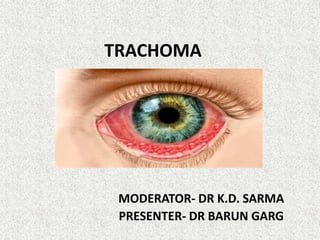
TRACHOMA
MODERATOR- DR K.D. SARMA
PRESENTER- DR BARUN GARG
INTRODUCTION
• Leading cause of infective blindness globally
• >150 million people have been affected
• Associated with poor hygiene and inadequate
sanitation
• Recent estimates show 59 countries are
endemic and India has high burden
• Egyptian ophthalmia , north western belt of
india
DEFINITION
• Chronic granulomatous kerato-conjunctivitis
• Caused by Chlamydia trachomatis (A,B,Ba,C)
• Mainly affects children at early age who
develop blindness later
• Cause of 3.6% of global blindness(WHO)
• Highly contagious
• Spread by transfer of conjunctival secretions
through fingers, towels, flies etc
PATHOLOGY
• C. Trachomatis- prokaryotic, obligatory
intracellular parasite
• Halberstaedter-Prowazek inclusion bodies– in
epithelial cells of conjunctiva( not pathognomic)
• Primary infection- epithelia of conjunctiva &
cornea
• Diffuse inflammation- congestion, papillary
enlargement, follicles
• Recurrent infection- type IV hypersensitivity to Ag
• Lymphocytic infiltration of adenoid layer
• LEBER cells – necrosed and multinucletaed
giant cells
• Cicatricial bands- in late stages, characteristic
• Arlt line- white conjunctival scar at junction of
lower third and upper two-third of superior
tarus, characteristic
RISK FACTORS
6 D’s
• Dry
• Dusty
• Dirty
• Dung
• Discharge
• Density ( crowding)
TRANSMISSION OF TRACHOMA
 5 F’s
• Fingers
• Flies
• Face
• Faeces
• Fomites
PREDISPOSING FACTORS
• Age- more in infancy/ childhood
• Sex- commoner in females
• Dry and dusty environment
• Low socio economic status, unhygienic
conditions, lack of sanitation
SPREAD OF INFECTIONS
• DIRECT- contact with airborne or waterborne
infections
• VECTOR- flies ( Musca domestica)
• MATERIAL- most important
CLINICAL FEATURES
• Incubation period- 5 to 21 days
• Onset – subacute , but on massive outbreaks
can be acute
• Symptoms – watering, fb sensation, redness,
mucopurulent discharge, photophobia,
blurring, mild pain
Signs –
• Upper tarsal conjunctiva – mc affected , appears
red velvety, congested
• Trachomatous follicle- essential lesion, upto 5mm
size
- characteristic distribution- upper fornix(mc),
upper margin of tarsus, palprebral conjunctiva
• Scarring of conjunctiva
• Arlt’s line
• Limbal follicles
• Herbert pits- oval/pitted scars in limbus
Cornea-
• Early- superficial keartitis on SLE( flourescence
staining), in upper part due to erosion
• Later- trachomatous pannus, starts in upper half
then spreads centrally to involve whole cornea
• Vascularisation- in between BM and epithelium
• Pannus- a) progressive- vessels parallel, directed
vt downwards, infiltration ahead of vessels
b) regressive- vessels ahead of infiltartion
• Ulcers- mc at advancing edge of pannus
• Corneal opacity
Lids-
• Edema
• Trichiasis
• Distiachsis
• Entropion
• Scarring
• Trachomatous ptosis
• TWO STAGES- a) active
b) cicatrical
WHO CLASSIFICATION(FISTO)
• developed for use by trained personnel other
than ophthalmologists to assess the prevalence
and severity of trachoma in population-based
surveys in endemic areas.
TRACHOMATOUS FOLLICULAR(TF)
• Active disease
• 5 or more follicles of > 0.5mm on upper tarsus
• Deep conjunctival vessels seen
• If treated properly- no scarring
TRACHOMA INTENSE
• Severe disease, needs urgents rx
• diffuse involvement of the tarsal conjunctiva,
obscuring 50% or more of the normal deep
tarsal vessels; papillae are present
TRACHOMATOUS SCARRING-
• Inactive infection
• conjunctival scarring
• visible fibrous white bands on tarsal
conjunctiva
 TRACHOMATOUS TRICHIASIS
• at least one lash touching the globe
• Needs corrective surgery
 CORNEAL OPACITY
• sufficient to blur details of at least part of the pupillary margin
Mc CALLANS CLASSIFICTION
STAGE 1- incipient trachoma/ stage of
infiltration
• Hyperemia of palpebral conjunctiva &
immature follicles
STAGE 2- stage of florid infiltration
• mature follicles, papillae, progressive pannus
STAGE 3- cicatarizing trachoma/ stage of
scarring
STAGE 4- healed trachoma/ stage of sequale
DIAGNOSIS
Requires at least 2 of the following clinical
features:
• follicles on the upper tarsal conjunctiva
• limbal follicles and their sequelae (Herbert
pits)
• typical tarsal conjunctival scarring
• vascular pannus most marked on the superior
limbus
SEQUELAE
• Lids- trichiasis, entropion, tylois, ptosis,
madarosis
• Conjunctiva- concretions, pseudocyst, xerosis,
symblepharon
• Cornea – opacity, ectasia, xerosis, total
corneal pannus( blinding sequale)
• Punctal stenosis, fibrosis of canaliculi
LAB INVESTIGATIONS
• Conjunctival inclusion bodies- giemsa/ iodine/
immunofluorescence staining
• Conjunctival cytology- giemsa stain- pmn with
leber cells
• ELISA- chlamydial Ags
• PCR
• Microimmunoflourescence (microIF)- for Abs
• Direct monoclonal ab microscopy- rapid and
inexpensive
• McCoy cell cultures
DIFFERENTIAL DIAGNOSIS
• Allergic/ vernal conjunctivitis
• Bacterial conjunctivitis
• Follicular conjunctivitis
MANAGEMENT
A)Treatment – of active disease and sequalae
B) Prevention
 Rx of active disease
Antibiotics- main stay
• oral- Azithromycin 1gm stat(20mg/kg) – DOC
Tetracycline or erythromycin 250mg
QID for 4 weeks
Doxycycline 100mg BD for 4 weeks
• Topical – best for indiviual cases, cheaper, no
systemic side effects
Regimes – 1% tetracyclines/ erythtromycin
eye ointment QID for 6 weeks
20% sulfacetamide eye drops thrice daily with
1% tetracycline oint at bedtime for 6 weeks
• Other topical antibiotics for secondary
bacterial infections
• Lubricants
• Analgescics
Prevention
SAFE STRATEGY was devised
• Surgery- correction of entropion
trichaisis rx- epilation, cryolysis, electrolysis
• Antibiotics
• Facial cleanliness
• Envioronmental improvements
PROPHYLAXIS
• Good personal hygeine and environmental
sanitation
• Health education
• Use of common towels, hankerchiefs are
discouraged
• Clean water supply for washing
• Flies control- insecticides, good sewerage,
garbage disposal, window screen protectors
• Prevention of recurrent infections
• Early detection and rx
Blanket antibiotic therapy/intermittent
therapy( WHO)
• In endemic areas to control intensity and
severity
• Regimen- 1% tetracycline oint BD 7 days/
month X 6 months
National trachoma control program-
• Launched in 1963
• Under NPCB
• Centrally sponsored
• SAFE strategy
• Training at root level
• Health education
GET 2020
• Global Elimination of Trachoma by 2020
• Launched by WHO
• Objective- to eliminate trachoma as blinding
disease
• ICTC- international coalation of trachoma control
• WHO defines blinding trachoma elimination as:
–TF prevalence <5% in 1-9 year old children
–TT prevalence <1 per 1000 in total population
Trachoma
Trachoma

Trachoma

  • 1.
    TRACHOMA MODERATOR- DR K.D.SARMA PRESENTER- DR BARUN GARG
  • 2.
    INTRODUCTION • Leading causeof infective blindness globally • >150 million people have been affected • Associated with poor hygiene and inadequate sanitation • Recent estimates show 59 countries are endemic and India has high burden • Egyptian ophthalmia , north western belt of india
  • 3.
    DEFINITION • Chronic granulomatouskerato-conjunctivitis • Caused by Chlamydia trachomatis (A,B,Ba,C) • Mainly affects children at early age who develop blindness later • Cause of 3.6% of global blindness(WHO) • Highly contagious • Spread by transfer of conjunctival secretions through fingers, towels, flies etc
  • 4.
    PATHOLOGY • C. Trachomatis-prokaryotic, obligatory intracellular parasite • Halberstaedter-Prowazek inclusion bodies– in epithelial cells of conjunctiva( not pathognomic) • Primary infection- epithelia of conjunctiva & cornea • Diffuse inflammation- congestion, papillary enlargement, follicles • Recurrent infection- type IV hypersensitivity to Ag
  • 6.
    • Lymphocytic infiltrationof adenoid layer • LEBER cells – necrosed and multinucletaed giant cells • Cicatricial bands- in late stages, characteristic • Arlt line- white conjunctival scar at junction of lower third and upper two-third of superior tarus, characteristic
  • 8.
    RISK FACTORS 6 D’s •Dry • Dusty • Dirty • Dung • Discharge • Density ( crowding)
  • 9.
    TRANSMISSION OF TRACHOMA 5 F’s • Fingers • Flies • Face • Faeces • Fomites
  • 10.
    PREDISPOSING FACTORS • Age-more in infancy/ childhood • Sex- commoner in females • Dry and dusty environment • Low socio economic status, unhygienic conditions, lack of sanitation
  • 11.
    SPREAD OF INFECTIONS •DIRECT- contact with airborne or waterborne infections • VECTOR- flies ( Musca domestica) • MATERIAL- most important
  • 12.
    CLINICAL FEATURES • Incubationperiod- 5 to 21 days • Onset – subacute , but on massive outbreaks can be acute • Symptoms – watering, fb sensation, redness, mucopurulent discharge, photophobia, blurring, mild pain
  • 13.
    Signs – • Uppertarsal conjunctiva – mc affected , appears red velvety, congested • Trachomatous follicle- essential lesion, upto 5mm size - characteristic distribution- upper fornix(mc), upper margin of tarsus, palprebral conjunctiva • Scarring of conjunctiva • Arlt’s line • Limbal follicles • Herbert pits- oval/pitted scars in limbus
  • 16.
    Cornea- • Early- superficialkeartitis on SLE( flourescence staining), in upper part due to erosion • Later- trachomatous pannus, starts in upper half then spreads centrally to involve whole cornea • Vascularisation- in between BM and epithelium • Pannus- a) progressive- vessels parallel, directed vt downwards, infiltration ahead of vessels b) regressive- vessels ahead of infiltartion • Ulcers- mc at advancing edge of pannus • Corneal opacity
  • 18.
    Lids- • Edema • Trichiasis •Distiachsis • Entropion • Scarring • Trachomatous ptosis • TWO STAGES- a) active b) cicatrical
  • 19.
    WHO CLASSIFICATION(FISTO) • developedfor use by trained personnel other than ophthalmologists to assess the prevalence and severity of trachoma in population-based surveys in endemic areas. TRACHOMATOUS FOLLICULAR(TF) • Active disease • 5 or more follicles of > 0.5mm on upper tarsus • Deep conjunctival vessels seen • If treated properly- no scarring
  • 20.
    TRACHOMA INTENSE • Severedisease, needs urgents rx • diffuse involvement of the tarsal conjunctiva, obscuring 50% or more of the normal deep tarsal vessels; papillae are present
  • 21.
    TRACHOMATOUS SCARRING- • Inactiveinfection • conjunctival scarring • visible fibrous white bands on tarsal conjunctiva
  • 22.
     TRACHOMATOUS TRICHIASIS •at least one lash touching the globe • Needs corrective surgery  CORNEAL OPACITY • sufficient to blur details of at least part of the pupillary margin
  • 23.
    Mc CALLANS CLASSIFICTION STAGE1- incipient trachoma/ stage of infiltration • Hyperemia of palpebral conjunctiva & immature follicles STAGE 2- stage of florid infiltration • mature follicles, papillae, progressive pannus STAGE 3- cicatarizing trachoma/ stage of scarring STAGE 4- healed trachoma/ stage of sequale
  • 24.
    DIAGNOSIS Requires at least2 of the following clinical features: • follicles on the upper tarsal conjunctiva • limbal follicles and their sequelae (Herbert pits) • typical tarsal conjunctival scarring • vascular pannus most marked on the superior limbus
  • 25.
    SEQUELAE • Lids- trichiasis,entropion, tylois, ptosis, madarosis • Conjunctiva- concretions, pseudocyst, xerosis, symblepharon • Cornea – opacity, ectasia, xerosis, total corneal pannus( blinding sequale) • Punctal stenosis, fibrosis of canaliculi
  • 26.
    LAB INVESTIGATIONS • Conjunctivalinclusion bodies- giemsa/ iodine/ immunofluorescence staining • Conjunctival cytology- giemsa stain- pmn with leber cells • ELISA- chlamydial Ags • PCR • Microimmunoflourescence (microIF)- for Abs • Direct monoclonal ab microscopy- rapid and inexpensive • McCoy cell cultures
  • 27.
    DIFFERENTIAL DIAGNOSIS • Allergic/vernal conjunctivitis • Bacterial conjunctivitis • Follicular conjunctivitis
  • 28.
    MANAGEMENT A)Treatment – ofactive disease and sequalae B) Prevention  Rx of active disease Antibiotics- main stay • oral- Azithromycin 1gm stat(20mg/kg) – DOC Tetracycline or erythromycin 250mg QID for 4 weeks Doxycycline 100mg BD for 4 weeks
  • 29.
    • Topical –best for indiviual cases, cheaper, no systemic side effects Regimes – 1% tetracyclines/ erythtromycin eye ointment QID for 6 weeks 20% sulfacetamide eye drops thrice daily with 1% tetracycline oint at bedtime for 6 weeks • Other topical antibiotics for secondary bacterial infections • Lubricants • Analgescics
  • 30.
    Prevention SAFE STRATEGY wasdevised • Surgery- correction of entropion trichaisis rx- epilation, cryolysis, electrolysis • Antibiotics • Facial cleanliness • Envioronmental improvements
  • 33.
    PROPHYLAXIS • Good personalhygeine and environmental sanitation • Health education • Use of common towels, hankerchiefs are discouraged • Clean water supply for washing • Flies control- insecticides, good sewerage, garbage disposal, window screen protectors • Prevention of recurrent infections • Early detection and rx
  • 35.
    Blanket antibiotic therapy/intermittent therapy(WHO) • In endemic areas to control intensity and severity • Regimen- 1% tetracycline oint BD 7 days/ month X 6 months
  • 36.
    National trachoma controlprogram- • Launched in 1963 • Under NPCB • Centrally sponsored • SAFE strategy • Training at root level • Health education
  • 37.
    GET 2020 • GlobalElimination of Trachoma by 2020 • Launched by WHO • Objective- to eliminate trachoma as blinding disease • ICTC- international coalation of trachoma control • WHO defines blinding trachoma elimination as: –TF prevalence <5% in 1-9 year old children –TT prevalence <1 per 1000 in total population