RATINAL DETACHMENT
Mr. Manikandan.T,
RN., RM., M.Sc(N)., D.C.A .,(Ph.D)
Assistant Professor,
Dept. of Medical Surgical Nursing,
VMCON, Puducherry.
RATINALDETACHMENT
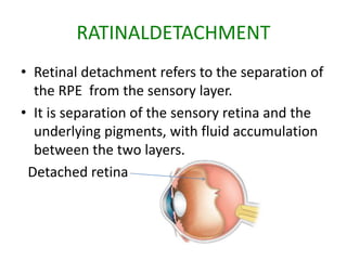
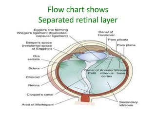
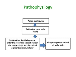
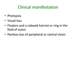
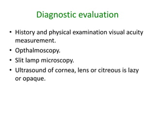
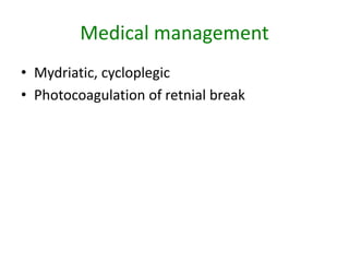
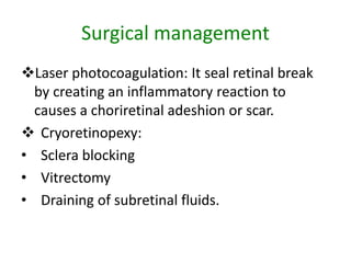
• Retinal detachment refers to the separation of
the RPE from the sensory layer.
• It is separation of the sensory retina and the
underlying pigments, with fluid accumulation
between the two layers.
Detached retina
Types
• Rhegmatogenous detachment .
• Traction retinal detachment .
• Exudative retinal detachments .
• Combination of rhegmatogenous and traction,
and exudative.
Rhegmatogenous detachment
• In this condition, a hole or tear develops in the
sensory retina, allowing some of the liquid
vitreous to seep through the sensory retina
and detach it from the RPE
Traction retinal detachment .
• Tension, or a pulling force, is responsible for
traction retinal detachment, patients develop
scar tissue.
Exudative retinal detachments
• It occurs as a result of the production of a
serous fluid under the retina from the choroid.
Conditions such as uveitis and macular
degeneration may cause the production of
this serous fluid.
Risk factors
• Increasing age.
• Severe myopia.
• Eye trauma.
• Retinopathy (diabetic).
• Cataract or glaucoma surgery.
• Family history .
• Personal history .
Flow chart shows
Separated retinal layer
Pathophysilogy
Aging, eye trauma
Retina tears and pulls
retina
Break retina, liquid viteous can
enter the subretinal space between
the sensory layer and the retinal
pigment enithelium layer
Rhegmatogenous retinal
detachment.
Clinical manifestation
• Photopsia
• Visual loss
• Floaters and a cobweb hairnet or ring in the
field of vision
• Painless loss of peripheral or central vision
Diagnostic evaluation
• History and physical examination visual acuity
measurement.
• Opthalmoscopy.
• Slit lamp microscopy.
• Ultrasound of cornea, lens or citreous is lazy
or opaque.
Medical management
• Mydriatic, cycloplegic
• Photocoagulation of retnial break
Surgical management
Laser photocoagulation: It seal retinal break
by creating an inflammatory reaction to
causes a choriretinal adeshion or scar.
 Cryoretinopexy:
• Sclera blocking
• Vitrectomy
• Draining of subretinal fluids.
Preoperative nursing inteventions
• Provide emotional support to the patient who may be
distraught at the potential loss of vision
• Prepare the patient for surgery by cleaning his face and
giving him antibiotics and eyedrops, as ordered
• Teach the patient for surgery by cleaning his face and
giving him antibiotics and eyedrops, as ordered
• Teach the patient about the role of the retina and why
floaters, flashes of light, and decreased vision occur.
• Allow the patient and family to discuss their concerns

Retinal detachment

  • 1.
    RATINAL DETACHMENT Mr. Manikandan.T, RN.,RM., M.Sc(N)., D.C.A .,(Ph.D) Assistant Professor, Dept. of Medical Surgical Nursing, VMCON, Puducherry.
  • 2.
    RATINALDETACHMENT • Retinal detachmentrefers to the separation of the RPE from the sensory layer. • It is separation of the sensory retina and the underlying pigments, with fluid accumulation between the two layers. Detached retina
  • 3.
    Types • Rhegmatogenous detachment. • Traction retinal detachment . • Exudative retinal detachments . • Combination of rhegmatogenous and traction, and exudative.
  • 4.
    Rhegmatogenous detachment • Inthis condition, a hole or tear develops in the sensory retina, allowing some of the liquid vitreous to seep through the sensory retina and detach it from the RPE
  • 5.
    Traction retinal detachment. • Tension, or a pulling force, is responsible for traction retinal detachment, patients develop scar tissue.
  • 6.
    Exudative retinal detachments •It occurs as a result of the production of a serous fluid under the retina from the choroid. Conditions such as uveitis and macular degeneration may cause the production of this serous fluid.
  • 7.
    Risk factors • Increasingage. • Severe myopia. • Eye trauma. • Retinopathy (diabetic). • Cataract or glaucoma surgery. • Family history . • Personal history .
  • 8.
  • 9.
    Pathophysilogy Aging, eye trauma Retinatears and pulls retina Break retina, liquid viteous can enter the subretinal space between the sensory layer and the retinal pigment enithelium layer Rhegmatogenous retinal detachment.
  • 10.
    Clinical manifestation • Photopsia •Visual loss • Floaters and a cobweb hairnet or ring in the field of vision • Painless loss of peripheral or central vision
  • 11.
    Diagnostic evaluation • Historyand physical examination visual acuity measurement. • Opthalmoscopy. • Slit lamp microscopy. • Ultrasound of cornea, lens or citreous is lazy or opaque.
  • 12.
    Medical management • Mydriatic,cycloplegic • Photocoagulation of retnial break
  • 13.
    Surgical management Laser photocoagulation:It seal retinal break by creating an inflammatory reaction to causes a choriretinal adeshion or scar.  Cryoretinopexy: • Sclera blocking • Vitrectomy • Draining of subretinal fluids.
  • 14.
    Preoperative nursing inteventions •Provide emotional support to the patient who may be distraught at the potential loss of vision • Prepare the patient for surgery by cleaning his face and giving him antibiotics and eyedrops, as ordered • Teach the patient for surgery by cleaning his face and giving him antibiotics and eyedrops, as ordered • Teach the patient about the role of the retina and why floaters, flashes of light, and decreased vision occur. • Allow the patient and family to discuss their concerns