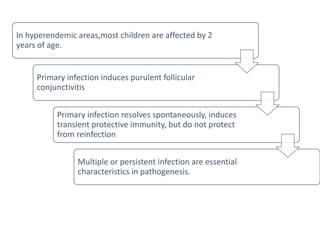
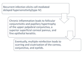
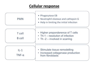
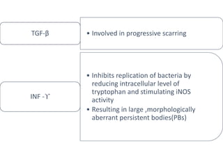
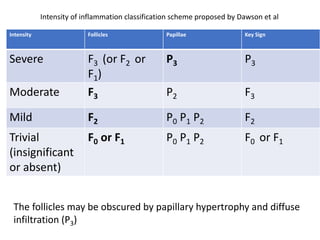
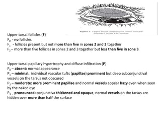
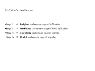
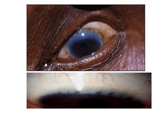
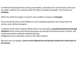
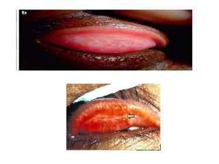
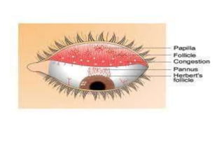
2. After the adoption of Global Elimination of Trachoma (GET)
2020 program by the WHO in 1996 , the prevalence of
trachoma has substansially reduced. In 2016, as per the
WHO report, trachoma is restricted to 42 countries, causing
blindness/visual impairment in ~1.9 million people. 1
India is one of the five countries with nearly half of total
active trachoma patients. 1
A 2006-2007 study estimates proportion of active trachoma
to be 15.2 % in Uttaranchal, 7.6 % in Rajasthan 5.9% in
Uttar Pradesh, 5.5% in Punjab, 4% in Haryana and 0.9% in
Gujarat. 2
1Satpathy G, Behera HS, Ahmed NH. Chlamydial eye infections : Current perspectives. Indian J
Ophthalmol 2017; 65:97-102.
2Rapid Assessment of Trachoma in India. A Report 2006-2007. National Program for Control of
Blindness in India. National Program for Control of Blindness in India, Directorate General of
Health Services, Ministry of Health & Family Welfare, Government of India, New Delhi.
3. A specific communicable keratoconjunctivitis
caused by the Chlamydia trachomatis,
primarily affecting the superficial epithelium,
characterized by formation of follicles, papillary hyperplasia
and pannus, the natural evolution of which is by cicatrization
involving potentially considerable visual disability.
It is mostly a disease of children.
It means rough (Greek)
Trachoma is the most common preventable cause of blindness .
4. Chlamydia trachomatis
Obligate intracellular gram negative organism
Serotypes- A, B, Ba & C (commonest is C)
D – K - Adult Inclusion conjunctivitis or Swimming Pool
Conjunctivitis
L1, L2 & L3 - Lymphogranuloma venereum
5.
6. Transmission
The main source of C. trachomatis infection -- elementary bodies
Routes of infection:
•Direct contact -> eye-to-eye transmission with fingers, such as by touching
the eyes or ocular and nasal secretions of infected individuals, and then
touching one's own eyes
•Indirect contact -> transmission via shared towels, handkerchiefs, bedclothes,
etc.
•Eye-seeking flies
•Coughing or sneezing
7. Matthew J. Burton. Trachoma. In: G.K. Krieglstein, R.N. Weinreb, editors. Cornea and
external eye disease: Corneal Allotransplantation, Allergic disease and Trachoma
8. PATHOGENESIS
Risk factors
- poverty
-over crowding
-poor hygiene
Causes re-infection cycles in the communities
- HLA-A28, haplotype is significantly associated with advanced trachoma.
Pathophysiology
Depends on both parasite and host factors
Parasite factors- presence of cytotoxin for epithelial cells in the
elementary bodies,
provides a mechanism for immune invasion.
Host factors - infection with C.trachomatis leads to upregulaton
of genes like IL-11, LIF, chemokine gene MIP2a,
transcription factor genes, apoptosis related genes
and adhesion molecule genes as ICAM-1.
9. In hyperendemic areas,most children are affected by 2
years of age.
Primary infection induces purulent follicular
conjunctivitis
Primary infection resolves spontaneously, induces
transient protective immunity, but do not protect
from reinfection
Multiple or persistent infection are essential
characteristics in pathogenesis.
10. Recurrent infection elicits cell mediated
delayed hypersensitivity(type IV)
Chronic inflammation leads to follicular
conjunctivitis and papillary hypertrophy
of the upper palpebral conjunctiva, a
superior superficial corneal pannus, and
fine epithelial keratitis.
Eventually, multiple reinfection leads to
scarring and cicatrization of the cornea,
conjunctiva, and eyelids.
11. Both humoral and cell mediated immune responses are involved.
Humoral response
Target antigens
Major outer membrane protein (MOMP) and
Heat shock proteins (HSP60)
Polymorphic outer membrane proteins (POMPs)
IgM and IgG antibodies appear in serum
IgG and IgA in mucosal secretions
Antichlymadial antibodies
-Neutralize chlamydiae
-Block attachment
-Inhibit internalisaton of the organism
IMMMUNOLOGY
12. Cellular response
• Phagocytose EB
• Neutrophil elastase and cathepsin G
• Help in limiting the initial infection
PMN
• Higher preponderence of T cells
• Th-1 – resolution of infection
• Th -2 – involved in scarring
T cell
B cell
• Stimulate tissue remodelling
• Increased collagenase production
from fibroblasts
IL-1
TNF-α
13. • Involved in progressive scarring
TGF-β
• Inhibits replication of bacteria by
reducing intracellular level of
tryptophan and stimulating iNOS
activity
• Resulting in large ,morphologically
aberrant persistent bodies(PBs)
INF -ϒ
14. PATHOLOGY
Primary epithelial lesion of conjunctiva and cornea
Chronic inflammation characterized by papillary hypertrophy of epithelium
and lymphoid infiltration of subepithelial tissue.
Follicle
Mass of mononuclear cells surrounded by phagocytes, giant phagocytes
(Leber’s cells), polymorphs, mast cells and eosinophils.
May be large (upto 5 mm)
Central necrosis , mature(Sago grain), cicatrization
Many follicles may coalesce -Folliculoma of Pascheff
Papillae
Epithelium undergoes hypertrophy and is thrown in folds to form papillae.
Between adjacent papillae pseudoglands may form
15.
16. Pannus
Subepithelial infiltration and vascularization of peripheral cornea
contiguous with the limbus, first between epithelium and the Bowman’s
membrane followed by destruction of the latter.
In progressive pannus, infiltration of cornea is ahead of vascularisation
In regressive pannus, vessels extend a short distance beyond the area of infiltration.
18. Other changes
The desquamated conjunctival epithelium exhibits glycogen-rich
intracytoplasmic inclusion bodies and large macrophages containing
nuclear fragments (Leber cells)
Alteration of extracellular matrix formation of new collagen type I,II , III,IV
Increase in basement membrane collagen type IV and V and decrease in
types I,III (in scarred trachoma)(role of matrix metalloproteinase B)
Cellular infiltration of tarsus , thickening ,degeneration , softening
Lacrimal gland infiltration
Infiltration of lacrimal sac and dacryolith formation
Decrease Tear lysozyme
Increase C3 & Factor B in tears and corresponding decrease in serum.
Primary and secondary orbicularis oculi changes.
20. WHO Classification (1987)
Meant to be used by “less experienced observers” in “population based surveys or for the
simple assessment of the disease at the community level
TF : Trachomatous Inflammation (Follicular)
5 or more folicles (> 0.5 mm diameter)
on superior tarsal plate
TI : Trachomatous Inflammation (Intense)
diffuse involvement of the tarsal conjunctiva
Pronounced inflammatory thickening,
obscuring 50% or more of the normal deep
tarsal vessels; papillae are present
21. TS : Trachomatous Scarring
Obvious trachomatous scarring of upper tarsal
conjunctiva, easily visible fibrous
white tarsal bands
TT : Trachomatous Trichiasis
At least 1 trichiatic cilia rubbing on
the globe or evidence of its recent removal
CO : Corneal Opacity
Corneal opacity sufficient to blur details of
atleast part of the pupillary margin
22. Zone 1 : includes the entire upper tarsal border and adjacent lateral tarsal surface
Zone 2 : occupies the area between zones 1 and 3 and extends to the lateral quarters of
the lid margin
Zone 3 : includes the tarsal conjunctiva adjacent to the central half of the lid margin and,
at its center, covers just less than half the vertical extent of the tarsal surface
Modified WHO system or FPC system (1981)
23. Intensity Follicles Papillae Key Sign
Severe F3 (or F2 or
F1)
P3 P3
Moderate F3 P2 F3
Mild F2 P0 P1 P2 F2
Trivial
(insignificant
or absent)
F0 or F1 P0 P1 P2 F0 or F1
Intensity of inflammation classification scheme proposed by Dawson et al
The follicles may be obscured by papillary hypertrophy and diffuse
infiltration (P3)
24. Upper tarsal follicles (F)
F0 - no follicles
F1 - follicles present but not more than five in zones 2 and 3 together
F2 – more than five follicles in zones 2 and 3 together but less than five in zone 3
Upper tarsal papillary hypertrophy and diffuse infiltration (P)
P0 – absent: normal appearance
P1 – minimal: individual vascular tufts (papillae) prominent but deep subconjunctival
vessels on the tarsus not obscured
P2 – moderate: more prominent papillae and normal vessels appear hazy even when seen
by the naked eye
P3 - pronounced: conjunctiva thickened and opaque, normal vessels on the tarsus are
hidden over more than half the surface
25. Conjunctival scarring (C)
C0 – no scarring on the conjunctiva
C1 – mild, fine, scattered scars on the upper tarsal conjunctiva or scars on the other parts
of the conjunctiva
C2 – moderate, more severe scarring but without shortening or distortion of the upper
tarsus
C3 – severe scarring with distortion of upper tarsus
Trichiasis/ Entropion (T/E)
T/ E0 - no trichiasis or entropion
T/E1 - lashes deviated towards the eye but not touching the globe
T/E2 – lashes touching the globe but not rubbing the cornea
T/E3 – lashes constantly rubbing the cornea
Corneal Scarring
CC0 - absent
CC1 – minimal scarring or opacity not involving the visual axis and with clear central
cornea
CC2 – moderate scarring or opacity involving the visual axis, with the pupillary margin
visible through the opacity
CC3 – severe central scarring or opacity, with the pupillary margin not visible through the
opacity
26. McCallan's classification
•Stage I Incipient trachoma or stage of infiltration
•Stage II Established trachoma or stage of florid infiltration
•Stage III Cicatrizing trachoma or stage of scarring
•Stage IV Healed trachoma or stage of sequelae
28. Many infections are asymptomatic. In other cases, following an incubation period of 5 to
10 days, conjunctival infection produces irritatation, redness and scanty mucopurulent
discharge. Trachoma usually affects both the eyes.
Involvement of the cornea in the acute inflammatory process can cause pain and
photophobia.
The first sign of infection is a nonspecific vasodilation of conjunctival blood vessels.
Specific changes may be noted after infection of several weeks duration, with the
development of follicles subjacent to the conjunctivae of the fornices, the tarsal plates,
and the limbus.
Follicles are lymphoid germinal centers and are found immediately below the epithelial
cell layer. They are grey or creamy masses (sagograin-like) 0.2 to 3.0 mm or more in
diameter.
Because the superficial layer of the conjunctival stroma lacks lymphoid tissue until about
3 months after birth, newborns are unable to mount a follicular response to ocular
chlamydial infection.
29. Papillae may also be noted at this stage: in mild cases, a few
isolated, small red dots can be seen with the naked eye.
When inflammation is severe, an intense papillary reaction
on the tarsal conjunctiva is associated with a diffuse
thickening of the conjunctiva, obscuration of the deep tarsal
vessels, and, sometimes, eyelid edema.
Active trachoma in a child, characterised by mixed papillary (TI) and follicular
response (TF)
30. If the cornea is involved in the inflammatory process, a superficial
punctate keratitis may be noted upon instillation of fluorescein into
the conjunctival sac.
Superficial infiltrates or pannus (subepithelial infiltration of
fibrovascular tissue into the peripheral cornea, once thought to be
found to at least some degree in every case of
trachoma), also indicate corneal inflammation.
Follicles, papillae, and these corneal signs are features of active
disease.
31. Cicatricial trachoma
Resolution of follicles may be accompanied by scarring of the
subepithelial conjunctiva. Scar deposition is most prominent in the
upper tarsal plate, although the conjunctival fornices, the bulbar
conjunctiva, and the upper part of the cornea may also be involved.
In areas where trachoma is endemic, upper tarsal plate scars
derived from repeated episodes of infection can eventually
accumulate to such an extent that they becomes visible
macroscopically after eversion of the upper lid, appearing as white
bands against the erythematous background of the conjunctiva.
At the superior limbus, replacement of follicles by scar results in the
formation of translucent depressions called Herbert’s pits.
32.
33. If sufficient tarsoconjunctival scarring accumulates, contraction of it over the years will cause
the upper eyelid to turn inward so that the lashes rub against the globe. This is known as
trichiasis.
When the whole lid margin is turned in, the condition is known as entropion.
Scars around the bases of hair follicles can pull individual eyelashes into contact with the
cornea, even without entropion.
In addition to the direct abrasive effect of the in-turned lashes, secondary bacterial and fungal
infections of the cornea and corneal drying due to scarring of forniceal-mucous, lacrimal, and
meibomian glands accelerate epithelial damage.
Collagenous scar is laid down as part of the repair process.
Because scars are opaque, vision can be affected by scarring that involves the central part of
the cornea.