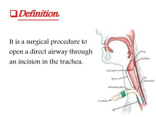
Tracheostomy
Trachea anatomy:
Cylindrical structure but is flattened posteriorly
Measures about 11 cm in length
Starts from the inferior part of the larynx(cricoid
cartilage) in the neck, opposite the C6 vertebra,
to the inter-vertebral disc between T4-T5
vertebrae in the thorax
In cross-section, it is D-shaped, with incomplete
cartilaginous rings anteriorly and laterally, and a
straight membranous wall posteriorly
(chondromembranous)
Definition:
It is a surgical procedure to
open a direct airway through
an incision in the trachea.
Procedure:
The operation should be carried out under
general anesthesia and endotracheal
intubation.
The neck should be extended, the head should
be straight (not turned to one side).
Transverse incision is preferable to vertical
incision.
incision
Transverse
Elective
procedures
Vertical
Emergency
Pediatrics
 The incision should be centered midway
between the cricoid cartilage and sternal notch .
The strap muscles are identified and retracted
laterally and the thyroid isthmus is divided.
 The cricoid must be identified by palpation and
the tracheal rings counted.
An opening is made into the trachea and stitch is
made in place.
Indications:
 Obstruction of the upper airway like foreign body,
trauma, infection, laryngeal tumors and facial
fractures.
 Impaired respiratory function (head trauma lead to
unconsciousness, bulbar poliomyelitis).
 To assist weaning from ventilation support in patient
in the ICU.
 To help clear secretions in the upper airway.
 Prophylaxis (as in preparation for extensive head and
neck procedures and the convalescent period)
Complications:
Immediate complications:
Hemorrhage.
Apnea.
Trauma to recurrent laryngeal nerve and
great vessels.
Damage to esophagus.
Pneumothorax.
Subcutaneous emphysema.
Early complications:
The critical period is the first 48 hrs.
Tube obstruction or displacement.
Bleeding from tracheostomy site.
Infection (Tracheitis, Cellulites).
Aspiration.
Plugging with mucus .
Subcutaneous emphysema .
Atelectasis.
Late complicatios:
Tracheal stenosis.
Fistula formation (tracheo-cutaneous or
trachea-esophagus or trachea-innominate).
Airway obstruction with aspiration.
Bleeding.
Scarring.
Tracheomalacia.
Decannulation:
When ventilation or suctioning no longer
needed
Patient can control their own airway
Not be at risk for aspiration.
This can be expected when patient
has :
Good cough.
Good ABGs (relative, for the patient).
Clear lungs.
No pathogens in sputum.
Contraindications:
A strong relative contraindication to discrete
surgical access to the airway is the
anticipation that the blockage is a laryngeal
carcinoma.
No absolute contraindications
Tracheostomy speaking valve:

Tracheostomy

  • 1.
  • 2.
    Trachea anatomy: Cylindrical structurebut is flattened posteriorly Measures about 11 cm in length Starts from the inferior part of the larynx(cricoid cartilage) in the neck, opposite the C6 vertebra, to the inter-vertebral disc between T4-T5 vertebrae in the thorax In cross-section, it is D-shaped, with incomplete cartilaginous rings anteriorly and laterally, and a straight membranous wall posteriorly (chondromembranous)
  • 5.
    Definition: It is asurgical procedure to open a direct airway through an incision in the trachea.
  • 6.
    Procedure: The operation shouldbe carried out under general anesthesia and endotracheal intubation. The neck should be extended, the head should be straight (not turned to one side). Transverse incision is preferable to vertical incision.
  • 7.
  • 8.
     The incisionshould be centered midway between the cricoid cartilage and sternal notch . The strap muscles are identified and retracted laterally and the thyroid isthmus is divided.  The cricoid must be identified by palpation and the tracheal rings counted. An opening is made into the trachea and stitch is made in place.
  • 10.
    Indications:  Obstruction ofthe upper airway like foreign body, trauma, infection, laryngeal tumors and facial fractures.  Impaired respiratory function (head trauma lead to unconsciousness, bulbar poliomyelitis).  To assist weaning from ventilation support in patient in the ICU.  To help clear secretions in the upper airway.  Prophylaxis (as in preparation for extensive head and neck procedures and the convalescent period)
  • 11.
    Complications: Immediate complications: Hemorrhage. Apnea. Trauma torecurrent laryngeal nerve and great vessels. Damage to esophagus. Pneumothorax. Subcutaneous emphysema.
  • 12.
    Early complications: The criticalperiod is the first 48 hrs. Tube obstruction or displacement. Bleeding from tracheostomy site. Infection (Tracheitis, Cellulites). Aspiration. Plugging with mucus . Subcutaneous emphysema . Atelectasis.
  • 13.
    Late complicatios: Tracheal stenosis. Fistulaformation (tracheo-cutaneous or trachea-esophagus or trachea-innominate). Airway obstruction with aspiration. Bleeding. Scarring. Tracheomalacia.
  • 14.
    Decannulation: When ventilation orsuctioning no longer needed Patient can control their own airway Not be at risk for aspiration.
  • 15.
    This can beexpected when patient has : Good cough. Good ABGs (relative, for the patient). Clear lungs. No pathogens in sputum.
  • 16.
    Contraindications: A strong relativecontraindication to discrete surgical access to the airway is the anticipation that the blockage is a laryngeal carcinoma. No absolute contraindications
  • 17.