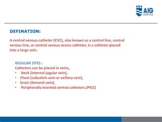
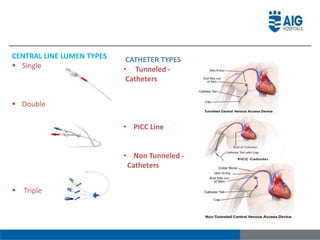
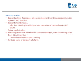
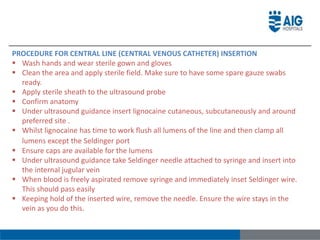
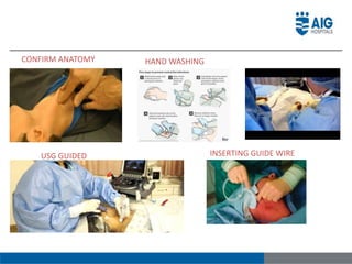
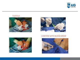
MOHMED IRFAN
CENTRAL VENOUS CATHETER (CVC)
DEFINATION:
A central venous catheter (CVC), also known as a central line, central
venous line, or central venous access catheter, is a catheter placed
into a large vein.
REGULAR SITES :
Catheters can be placed in veins,
• Neck (internal jugular vein),
• Chest (subcalvin vein or axillary vein),
• Groin (femoral vein),
• Peripherally inserted central catheters.(PICC)
MEDICAL USES
• Administer medication
• Fluids that are unable to be taken by mouth
• Would harm a smaller peripheral lines
• Obtain blood tests
• Measure central venous pressure
INDICATIONS:
• Long-term intravenous antibiotics, parenteral nutrition in chronically
ill persons
• Long-term pain medications &Chemotherapy
• Drugs that are prone to cause phlebitis in peripheral veins such as:
 Calcium chloride Chemotherapy
 Hypertonic saline Potassium chloride (KCl)
• Frequent blood draws
• Monitoring of the central venous pressure (CVP).
CENTRAL LINE LUMEN TYPES
 Single
 Double
 Triple
CATHETER TYPES
• Tunneled -
Catheters
• PICC Line
• Non Tunneled -
Catheters
• TUNNELED – CATHETERS:
 PREFERABLE SITES:
• Neck (internal jugular
• Groin (femoral),
• Liver (transhepatic),
• Back (translumbar). The catheter
is tunneled under the skin.
 INDICATIONS:
 Chemotherapy
 Nutrition and
 Fluids
 Blood samples
 NON TUNNELED – CATHETERS:
 PREFERABLE SITES:
 First choice: right internal jugular vein
 Second choice: femoral vein
 Third choice: left internal jugular vein
 Last choice: subclavian vein with preference
for the dominant side.
 INDICATIONS:
 Increasing age of patients initiating
hemodialysis
 Increasing number of comorbid conditions
including significant vascular disease.
 Urgent renal replacement therapy (RRT).
 CONTRA INDICATIONS FOR TUNNELED AND NON- TUNNELED CATHETER.
 Local cellulitis
 Low platelet counts
 Local infection
 Avoid in raised intracranial pressure- aim for a femoral approach if required
 Patient non-compliance
 Systemic sepsis is an absolute contraindication for central venous access via
tunneled catheter because it can lead to line infection.
 COMPLICATIONS :
 Bleeding
 Infection
 Puncture of adjacent structures (such as other veins or arteries)
 Air embolism (air in the veins)
 Collapse of the lung (pneumothorax)
 Bleeding into the chest (hemothorax)
 Catheter breakage (when it is being removed)
 PATIENT EDUCATION :
• Explain the procedure to the patient and family members.
• Explain the need of central line and benefits, risk and complications.
 Equipment required for central line (central
venous catheter) insertion
• Sterile trolley( cvp tray )
• Sterile field, gloves, gown and mask
• Central line kit
• Saline flush
• Chlorhexidine
• Lignocaine (4ml (2 vials) of 2% is reasonable)
• Suture
• Scalpel
• Central line fix
SELDINGER TECHNIQUE:
The Seldinger technique, also known as Seldinger wire technique, is
a medical procedure to obtain safe access to blood vessels and other
hollow organs
PRE-PROCEDURE
 Consent patient if conscious otherwise document why the procedure is in the
patient’s best interests.
 Consent should include.
Infection, bleeding (arterial puncture, haematoma, haemothorax), pain,
failure,
 Set up sterile trolley.
 Position patient with head down if they can tolerate it, with head facing away
from side of insertion
This ensures maximum venous filling
 Having a nurse or assistant is helpful.
PROCEDURE FOR CENTRAL LINE (CENTRAL VENOUS CATHETER) INSERTION
 Wash hands and wear sterile gown and gloves
 Clean the area and apply sterile field. Make sure to have some spare gauze swabs
ready.
 Apply sterile sheath to the ultrasound probe
 Confirm anatomy
 Under ultrasound guidance insert lignocaine cutaneous, subcutaneously and around
preferred site .
 Whilst lignocaine has time to work flush all lumens of the line and then clamp all
lumens except the Seldinger port
 Ensure caps are available for the lumens
 Under ultrasound guidance take Seldinger needle attached to syringe and insert into
the internal jugular vein
 When blood is freely aspirated remove syringe and immediately inset Seldinger wire.
This should pass easily
 Keeping hold of the inserted wire, remove the needle. Ensure the wire stays in the
vein as you do this.
CONFIRM ANATOMY HAND WASHING
USG GUIDED INSERTING GUIDE WIRE
Continue….
CONFRIM WITH BLOOD DRAW
FLUSH THE LINE
SUTURING SECURING THE LINE
• Use scalpel to make an small incision in the skin (approx 3mm). This should be
done cutting away from the wire so as not to damage it
• Pass the dilator over the wire and gently but firmly dilate a tract through to the
internal jugular.
• At this stage there may be some bleeding so ensure to have some swabs ready
• Remove the dilator and pass the central line over the Seldinger wire. Do not
advance the line until you have hold of the end of the wire
• Once the central line is in place, remove the wire
• Aspirate and flush all lumens and re clamp and apply lumen caps
• Suture the line.
• Dress with a clear dressing so the insertion point can be clearly seen
 DOCUMENTATION
• Patient is educated about the need
• Site assessed and marked
• All lumens clamped
• Inserted by Physician , assisted by
• Tip position confirmation via fluoroscopy OR chest X-ray
• Date and time of insertion , assess the site for extra
bleeding.
• Anatomical location .
• Catheter depth according to catheter reference.
 CVP MONITORING
Central venous pressure (CVP) is the blood pressure in the venae cavae, near the right
atrium of the heart. CVP reflects the amount of blood returning to the Heart and the ability
of the heart to pump the blood back into the arterial system.
• The normal range for CVP is 0 to 5 mm H2O
CARE OF CVP LINE :
 CARE AND MAINTENANCE
 Assessment: - Insertion Site - Catheter Tract - Adjacent Skin
• Site Care: - Skin Disinfectant - Clean, dry, and occlusive dressings
• Dressing Gauze or Transparent Semi-Permeable
 PROCEDURE FOR SITE CARE AND DRESSING CHANGE
• DURING PROCEDURE
1. Remove dressing from VAD insertion site. 2. Inspect Site and catheter 3. Disinfect the
catheter-skin junction using antiseptic solution 4. Dress access site
 AFTER PROCEDURE
1. Discard used supplies 2. Remove gloves 3. Wash hands 4. Label new dressing
5. Document
Central line

Central line

  • 1.
  • 2.
    DEFINATION: A central venouscatheter (CVC), also known as a central line, central venous line, or central venous access catheter, is a catheter placed into a large vein. REGULAR SITES : Catheters can be placed in veins, • Neck (internal jugular vein), • Chest (subcalvin vein or axillary vein), • Groin (femoral vein), • Peripherally inserted central catheters.(PICC)
  • 3.
    MEDICAL USES • Administermedication • Fluids that are unable to be taken by mouth • Would harm a smaller peripheral lines • Obtain blood tests • Measure central venous pressure INDICATIONS: • Long-term intravenous antibiotics, parenteral nutrition in chronically ill persons • Long-term pain medications &Chemotherapy • Drugs that are prone to cause phlebitis in peripheral veins such as:  Calcium chloride Chemotherapy  Hypertonic saline Potassium chloride (KCl) • Frequent blood draws • Monitoring of the central venous pressure (CVP).
  • 4.
    CENTRAL LINE LUMENTYPES  Single  Double  Triple CATHETER TYPES • Tunneled - Catheters • PICC Line • Non Tunneled - Catheters
  • 5.
    • TUNNELED –CATHETERS:  PREFERABLE SITES: • Neck (internal jugular • Groin (femoral), • Liver (transhepatic), • Back (translumbar). The catheter is tunneled under the skin.  INDICATIONS:  Chemotherapy  Nutrition and  Fluids  Blood samples  NON TUNNELED – CATHETERS:  PREFERABLE SITES:  First choice: right internal jugular vein  Second choice: femoral vein  Third choice: left internal jugular vein  Last choice: subclavian vein with preference for the dominant side.  INDICATIONS:  Increasing age of patients initiating hemodialysis  Increasing number of comorbid conditions including significant vascular disease.  Urgent renal replacement therapy (RRT).
  • 6.
     CONTRA INDICATIONSFOR TUNNELED AND NON- TUNNELED CATHETER.  Local cellulitis  Low platelet counts  Local infection  Avoid in raised intracranial pressure- aim for a femoral approach if required  Patient non-compliance  Systemic sepsis is an absolute contraindication for central venous access via tunneled catheter because it can lead to line infection.  COMPLICATIONS :  Bleeding  Infection  Puncture of adjacent structures (such as other veins or arteries)  Air embolism (air in the veins)  Collapse of the lung (pneumothorax)  Bleeding into the chest (hemothorax)  Catheter breakage (when it is being removed)
  • 7.
     PATIENT EDUCATION: • Explain the procedure to the patient and family members. • Explain the need of central line and benefits, risk and complications.  Equipment required for central line (central venous catheter) insertion • Sterile trolley( cvp tray ) • Sterile field, gloves, gown and mask • Central line kit • Saline flush • Chlorhexidine • Lignocaine (4ml (2 vials) of 2% is reasonable) • Suture • Scalpel • Central line fix
  • 8.
    SELDINGER TECHNIQUE: The Seldingertechnique, also known as Seldinger wire technique, is a medical procedure to obtain safe access to blood vessels and other hollow organs
  • 9.
    PRE-PROCEDURE  Consent patientif conscious otherwise document why the procedure is in the patient’s best interests.  Consent should include. Infection, bleeding (arterial puncture, haematoma, haemothorax), pain, failure,  Set up sterile trolley.  Position patient with head down if they can tolerate it, with head facing away from side of insertion This ensures maximum venous filling  Having a nurse or assistant is helpful.
  • 10.
    PROCEDURE FOR CENTRALLINE (CENTRAL VENOUS CATHETER) INSERTION  Wash hands and wear sterile gown and gloves  Clean the area and apply sterile field. Make sure to have some spare gauze swabs ready.  Apply sterile sheath to the ultrasound probe  Confirm anatomy  Under ultrasound guidance insert lignocaine cutaneous, subcutaneously and around preferred site .  Whilst lignocaine has time to work flush all lumens of the line and then clamp all lumens except the Seldinger port  Ensure caps are available for the lumens  Under ultrasound guidance take Seldinger needle attached to syringe and insert into the internal jugular vein  When blood is freely aspirated remove syringe and immediately inset Seldinger wire. This should pass easily  Keeping hold of the inserted wire, remove the needle. Ensure the wire stays in the vein as you do this.
  • 11.
    CONFIRM ANATOMY HANDWASHING USG GUIDED INSERTING GUIDE WIRE
  • 12.
  • 13.
    FLUSH THE LINE SUTURINGSECURING THE LINE
  • 14.
    • Use scalpelto make an small incision in the skin (approx 3mm). This should be done cutting away from the wire so as not to damage it • Pass the dilator over the wire and gently but firmly dilate a tract through to the internal jugular. • At this stage there may be some bleeding so ensure to have some swabs ready • Remove the dilator and pass the central line over the Seldinger wire. Do not advance the line until you have hold of the end of the wire • Once the central line is in place, remove the wire • Aspirate and flush all lumens and re clamp and apply lumen caps • Suture the line. • Dress with a clear dressing so the insertion point can be clearly seen
  • 15.
     DOCUMENTATION • Patientis educated about the need • Site assessed and marked • All lumens clamped • Inserted by Physician , assisted by • Tip position confirmation via fluoroscopy OR chest X-ray • Date and time of insertion , assess the site for extra bleeding. • Anatomical location . • Catheter depth according to catheter reference.  CVP MONITORING Central venous pressure (CVP) is the blood pressure in the venae cavae, near the right atrium of the heart. CVP reflects the amount of blood returning to the Heart and the ability of the heart to pump the blood back into the arterial system. • The normal range for CVP is 0 to 5 mm H2O
  • 16.
    CARE OF CVPLINE :  CARE AND MAINTENANCE  Assessment: - Insertion Site - Catheter Tract - Adjacent Skin • Site Care: - Skin Disinfectant - Clean, dry, and occlusive dressings • Dressing Gauze or Transparent Semi-Permeable  PROCEDURE FOR SITE CARE AND DRESSING CHANGE • DURING PROCEDURE 1. Remove dressing from VAD insertion site. 2. Inspect Site and catheter 3. Disinfect the catheter-skin junction using antiseptic solution 4. Dress access site  AFTER PROCEDURE 1. Discard used supplies 2. Remove gloves 3. Wash hands 4. Label new dressing 5. Document