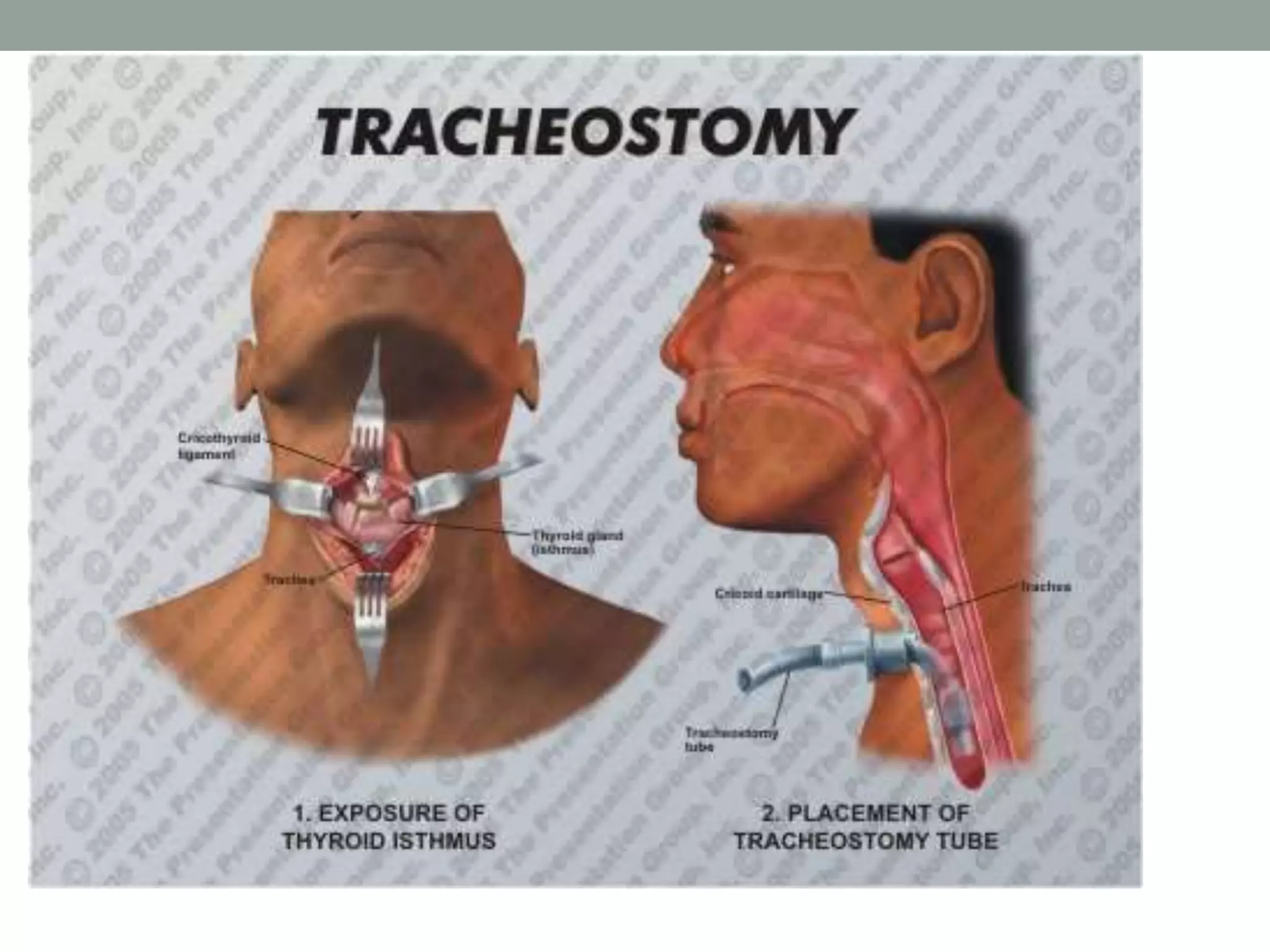
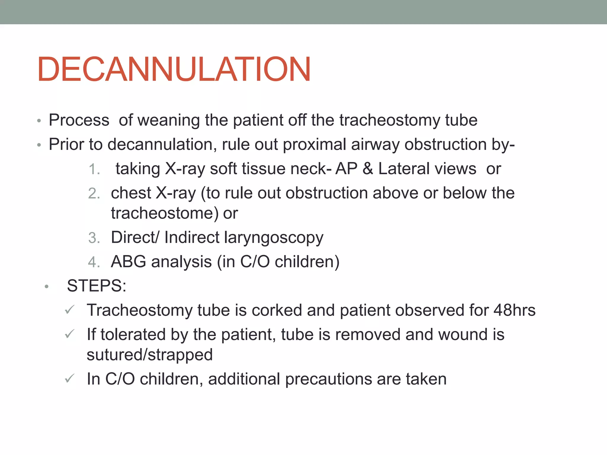
TRACHEOSTOMY
Tracheostomy is making an opening in the anterior
wall of trachea and converting it into a stoma on the
skin surface.
Functions of tracheostomy-
• Alternate pathway for breathing.
• Improves alveolar ventilation.
• Protects the airways.
• Permits removal of tracheobronchial secretions.
• Intermittent positive pressure respiration.
• To administer anaesthesia.
INDICATIONS-
1. Respiratory obstruction-
• Infections -Acute laryngo-tracheo-bronchitis, acute epiglottitis, diptheria, ludwing’s
angina, peritonsillar abscess.
• Trauma – external injury of larynx and trachea, fracture of mandible or maxillofacial
injuries.
• Neoplasms.
• Foreign body larynx.
• Oedema larynx due to steam, irritant fumes or gases , allergy
.
• Bilateral abductor paralysis.
• Congenital anomalies – Laryngeal web,cysts,tracheo-oesophageal fistula,bilateral
choanal atresia
2. Retained secretions
• Inability to cough –
• 1. coma of any cause like head injuries, CVA narcotic abuse.
• 2. Paralysis of respiratory muscles in spinal injuries,polio, GB
syndrome.
• 3.spasm of respiratory muscles, tetanus, eclampsia, strychnine
poisoning.
• Painful cough – chest injuries , multiple rib fractures , pneumonia.
• Aspiration of pharyngeal secretions – bulbar polio ,polyneuritis, bilateral
laryngeal paralysis.
3. Respiratory insufficiency
Chronic lung conditions , emphysema , chronic bronchitis,
bronchiectasis, atelectasis
TYPES
1. Emergency tracheostomy
2. Elective or tranquil tracheostomy
3. Permanent tracheostomy
4. Percutaneous dilatational tracheostomy
5. Mini tracheostomy (cricothyroidotomy)
TECHNIQUE
Whenever possible endotracheal intubation should be
done before tracheostomy , especially in infants and
children.
POSITION - Patient lies in supine with a pillow under the
shoulder so that neck is extended. This brings the trachea
forward.
ANAESTHESIA – No anaesthesia is required in
unconcious patients or when in an emergency procedure.
In concious patients, 1-2%lignocaine with epinephrine is
used. Sometimes, general anaesthesia with intubation is
used.
STEPS
1. A vertical incision is made in the middle of the neck , extending
from cricoid cartilage to just above the sternal notch.( It gives
rapid access with minimum of bleeding and tissue dissection.)
2.Tissues are dissected in the midline. Dilated veins are either
displaced or ligated.
3. Strap muscles are separated in the midline and retracted laterally.
4. Thyroid isthmus is displaced upwards or divided between the
clamps , and suture ligated.
5. A few drops of 4% lignocaine are injected into trachea to suppress
the cough when trachea is incised.
6. Trachea is fixed with a hook and opened with a vertical incision in
the region of 2nd and 3rd rings . This is then converted into a circular
opening.
7. Tracheostomy tube of appropriate size is inserted and
secured by tapes.
8. Skin incision should not be sutured or packed tightly as
it may lead to development of subcutaneous
emphysema.
9. Gauze dressing is placed between the skin and flange
of the tube around the stoma.
POST-OP CARE
• Periodic check of tube patency and position.
• Cuff Management- periodic deflation to prevent pressure
necrosis/stenosis of trachea.
• Care of Tube- change on 3rd day (after the tract has
formed) and subsequently, once in a week (to prevent
granulation tissue formation)
• Wound Dressing (to prevent maceration and skin
erosion)
• Periodic tracheo-bronchial toilet using suction tube with
Y-connector
• Supportive care such as-
 prevention of crusting/tracheitis by humidification or instillation of
RL/NS/mucolytic agents
Analgesics and antibiotics
COMPLICATIONS
1. Immediate (at the time of operation)
• Aspiration
• Apnea (due to sudden CO2 wash out)
• Bleeding
• Collapse of lungs/Pneumothorax
• Damage to surrounding structures
• Embolism- Air
2. Intermediate (within first few hours/days)
• Hemorrhage
• Displacement or blocking of tube
• Subcutaneous emphysema
• Tracheitis and tracheobronchitis with crusting in trachea
• Lung abscess
• Local wound infection and granulations
• Dysphagia
3. Late (after prolonged use of tube for weeks/months)
• Hemorrhage (due to erosion of major vessels)
• Laryngeal stenosis (due to perichondritis of cricoid cartilage).
• Tracheal stenosis (due to tracheal ulceration and infection).
• Tracheo-oesophageal fistula.
• Problems of decannulations.
• Persistent tracheo-cutaneous fistula.
• Problems of tracheostomy scar.
• Corrosion of tracheostomy tube and aspiration of its fragments into
the tracheo-bronchial tree (FB).
DECANNULATION
• Process of weaning the patient off the tracheostomy tube
• Prior to decannulation, rule out proximal airway obstruction by-
1. taking X-ray soft tissue neck- AP & Lateral views or
2. chest X-ray (to rule out obstruction above or below the
tracheostome) or
3. Direct/ Indirect laryngoscopy
4. ABG analysis (in C/O children)
• STEPS:
 Tracheostomy tube is corked and patient observed for 48hrs
 If tolerated by the patient, tube is removed and wound is
sutured/strapped
 In C/O children, additional precautions are taken
THANK YOU

Tracheostomy

  • 1.
  • 2.
    Tracheostomy is makingan opening in the anterior wall of trachea and converting it into a stoma on the skin surface. Functions of tracheostomy- • Alternate pathway for breathing. • Improves alveolar ventilation. • Protects the airways. • Permits removal of tracheobronchial secretions. • Intermittent positive pressure respiration. • To administer anaesthesia.
  • 3.
    INDICATIONS- 1. Respiratory obstruction- •Infections -Acute laryngo-tracheo-bronchitis, acute epiglottitis, diptheria, ludwing’s angina, peritonsillar abscess. • Trauma – external injury of larynx and trachea, fracture of mandible or maxillofacial injuries. • Neoplasms. • Foreign body larynx. • Oedema larynx due to steam, irritant fumes or gases , allergy . • Bilateral abductor paralysis. • Congenital anomalies – Laryngeal web,cysts,tracheo-oesophageal fistula,bilateral choanal atresia
  • 4.
    2. Retained secretions •Inability to cough – • 1. coma of any cause like head injuries, CVA narcotic abuse. • 2. Paralysis of respiratory muscles in spinal injuries,polio, GB syndrome. • 3.spasm of respiratory muscles, tetanus, eclampsia, strychnine poisoning. • Painful cough – chest injuries , multiple rib fractures , pneumonia. • Aspiration of pharyngeal secretions – bulbar polio ,polyneuritis, bilateral laryngeal paralysis. 3. Respiratory insufficiency Chronic lung conditions , emphysema , chronic bronchitis, bronchiectasis, atelectasis
  • 5.
    TYPES 1. Emergency tracheostomy 2.Elective or tranquil tracheostomy 3. Permanent tracheostomy 4. Percutaneous dilatational tracheostomy 5. Mini tracheostomy (cricothyroidotomy)
  • 6.
    TECHNIQUE Whenever possible endotrachealintubation should be done before tracheostomy , especially in infants and children. POSITION - Patient lies in supine with a pillow under the shoulder so that neck is extended. This brings the trachea forward. ANAESTHESIA – No anaesthesia is required in unconcious patients or when in an emergency procedure. In concious patients, 1-2%lignocaine with epinephrine is used. Sometimes, general anaesthesia with intubation is used.
  • 7.
    STEPS 1. A verticalincision is made in the middle of the neck , extending from cricoid cartilage to just above the sternal notch.( It gives rapid access with minimum of bleeding and tissue dissection.) 2.Tissues are dissected in the midline. Dilated veins are either displaced or ligated. 3. Strap muscles are separated in the midline and retracted laterally. 4. Thyroid isthmus is displaced upwards or divided between the clamps , and suture ligated. 5. A few drops of 4% lignocaine are injected into trachea to suppress the cough when trachea is incised. 6. Trachea is fixed with a hook and opened with a vertical incision in the region of 2nd and 3rd rings . This is then converted into a circular opening.
  • 8.
    7. Tracheostomy tubeof appropriate size is inserted and secured by tapes. 8. Skin incision should not be sutured or packed tightly as it may lead to development of subcutaneous emphysema. 9. Gauze dressing is placed between the skin and flange of the tube around the stoma.
  • 10.
    POST-OP CARE • Periodiccheck of tube patency and position. • Cuff Management- periodic deflation to prevent pressure necrosis/stenosis of trachea. • Care of Tube- change on 3rd day (after the tract has formed) and subsequently, once in a week (to prevent granulation tissue formation) • Wound Dressing (to prevent maceration and skin erosion) • Periodic tracheo-bronchial toilet using suction tube with Y-connector • Supportive care such as-  prevention of crusting/tracheitis by humidification or instillation of RL/NS/mucolytic agents Analgesics and antibiotics
  • 11.
    COMPLICATIONS 1. Immediate (atthe time of operation) • Aspiration • Apnea (due to sudden CO2 wash out) • Bleeding • Collapse of lungs/Pneumothorax • Damage to surrounding structures • Embolism- Air 2. Intermediate (within first few hours/days) • Hemorrhage • Displacement or blocking of tube • Subcutaneous emphysema • Tracheitis and tracheobronchitis with crusting in trachea • Lung abscess • Local wound infection and granulations • Dysphagia
  • 12.
    3. Late (afterprolonged use of tube for weeks/months) • Hemorrhage (due to erosion of major vessels) • Laryngeal stenosis (due to perichondritis of cricoid cartilage). • Tracheal stenosis (due to tracheal ulceration and infection). • Tracheo-oesophageal fistula. • Problems of decannulations. • Persistent tracheo-cutaneous fistula. • Problems of tracheostomy scar. • Corrosion of tracheostomy tube and aspiration of its fragments into the tracheo-bronchial tree (FB).
  • 13.
    DECANNULATION • Process ofweaning the patient off the tracheostomy tube • Prior to decannulation, rule out proximal airway obstruction by- 1. taking X-ray soft tissue neck- AP & Lateral views or 2. chest X-ray (to rule out obstruction above or below the tracheostome) or 3. Direct/ Indirect laryngoscopy 4. ABG analysis (in C/O children) • STEPS:  Tracheostomy tube is corked and patient observed for 48hrs  If tolerated by the patient, tube is removed and wound is sutured/strapped  In C/O children, additional precautions are taken
  • 14.

Editor's Notes