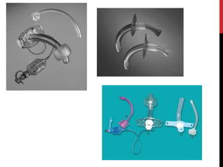
1) Tracheostomy is a surgical opening into the trachea through the neck that allows for an alternative airway. It has several purposes including bypassing upper airway obstruction and making it easier to clear secretions.
2) The procedure involves making incisions through the skin and trachea rings to insert a tracheostomy tube. It can be done via open or percutaneous dilational tracheostomy techniques.
3) Tracheostomy is indicated when normal breathing is compromised due to conditions like upper airway infections/injuries or an inability to cough effectively. It provides benefits like improved ventilation and protection against aspiration.
A Tracheostomy is a medical procedure either temporary or permanent that involves creating an opening in the neck in order to place a tube into a person's windpipe.
detailed information about tracheostomy for the medical students , includes difinition, causes, indications, care provided, management, medical and nursing management of opening , complete care of the patient , patient teaching, family teaching and contained other detailled explanation of tracheostomy
A Brief description of Tracheotomy.. Good enough for Undergraduate MBBS Students. . You can staright away download this and present in your class seminars.. ;)
Bag and Mask Ventilation By Sakun Rasaily @Ram K Dhamalaramdhamala11
Bag and mask Ventilation Presented by Sakun Rasaily,
(Pediatric Nurse, Pediatric ward , B.P. Koirala Institute of Health Science
Dharan, Sunsari (Nepal)
At the end of the lecture, the students will be able to:
Define tracheostomy
State two reasons why tracheostomy tubes are inserted
Discuss types of tracheostomy tubes
Discuss the procedure for cleaning a tracheostomy tube
a. Single
b. Double
Discuss the procedure for suctioning an established tracheostomy
The Indian Dental Academy is the Leader in continuing dental education , training dentists in all aspects of dentistry and
offering a wide range of dental certified courses in different formats.for more details please visit
www.indiandentalacademy.com
TRACHEOSTOMY IN FULL DETAILS......
TRACHEOSTOMY, EMERGENCY TRACHEOSTOMY, SECURE AIRWAY, PERCUTANEOUS TRACHEOSTOMY, HISTORY OF TRACHEOSTOMY, PROCEDURES, GIGGS TECHNIQUE,VARIOUS TYPES OF TRACHEOSTOMY, TRACHEOSTOMA, DECANNULATION, INDICATIONS, CONTRAINDICATIONS, CRICOTHYROIDOTOMY, MINITRACHEOSTOMY, TRACHEOSTOMY TUBES, COMPLICATIONS OF TRACHEOSTOMY.
TRACHEOSTOMY is a surgical procedure to maintain a patent airway to the person who is in airway distress or electively in certain surgical procedures like oncological resections to maintain an adequate oxygenation to the patient by creating a stoma on the trachea
A tracheostomy is an opening (made by an incision) through the neck into the trachea (windpipe). A tracheostomy opens the airway and aids breathing.
A tracheostomy may be done in an emergency, at the patient’s bedside or in an operating room. Anesthesia pain relief medication may be used before the procedure. Depending on the person’s condition, the tracheostomy may be temporary or permanent
A tracheostomy (also called a tracheotomy) is an opening surgically created through the neck into the trachea (windpipe) to allow air to fill the lungs. After creating the tracheostomy opening in the neck, surgeons insert a tube through it to provide an airway and to remove secretions from the lungs.
Operation “Blue Star” is the only event in the history of Independent India where the state went into war with its own people. Even after about 40 years it is not clear if it was culmination of states anger over people of the region, a political game of power or start of dictatorial chapter in the democratic setup.
The people of Punjab felt alienated from main stream due to denial of their just demands during a long democratic struggle since independence. As it happen all over the word, it led to militant struggle with great loss of lives of military, police and civilian personnel. Killing of Indira Gandhi and massacre of innocent Sikhs in Delhi and other India cities was also associated with this movement.
Macroeconomics- Movie Location
This will be used as part of your Personal Professional Portfolio once graded.
Objective:
Prepare a presentation or a paper using research, basic comparative analysis, data organization and application of economic information. You will make an informed assessment of an economic climate outside of the United States to accomplish an entertainment industry objective.
Read| The latest issue of The Challenger is here! We are thrilled to announce that our school paper has qualified for the NATIONAL SCHOOLS PRESS CONFERENCE (NSPC) 2024. Thank you for your unwavering support and trust. Dive into the stories that made us stand out!
2024.06.01 Introducing a competency framework for languag learning materials ...Sandy Millin
http://sandymillin.wordpress.com/iateflwebinar2024
Published classroom materials form the basis of syllabuses, drive teacher professional development, and have a potentially huge influence on learners, teachers and education systems. All teachers also create their own materials, whether a few sentences on a blackboard, a highly-structured fully-realised online course, or anything in between. Despite this, the knowledge and skills needed to create effective language learning materials are rarely part of teacher training, and are mostly learnt by trial and error.
Knowledge and skills frameworks, generally called competency frameworks, for ELT teachers, trainers and managers have existed for a few years now. However, until I created one for my MA dissertation, there wasn’t one drawing together what we need to know and do to be able to effectively produce language learning materials.
This webinar will introduce you to my framework, highlighting the key competencies I identified from my research. It will also show how anybody involved in language teaching (any language, not just English!), teacher training, managing schools or developing language learning materials can benefit from using the framework.
Francesca Gottschalk - How can education support child empowerment.pptxEduSkills OECD
Francesca Gottschalk from the OECD’s Centre for Educational Research and Innovation presents at the Ask an Expert Webinar: How can education support child empowerment?
Embracing GenAI - A Strategic ImperativePeter Windle
Artificial Intelligence (AI) technologies such as Generative AI, Image Generators and Large Language Models have had a dramatic impact on teaching, learning and assessment over the past 18 months. The most immediate threat AI posed was to Academic Integrity with Higher Education Institutes (HEIs) focusing their efforts on combating the use of GenAI in assessment. Guidelines were developed for staff and students, policies put in place too. Innovative educators have forged paths in the use of Generative AI for teaching, learning and assessments leading to pockets of transformation springing up across HEIs, often with little or no top-down guidance, support or direction.
This Gasta posits a strategic approach to integrating AI into HEIs to prepare staff, students and the curriculum for an evolving world and workplace. We will highlight the advantages of working with these technologies beyond the realm of teaching, learning and assessment by considering prompt engineering skills, industry impact, curriculum changes, and the need for staff upskilling. In contrast, not engaging strategically with Generative AI poses risks, including falling behind peers, missed opportunities and failing to ensure our graduates remain employable. The rapid evolution of AI technologies necessitates a proactive and strategic approach if we are to remain relevant.
2. DEFINITION
• Tracheostomy is an opening in the anterior
wall of trachea and converted into a stoma on
the skin surface.
• The terms tracheostomy and tracheotomy
have been used interchangeably.
• The tracheotomy means opening the trachea,
which is a step of tracheostomy operation.
3. ANATOMY OF TRACHEA
• The trachea is a tubular structure, about 15
cm long in adults
• Extending from the cricoid cartilage to the
bronchial bifurcation.
• It has an outer diameter of 2.5 cm.
• It consists of 16 to 20 C-shaped cartilages
joined by fibroelastic tissue and closed
posteriorly by the trachealis muscle.
• The anterolateral portion is made up of
incomplete rings of cartilage, and the
posterior aspect by a flat muscular wall.
4.
5. In the neck, the trachea has the following relations.
Anterior:
1. Isthmus of the thyroid gland covering the second and
third tracheal rings;
2. Inferior thyroid veins below the isthmus;
3. Pretracheal fascia enclosing the thyroid and inferior
thyroid veins
4. Sternohyoid and sternothyroid muscles;
5. Investing layer of the deep cervical fascia and the
suprasternal space;
6. The skin and superficial fascia;
In children, the left brachiocephalic vein extends into the
neck and then lies in front of the trachea.
6. Posterior:
(1). Oesophagus lies behind the cervical trachea,
separating it from the vertebral column and the
prevertebral fascia.
(2). Recurrent laryngeal nerve in the tracheo-
oesophageal groove.
On each side:
(1). The corresponding lobe of the thyroid glands;
(2). The common carotid artery within the carotid
sheath.
7.
8. VESSELS AND NERVES:
• The trachea is supplied by branches
from the inferior thyroid arteries.
• Its veins drain into the left
brachiocephalic vein.
• Lymphatics drain into the pretracheal
and paratracheal nodes.
9. • Parasymphathetic nerves (from the vagus
through the recurrent laryngeal nerve) are
secretomotor to the mucous membrane, and
motor to the trachealis muscle.
• Sympathetic nerves (from the cervical
ganglion) are vasomotor.
10. TYPES OF TRACHEOSTOMY
A) Timings
• Emergent Tracheostomy (slash trach)
• Urgent (awake) Tracheostomy
• Elective Tracheostomy
• Temporary Tracheostomy
• Permanent Tracheostomy
B) Level and Site
1. High, 2. Mid and 3. Low Tracheostomy
C) SURGICAL TECHNIQUES
1. Cricothyroidotomy/Minitracheostomy
2. Open tracheostomy
3. Percutaneous dilatational tracheostomy
4. Translaryngeal tracheostomy
11. FUNCTIONS OF TRACHEOSTOMY
1. Alternative pathway for breathing
2. Improves alveolar ventilation by decreasing
the dead space by 30 -50% and reducing
resistance to airflow.
3. Protects the airway against aspiration of
pharyngeal secretions in case of coma or
bulbar paralysis and blood, etc. in
haemorrhage from pharynx, larynx or maxillo-
facial injuries. Pharynx and larynx can also be
packed to control bleeding.
12. 4. Permits removal of tracheo-bronchial
secretions : when patient is unable to cough /
when cough is painful.
5. Intermittent positive pressure ventilation : if
required beyond 72 hours.
6. Anaesthesia : laryngo-pharyngeal growths or
trismus.
7. Reduces resistance to airflow – better total
compliance with more effective alveolar
ventilation.
8. Enabling pt to swallow without reflex apnoea.
13. INDICATIONS
1) Upper Respiratory Tract Obstruction.
Infection, Trauma, Neoplasm, foreign body,
oedema, Bilateral abductor paralysis, Congenital
anomalies.
2) Retained secretions
Inability to cough, Painful cough, Aspiration of
pharyngeal secretions.
3) Prolonged Ventilation
4) Part of another procedure
14. Emergent Tracheostomy- emergency airway distress is
accompanied with impending death. The complication rate of
emergency tracheostomy is as high as 21%. This emergent
situation is an ideal indication for cricothyrotomy.
Urgent Tracheostomy: This is done in the operation theater
under local anesthesia with minimal sedation. The patient has
respiratory distress, and needs immediate surgical
intervention.
Elective Tracheostomy: routine planned surgery. It is
performed where all operative surgical facilities such as
endotracheal intubation, local and general anesthesia, are
available.
Therapeutic & Prophylactic:
15. • „„Temporary Tracheostomy:
tracheostomy is usually temporary, and is closed
when causative disease is cured.
• „Permanent Tracheostomy:
It is indicated in cases of bilateral abductor
paralysis and laryngeal stenosis. In cases of
laryngectomy and laryngopharyngectomy,
A tracheostome is created where lower tracheal
stump is stitched to the surface skin.
16. High Tracheostomy:
It is done at the level of first tracheal ring.
The high tracheostomy is generally avoided because of the
postoperative risk of perichondritis of the cricoid cartilage and
subglottic stenosis.
In cases of carcinoma larynx with stridor when total
laryngectomy would be done, high tracheostomy is indicated.
Mid Tracheostomy:
It is done through the second and third tracheal rings, and
needs either division of the thyroid isthmus or its retraction
upwards.
„„Low Tracheostomy:
It is done below the level of isthmus where trachea becomes
deep, and lies close to large vessels.
17. SURGICAL TECHNIQUES
Cricothyroidotomy/Minitracheosto
my
In an emergency, rapid entry to the
airway can be achieved through the
cricothyroid membrane.
Once cricothyroid has been breached
the airway can be maintained either
with minitracheostomy tube or a
large bore cannula.
18. PROCEDURE
Patient lies in supine and the anatomical is
ascertained by palpation. The skin is infiltrated
with local anaesthesia.
Once the anatomy of the larynx is identified, the
procedure can be performed with 2
incisions:
● Incision through the skin
● Incision through the cricothyroid membrane
19. • The correct location for the incision is
easiest to find by identifying the thyroid
notch on the thyroid cartilage
• By sliding the examining finger in an inferior
direction, the groove between the thyroid
and cricoid cartilages can be located
• A 3-cm vertical incision is made through the
skin, and the thyrohyoid membrane is
located
• A small midline incision is made, and a
tracheostomy tube is inserted to establish an
airway
23. OPEN TRACHEOSTOMY
The procedure is most commonly performed
under general anesthesia in a previously
intubated patient.
Occasionally, when the patient presents in
acute distress, the procedure is performed in
the nonintubated patient under local
anesthesia.
24. PROCEDURE
The patient is placed on the operating table
with a rolled towel or sheet under the
shoulders to extend the neck unless the
patient has documented or suspected cervical
spine injuries. In such cases, extension of the
neck is contraindicated because of the risk of
spinal cord compression.
Adults with airway obstruction may not be
able to tolerate the supine position, and
tracheostomy may need to be performed with
the patient sitting up at 45 degrees.
25. Skin Incision:
• Lidocaine (Xylocaine) with 1:100,000 epinephrine
is injected into the skin and subcutaneous tissue
to aid haemostasis.
• A vertical midline cervical incision that extends
from cricoid cartilage to just above the sternal
notch is the most frequently used skin incision.
• A transverse incision (5 cm above the sternal
notch) has the advantage of a cosmetically better
scar.
• Sharp dissection is carried through the
subcutaneous tissue.
26. • The anterior jugular veins should be
identified and retracted laterally.
• The strap muscles are split in the midline
and retracted laterally.
• Thyroid Isthmus: The thyroid isthmus is
either displaced upwards or divided
between the clamps.
27. • Trachea: After injecting few drops of 4%
lignocaine into the trachea that suppresses
the cough.
• trachea is incised with a vertical incision in
the region anywhere between second to
fourth tracheal rings.
• The incision is then converted into a circular
opening.
• Trachea may be fixed with a hook before the
incision.
• The first tracheal ring should not be
damaged as it may result in perichondritis of
cricoid cartilage and stenosis.
28. In infants and children, a vertical incision is
made between the second and the third or the
third and the fourth tracheal rings without
removing any cartilage. Traction sutures are
then placed just lateral to the incision
Traction sutures are inserted to reduce the
possibility of creating a false passage in the
event that the tracheostomy tube becomes
displaced in the immediate postoperative
period before a tract has been formed.
29.
30. • Once the traction sutures have been
satisfactorily placed.
• Tracheostomy Tube: An appropriate size of
tracheostomy tube is inserted.
• The tracheostomy cannula is secured by
suturing the neck plate to the skin and by
tracheostomy tapes tied securely with a
square knot with the neck flexed. Only one
fingertip should be admitted between the tape
and the patient's neck.
34. THE STRAP MUSCLES
ARE CLEARLY SEEN
WITH THE STRAP MUSCLES
RETRACTED, THE THYROID
ISTHMUS IS SEEN.
35. THE THYROID ISTHMUS IS
DIVIDED BETWEEN TWO
HAEMOSTATS.
THE TRACHEAL RINGS ARE
SEEN WITH THE CRICOID
CARTILAGE SUPERIORLY.
36. THE TRACHEAL DILATOR IS
USED TO OPEN THE TRACHEA
AFTER INCISING VERTICALLY
THROUGH THE TRACHEAL
RINGS
TRACHEOSTOMY
TUBE WITH TAPE
37. PERCUTANEOUS DILATATIONAL
TRACHEOSTOMY (PDT)
PDT involves the placement of a tracheostomy
tube without direct visualization of the trachea.
The general consensus is that PDT should only
be performed on intubated patients.
It is considered to be a minimally invasive
procedure that can be performed at the bedside
in monitored settings. Bronchoscopic guidance
is considered the standard of care.
38. The patient is positioned and draped as for
standard, open tracheostomy. A skin incision
is made and the pretracheal tissue cleared with
minimal blunt dissection.
The endotracheal tube is withdrawn until the
cuff is just at the level of the glottis.
The endoscopist can place the tip of the
bronchoscope such that the light from its tip is
visible through the surgical wound, thus
highlighting the target area.
39. The operator then enters the tracheal lumen
below the second tracheal ring with a needle
introducer. A guide wire is then inserted
through the needle .
The track extending from the skin to the
tracheal lumen is then serially dilated over a
guide wire.
40. A tracheostomy tube is then introduced under
direct bronchoscopic view over the dilator.
Proper placement is then confirmed by
viewing the tracheobronchial tree through the
tracheostomy tube, and the tube is secured
into place with sutures and ties.
41. CONTRAINDICATIONS OF PDT
Absolute contraindications:
1. Need for urgent surgical airway.
2. Prothrombin time or partial thromboplastin
time greater than 1.5 times control.
3. Limited ability to extend the neck, especially
in the unstable spine.
4. History of difficult intubation.
42. RELATIVE CONTRAINDICATIONS:
1. Children under 12 years of age.
2. Anatomic abnormalities of the trachea, including
tracheomalacia.
3. Palpable pulses over tracheotomy site.
4. Active infection over tracheotomy site.
5. Thyroid mass or goiter over tracheotomy site.
6. Obese neck or nonpalpable laryngotracheal
landmarks.
7. PEEP greater than 15 cm H2O.
8. Platelet count less than 40,000/mm3.
9. Bleeding time greater than 10 min.
43. TRANSLARYNGEAL TRACHEOSTOMY
In children and young adults, percutaneous
tracheostomy is not advised. The increased
elasticity of the tracheal cartilages means that
they are easily compressed and this can lead to
temporary loss of oxygenation as well as trauma
to the posterior tracheal wall.
To counteract these problems a technique of
translaryngeal tracheostomy has been
described.
44. TRACHEOSTOMY TUBES
The purpose of a tracheostomy tube is
(1) to provide an airway,
(2) to provide for the possibility of artificial
positive pressure ventilation if needed,
(3)to seal the trachea to reduce aspiration of
material from above the tube or in the
hypopharynx,
(4)to provide a means of suctioning the
tracheobronchial tree.
46. TRACHEOSTOMY TUBES CAN BE
CLASSIFIED INTO FOUR MAJOR GROUPS:
1. dual-cannula, cuffed;
2. dual-cannula, uncuffed;
3. single-cannula, cuffed;
4. single-cannula, uncuffed.
Certain types of tubes with unique features
are metal tracheostomy tubes, fenestrated
tubes, and extra-length tubes.
Accessories- Speaking valve, Occlusion cap.
47.
48.
49. CHOOSING OF TRACHEOSTOMY TUBE
Cuffed versus uncuffed tube:
• The first decision when choosing the type of
tracheotomy tube is whether or not the
patient requires a cuffed tube.
• Patients who require positive-pressure
ventilation almost always require a cuffed
tube.
• If the patient is ventilator dependent, a tube
with a low-pressure cuff would minimize
pressure against the tracheal wall.
50. Dual versus single cannula tracheostomy
tube:
• The primary advantage of a dual-cannula tube
is that the inner cannula can be removed,
inspected, and cleaned or replaced if
necessary. Single-cannula tubes do not have
this feature.
• If other factors (such as the desire to speak,
ventilator dependency, or altered anatomy) are
considered, a single-cannula tracheostomy
tube may be the better choice.
51. CHOOSING OPTIMAL TUBE SIZE
The appropriate size and type of tube for
each patient are determined by the goal of
the care plan, which takes the following
factors into consideration: need for
positive-pressure ventilation, phonation,
amount and viscosity of secretions,
hemodynamic stability, airway anatomy,
and coexisting medical disorders.
52. SIZE OF TRACHEOSTOMY TUBE AND THE
AGE OF PATIENT
Age group Tracheostomy tube size lumen (mm)
preterm neonates 2.5–3.0
1–2 years 3.5–4
3–6 years 4.5–5
6–12 years 5.5–6
12–14 years 7
Adults 8–9
(Roughly calculated with the following formula in
Children:
Size (number) of tube = (Age/4) + 4. It indicates internal
diameter in mm.)
53. POSTOPERATIVE CARE
• Watch for bleeding and displacement, and
blocking of tube.
• „„Paper pad and a pencil for patient’s
communication as these patients cannot
speak.
• Regular suction (hourly or half-hourly)
depending on the amount of secretion for their
removal.
• „„Proper humidification that prevents crusting.
54. • Tracheostomy tube: Inner cannula is removed,
and cleaned regularly for the first 3 days to
prevent respiratory distress.
• Outer tube is changed daily after 3–4 days of
tracheostomy when a track is formed that
facilitates easy tube placement.
• „„Periodical deflation of cuffed tube prevents
pressure necrosis and dilatation of trachea.
55. CARE OF THE TRACHEOSTOMY TUBE
CUFF
Proper inflation of the cuff can ensure an
adequate delivery of tidal volume and prevent
loss of air around the cuff, thus preventing
hypoxemia.
Inadequate cuff inflation has also been
implicated in the development of ventilator-
associated pneumonia.
Alternatively, hyperinflation of the cuff can
result in ischemia of the trachea, which can lead
to tracheal necrosis, tracheomalacia, and
tracheal stenosis.
56. CHANGING THE TRACHEOSTOMY TUBE
Indications for changing a tracheostomy tube
Elective:
• Facilitate weaning/speech production
• To increase patient comfort
• To allow non-routine cleaning and dressing of a tracheostomy wound
• To allow treatment of granulation tissue at stoma site and/or
fenestration
Emergency:
• Blocked tube
• Misplaced or displaced tube
• Cuff failure Faulty tube
• Resuscitation
57. CARE OF THE STOMA
• A healthy stoma should be clean and dry with
pink edges, though in the early postoperative
period it is normal to see dried blood around the
stoma.
• Any redness, swelling, or pus is abnormal.
• A small amount of blood should be expected with
each tracheostomy tube change especially when
a cuffed tube is inserted or removed.
• The constant exposure of the stoma to
secretions can be very irritating to the skin so the
stoma must be cleansed regularly and kept dry.
58. KEY POINTS OF A TRACHEOSTOMY
DRESSING CHANGE ARE:
1. Remove old dressing and tapes.
2. Clean the stoma and surrounding area with
saline and gauze.
3. Dry the peri-stoma area.
4. Apply keyhole dressing around the
stoma/under flange of tracheostomy tube.
5. Apply transparent film dressing if required.
6. Inspect dressing frequently.
7. Change dressing when exudate visible
61. FREQUENCY OF SUCTIONING
It is commonly held that suctioning should be
done only as needed,
In order to prevent obstruction of the tube and
the accumulation of secretions.
In the early postoperative period the patient will
require frequent suction to clear secretions.
This need will gradually settle as the trachea
becomes accustomed to the presence of the
tracheostomy tube and the patient learns to
clear the secretions by coughing
62. SIZE OF SUCTION CATHETER.
The effectiveness of the catheter is directly
related to its size, with larger catheters being
more effective.
The catheter should not be more than half the
internal diameter of the tracheostomy tube. This
allows space around the outside of the suction
catheter for air to pass to the lungs during
suctioning .
(Size of tracheostomy tube divided by 2) x3
Eg. 8/2=4, 4x3=12 French gauge.
63. APPLICATION OF SUCTION
When suction is applied, secretions as well as
oxygen are removed from the tracheobronchial
tree.
The application of suction using high negative
pressure for a prolonged period of time could
result in trauma to the trachea in addition to
hypoxemia and cardiac arrhythmias.
64. It is recommended that suction be applied for
less than 12 seconds, only upon withdrawal of
the catheter, and with a suction pressure of
less than –80 to –120 mm Hg.
d). Oxygenation.
Oxygenation is part of the suctioning
procedure and is used to avoid hypoxia and its
sequelae. Oxygenation can be accomplished
prior to, during, and after the procedure even
though it is usually referred to as
preoxygenation.
65. DEPTH OF SUCTIONING
Shallow suctioning is placing the tip of the
suction catheter no further than the depth of the
airway, and deep suctioning is anything beyond
that point.
Numerous studies have recommended
introducing the suction catheter to the level of
the carina, and then withdrawing 1–2 cm before
applying suction.
However, in patients with large amounts of
secretions, deep suctioning may be necessary.
66. HUMIDIFICATION
Mucous membranes often require added
moisture because the tracheostomy tube
bypasses the upper airway.
Humidification can be provided by a
tracheostomy collar, or atomized saline.
Lack of adequate humidification can cause
the trachea to develop squamous metaplasia,
desiccation of the tracheal mucosa, and
impaired ciliary function.
67. THE PROPERTIES OF AN IDEAL HUMIDIFIER
• Provision of adequate levels of
humidification
• Maintenance of body temperature
• Safety
• Lack of microbiological risk to the patient
• Suitable physical properties
• Convenience
• Economy
70. COMPLICATIONS
The complications of tracheostomy are
categorised under
1). Immediate complications
2). Intermediate complications
3). Late complications
75. DECANNULATION
If the initial cuffed tube has been changed
for an uncuffed, fenestrated tube, there
should be enough airflow around the tube
to allow the patient to breath easily with the
tube lumen occluded.
In this case the tube can be blocked off
with some form of obturator , during the
daytime initially, and then for a full 24
hours, followed by decannulation.
76. CONCLUSION:
Over the course of centuries, tracheostomy
has evolved into a safe procedure.
Endoscopic percutaneous dilatational
tracheostomy is a safe and attractive bedside
alternative to open surgical tracheostomy in
intubated adult ICU patients.
Obese individuals are at an increased risk for
accidental decannulation regardless of the
technique used.
77. The use of bronchoscopy is mandatory with
PDT and markedly reduces or eliminates the
risk of life threatening complications such as
pneumothorax, pneumomediastinum, false
passage, and even death.
Consistently high standards of nursing care are
key in preventing complications regardless of
surgical technique used.
78. Timely changes of soiled tracheostomy ties,
frequent cleaning of surgical site, and attention
to neck plate-skin interface all minimize skin
maceration, breakdown, and wound infection.
Continuous high humidity, judicious suctioning,
and/or changing of inner cannula effectively
prevent the formation of mucous plugs.